Arq. Bras. Oftalmol. 2026;89 (6 )
:1-9
| DOI: 10.5935/0004-2749.2025-0294
Abstract
PURPOSE: To compare obstructive sleep apnea screening classifications, anthropometric characteristics, and mechanical behaviors between participants with keratoconus and controls and to explore their associations with disease severity.
METHODS: This prospective cross-sectional study included 74 participants with keratoconus and 49 controls. The paper-based Berlin and STOP-BANG questionnaires were administered face-to-face by a researcher and scored manually. A study-specific questionnaire assessed eye rubbing and sleep position, whereas a separate structured field recorded self-reported ocular compression during sleep. Both groups underwent corneal tomography. The primary logistic regression model included age, sex, body mass index (BMI), eye rubbing, and ocular compression.
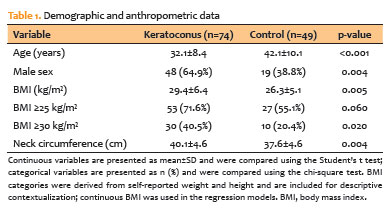
RESULTS: Participants with keratoconus had a higher BMI (29.4±6.4 vs. 26.3±5.1 kg/m²; p=0.005), larger neck circumference (40.1±4.6 vs. 37.6±4.6 cm; p=0.004), more frequent eye rubbing (71.6% vs. 51.0%; p=0.020), and more frequent ocular compression (43.2% vs. 20.4%; p=0.009) than controls. High-risk classifications did not differ significantly between groups according to either the Berlin Questionnaire (p=0.206) or STOP-BANG (p=0.139). In exploratory models adjusted for age and sex, positive associations were observed for the Berlin Questionnaire (OR, 2.58; 95% CI, 0.91-7.28) and STOP-BANG (OR, 3.30; 95% CI, 0.83-13.05). In the primary model, younger age (OR, 0.88; 95% CI, 0.83-0.93), male sex (OR, 2.91; 95% CI, 1.14-7.42), and higher BMI (OR, 1.15; 95% CI, 1.05-1.27) remained associated with keratoconus, whereas the estimates for behavioral factors were attenuated. No association with disease severity was detected.
CONCLUSIONS: Participants with keratoconus had higher anthropometric measurements and more frequent mechanical behaviors; however, the prevalence of high-risk obstructive sleep apnea classifications did not differ significantly between groups. Questionnaire-based and anthropometric findings are not independent because BMI contributes to both screening instruments, whereas neck circumference contributes to the STOP-BANG score. Ocular compression was assessed using a nonvalidated self-report measure rather than objective pressure measurements. Severity analyses were underpowered, and causality cannot be inferred from the study design.
Keywords: Keratoconus; Sleep apnea, obstructive; Risk factors; Body mass index; Sleep; Logistic models; Patient acuity; Tomography; Surveys and questionnaires
Arq. Bras. Oftalmol. 2026;89 (6 )
:1-9
| DOI: 10.5935/0004-2749.2026-0078
Abstract
PURPOSE: To compare standard single-step transepithelial photorefractive keratectomy, planned using a population-derived epithelial thickness profile, with MS-39 epithelial thickness map-guided single-step transepithelial photorefractive keratectomy in eyes with regular corneas, with a focus on early epithelial healing and short-term visual and refractive outcomes.
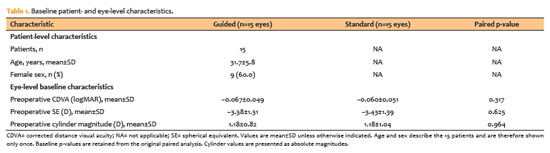
METHODS: This prospective, randomized, patient-masked, contralateral-eye clinical trial included 15 patients (30 eyes) with stable myopia, with or without regular astigmatism. Each patient underwent standard transepithelial photorefractive keratectomy in one eye and MS-39 epithelial map-guided transepithelial photorefractive keratectomy in the fellow eye using the same excimer laser platform and single-step transepithelial workflow. Early postoperative outcomes included pain scores and epithelial-defect area on postoperative day 2 and complete reepithelialization on day 7. Visual and refractive outcomes included uncorrected distance visual acuity at 30 and 90 days, corrected distance visual acuity at 90 days, residual spherical equivalent, and residual cylinder magnitude at 90 days. Paired analyses were performed.
RESULTS: All patients completed the 90-day follow-up. Early postoperative recovery did not differ significantly between groups. On day 2, the mean pain score was 2.33±0.82 in guided eyes and 2.40±0.63 in standard eyes, whereas the mean epithelial-defect area was 3.75±5.34 and 2.78±2.86 mm2, respectively. Complete reepithelialization by ay 7 occurred in all eyes. At 90 days, mean uncorrected distance visual acuity was −0.080±0.056 logMAR in guided eyes and −0.073±0.046 logMAR in standard eyes (p=0.564). Mean residual spherical equivalent was −0.03±0.35 D and 0.00±0.41 D, respectively (p=0.831). A total of 93.3% of guided eyes and 86.7% of standard eyes were within ±0.50 D of plano, and all eyes were within ±1.00 D.
CONCLUSIONS: In this randomized contralateral-eye pilot study, MS-39 epithelial map-guided single-step transepithelial photorefractive keratectomy did not demonstrate a statistically detectable short-term clinical advantage over standard population-based planning in eyes with regular corneas. Because the study was not designed as an equivalence or noninferiority trial, these findings should not be interpreted as evidence of equivalence.
Keywords: Photorefractive keratectomy/methods; Cornea/physiopathology; Corneal epithelium/surgery; Myopia/surgery; Astigmatism/surgery; Corneal topography; Refraction, ocular/physiology
Arq. Bras. Oftalmol. 2024;87 (4 )
:1-6
| DOI: 10.5935/0004-2749.2022-0035
Abstract
Objetivo: Comparar as diferenças entre a chord aparente µ e o chord real µ.
Métodos: Estudo prospectivo, comparativo, não randomizado e não intervencionista. Os exames de imagem (Pentacam e HD Analyzer) foram realizados na mesma sala e nas mesmas condições escotópicas. Os critérios de inclusão foram idade de 21 a 71 anos; compreensão do termo de consentimento; miopia até 4D e astigmatismo topográfico anterior até 1D. Os critérios de exclusão foram usuários de lentes de contato; pacientes com doenças oculares prévias ou cirurgias; opacidades da córnea; a presença de alterações tomográficas da córnea ou suspeita de ceratocone.
Resultados: Em nosso estudo foram analisados 116 olhos de 58 pacientes. A média de idade foi de 30,69 anos (± 7,85). Análises de correlação foram desenvolvidas e o coeficiente de correlação de Pearson (0,647) indica uma relação linear positiva moderada entre as variáveis. A média do chord µ real foi 226,21± 128,53 µm e a média do chord µ média foi 278,66 ± 123,90 µm, com diferença média de 52,45 µm (p=0,01).
A análise do diâmetro pupilar médio apresentou: 5,76mm no HD Analyzer e 3,31mm no Pentacam.
Conclusões: Entendemos a existência de uma diferença significativa entre os métodos e assim a medida de ambos os dispositivos com base em princípios diferentes devemos respeitar suas peculiaridades. Como encontramos correlação entre as duas medidas, acreditamos que ambas podem ser utilizadas na prática diária.
Keywords: Imagem óptica; Percepção visual; Pupila; Segmento anterior do olho; Córnea; Técnicas de diagnóstico oftalmológico
Arq. Bras. Oftalmol. 2026;89 (5 )
:1-8
| DOI: 10.5935/0004-2749.2025-0385
Abstract
PURPOSE:To investigate tomographic and biomechanical screening artifacts associated with primary pterygium that may mimic keratoconus-like patterns before and after surgical excision.
METHODS: Twenty-five eyes with primary pterygium underwent Scheimpflug corneal tomography and dynamic corneal response analysis before surgery and 3 months after excision. Tomographic and biomechanical (parameters were compared using paired analyses. The proportions of eyes exceeding established screening thresholds (Tomographic Biomechanical Index ≥0.29; Corvis Biomechanical Index ≥0.5) were compared using McNemar’s test.
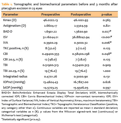
RESULTS: BAD-D decreased significantly after surgery (1.69±1.22 vs. 1.36±0.90; Wilcoxon, p=0.027). Pentacam irregularity indices also improved significantly, including ISV (51.16±44.21 vs. 36.88±42.94; p<0.001) and IVA (0.39±0.29 vs. 0.30±0.32; p=0.006). No significant changes were observed in maximum keratometry, corneal astigmatism. The mean CBI decreased significantly (0.494±0.353 vs. 0.384±0.298; p=0.048), and the proportion of CBI-positive eyes (≥0.5) declined from 48% (12/25) to 28% (7/25). In contrast, the mean tomographic Biomechanical Index did not change remained unchanged (0.519±0.313 vs. 0.534±0.323; p=0.909), and the proportion of Tomographic Biomechanical Index-positive eyes (≥0.29) remained 76% (19/25). The number of topographic keratoconus classification positive eyes decreased from 8 (32%) to 3 (12%). Integrated Radius showed no significant postoperative change (p=0.131).
CONCLUSIONS: Primary pterygium was associated with tomographic and biomechanical screening abnormalities that mimicked keratoconus-like patterns. Following surgical excision, anterior tomographic parameters improved, and the proportion of Corvis biomechanical index-positive eyes decreased. In contrast, Tomographic Biomechanical Index remained elevated in most eyes, and stiffness-related biomechanical parameters showed no significant change. These findings demonstrate a differential postoperative response among screening indices and suggest that pterygium-induced anterior corneal surface distortion may increase false-positive keratoconus screening rates, particularly for Tomographic Biomechanical Index, which may remain elevated even after lesion removal.
Keywords: Pterygium; Cornea; Corneal topography; Biomechanical phenomena; Tomography; Keratoconus; Refractive surgical procedures


 ABO is licensed under a Creative Commons Attribution-NonComercial 4.0 Internacional.