Arq. Bras. Oftalmol. 2015;78 (6 )
:356-358
| DOI: 10.5935/0004-2749.20150094
Abstract
Objetivo: Analisar o uso do PCR em tempo real (qPCR) na detecção do DNA do T. gondii no sangue periférico e no humor aquoso de pacientes com lesões de retinocoroidite focal, ativa por toxoplasmose. Métodos: Cinquenta e cinco pacientes com uveite infecciosa foram incluídos neste estudo. Os pacientes foram atendidos entre 2009 a 2013, no Departamento de Oftalmologia e Ciências Visuais da Universidade Federal de São Paulo. Quarenta e três pacientes tiveram o diagnóstico de lesões de retinocoroidite focal, ativa por toxoplasmose e, os outros 12 tiveram o diagnóstico de uveíte infecciosa não toxoplásmica e, por isso foram usados como grupo controle. A técnica de qPCR foi utilizada na detecção de DNA do T. gondii em amostras de sangue periférico e humor aquoso. Resultados: O qPCR foi positivo para o DNA do T. gondii em 37,21% (16/43) das amostras de humor aquoso, 2,33% (1/43) nas amostras de sangue periférico e, 16,27% (7/43) em ambas amostras simultaneamente. Conclusão: O qPCR foi capaz de detectar o DNA do T. gondii em pacientes com lesões de retinocoroidite focal, ativa por Toxoplasmose, no sangue bem como, no humor aquoso, podendo ajudar no diagnostico.
Keywords: Toxoplasmose ocular/diagnóstico; Toxoplasma; Sangue/parasitologia; Coriodite; Reação em cadeia da polimerase em tempo real; Humor aquoso
Arq. Bras. Oftalmol. 2017;80 (2 )
:84-87
| DOI: 10.5935/0004-2749.20170021
Abstract
Objetivo: Ceratites bacterianas ocorrem mundialmente e apesar dos novos desenvolvimentos permanece como uma condição que pode levar à cegueira. Avaliar a presença de herpes simples (-1 e -2) e vírus varicella zoster (VZV) por reação em cadeia quantitativa de polimerase em tempo real (qPCR) em raspados corneanos de pacientes com ceratite bacteriana. Métodos: Sessenta e cinco pacientes com ceratite infecciosa foram submetidos a raspados corneanos estudados para gram, Giemsa, cultura e qPCR (grupo de estudo). Foram avaliados fatores de risco e epidemiológicos. O grupo controle foi composto por 25 casos de úlcera dendrítica típica por herpes analisados por qPCR. Resultados: Do grupo de estudo (n=65), nove pacientes (13,8%) apresentaram cultura, qPCR e raspado negativos. Cinquenta e seis (86,2%) pacientes apresentaram cultura positiva, 51 para bacteria, 4 para fungo e 1 para ameba. A qPCR identificou 10 pacientes do grupo de cultura positiva para bactéria que também foram positivos para vírus, um VZV e 9 para HSV-1. Dos 25 pacientes que compunham o grupo controle, 21 apresentaram qPCR positivo para HSV-1. Conclusão: Herpes pode estar presente em pacientes com úlceras de córnea bacterianas e a qPCR pode ser útil na sua detecção.
Keywords: Herpes simples; Infecções por herpesviridae; Ceratite herpética; Reação em cadeia da polimerase
Arq. Bras. Oftalmol. 2022;85 (2 )
:158-165
| DOI: 10.5935/0004-2749.20220071
Abstract
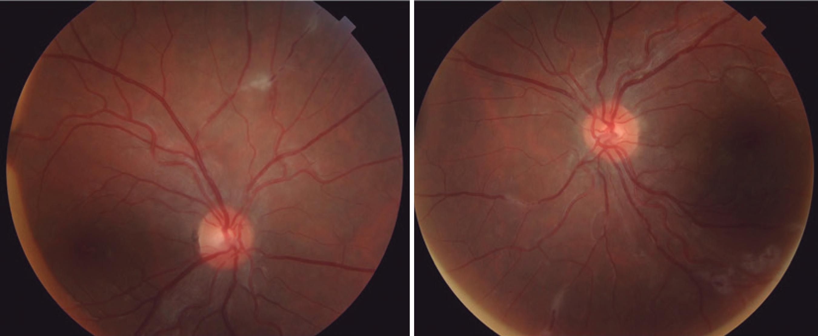
OBJETIVOS: o principal objetivo deste estudo foi descrever pacientes com achados vasculares retinianos temporalmente relacionados à vacinação contra COVID-19. Com maior notificação de possíveis eventos adversos similares, esperamos compreender a real dimensão e relevância do que foi apresentado.
MÉTODOS: Onze pacientes com queixas visuais após vacinação contra COVID-19 foram estudados. Os dados analisados foram: idade, gênero, tipo de vacinação, tempo de aparecimento de sintomas, achados sistêmicos, antecedentes pessoais, acuidade visual com melhor correção, biomicroscopia e imagem retiniana multimodal (retinografia colorida, red-free, SD-OCT, OCTA e angiofluoresceinografia). Os critérios de inclusão foram a presença de alterações oftalmológicas ocorridas dentro de 30 dias após a primeira ou segunda dose de qualquer vacina contra COVID-19.
RESULTADOS: Onze pacientes foram incluídos: 5 com oclusão arterial (45,4%), 4 com oclusão venosa (36,4%) e 2 (18,2%) com alterações não específicas vasculares sugestivas de isquemia retiniana como exsudatos algodonosos. A idade média dos pacientes foi de 57 anos (DP=16; com intervalo de 27 a 84 anos). A média de tempo de aparecimento de sintomas após a vacinação foi de 10 dias (DP=5,4; com intervalo de 3 a 16 dias). Nove dos onze pacientes eram do sexo feminino (81,8%). Fatores de risco sistêmicos foram observados em 36,4% dos pacientes. Dois pacientes tiveram sintomas neurológicos e visuais, com oclusão arterial. 36,4% dos pacientes tiveram infecção prévia por COVID-19 no último ano. Sete pacientes (63,6%) receberam a vacina ChAdOx1 nCoV-19 (AZD1222).
CONCLUSÕES: nossos dados sugerem que eventos retinianos temporalmente relacionados à vacinação contra COVID-19 são possíveis, porém raros. A relação entre estes eventos pós-vacinais exigem futura atenção antes de maiores conclusões.
Keywords: COVID-19; Infecções por coronavírus; Vacina; Oclusão arterial; Oclusão venosa; Síndrome de Susac
Arq. Bras. Oftalmol. 2025;88 (1 )
:1-63
| DOI: 10.5935/0004-2749.2023-0037
Abstract
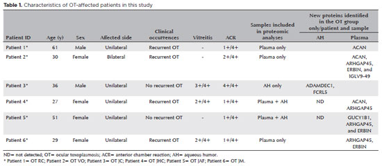
PURPOSE: To characterize the extracellular vesicle protein cargo in the aqueous humor and plasma of patients with ocular toxoplasmosis.
METHODS: Aqueous humor and plasma were collected from six patients with active ocular toxoplasmosis and six patients with cataract. Extracellular vesicles were isolated, and western blotting and mass spectrometry were performed for protein analysis.
RESULTS: All plasma samples from patients with ocular toxoplasmosis and cataract were positive for the tetraspanins CD63 and TSG101. However, the aqueous humor from patients with ocular toxoplasmosis was positive only for CD63. Sixty-seven new unreported proteins were identified in the aqueous humor and plasma of patients with the ocular toxoplasmosis and cataract. Of the 67 proteins, 10 and 7 were found only in the cataract and ocular toxoplasmosis groups, respectively. In general, these proteins were involved in immune system activation and retina homeostasis and were related to infections and retina-associated diseases. Conclusion: The distinct protein signatures between ocular toxoplasmosis and cataract may be helpful in the differential diagnosis of ocular toxoplasmosis. However, more studies are needed to better understand the role of these proteins in the pathogenesis of ocular toxoplasmosis.
Keywords: Extracellular vesicles; Proteomics; Toxoplasma gondii; Ocular toxoplasmosis, Aqueous humor; Plasma; Liquid biopsy
Arq. Bras. Oftalmol. 2025;88 (3 )
:1-7
| DOI: 10.5935/0004-2749.2023-0309
Abstract
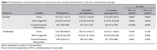
PURPOSE: Keratoconus presents certain peculiarities in pediatric patients when compared with adults. The greatest challenge in children is that the disease is more severe and faster in progression. In this retrospective study, we aimed to compare the accelerated and Dresden protocols for corneal crosslinking in patients aged <18 years who were followed-up for at least 12 months.
METHODS: A total of 36 eyes from 27 patients were included in the study. The best corrected and uncorrected visual acuity, maximal keratometry, corneal thickness, foveal thickness, and endothelial microscopy findings were evaluated at baseline and during the postoperative period at one, three, and six months. Thereafter, the patients were evaluated at one, three, six and twelve months postoperative. Corneal crosslinking was performed in all patients via the Dresden protocol (n=21 eyes) or the accelerated protocol (n=15 eyes). Data between the two groups were compared and XY statistical analysis was used.
RESULTS: Both protocols were effective in halting keratoconus progression. No patient had progression at the 12-month follow-up. A significant reduction in Kmax and improvement in the corrected distance visual acuity were observed only in the Dresden protocol group. Although the Dresden protocol was superior to the accelerated protocol in reducing Kmax (p=0.002), there was no significant difference in corrected distance visual acuity between the two groups.
CONCLUSION: The accelerated protocol is as efficient as the Dresden protocol in stabilizing keratoconus progression. Although the Dresden protocol was superior to the accelerated protocol in reducing the Kmax, it did not produce better clinical results. Thus, the accelerated protocol is an efficient option. Furthermore, considering the advantages of reduced surgical time, the accelerated protocol is effective in halting keratoconus progression in the pediatric age group.
Keywords: Keratoconus; Corneal diseases; Ultraviolet rays; Cross-linking reagents; Visual acuity
Arq. Bras. Oftalmol. 2024;87 (3 )
:1-7
| DOI: 10.5935/0004-2749.2022-0374
Abstract
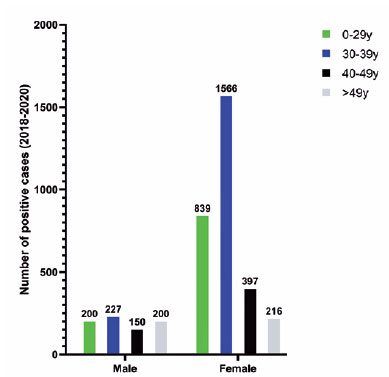
PURPOSE: To describe a 2019 acute toxoplasmosis outbreak in the city of São Paulo, Brazil, and to evaluate the laboratory serological profile for toxoplasmosis for three consecutive years. The ophthalmological manifestations of the patients involved in the outbreak were also studied.
METHODS: A cross-sectional descriptive study of a toxoplasmosis outbreak in São Paulo, Brazil, between February and May 2019. Epidemiological data were described, as were the observed ocular manifestations. As part of this study the number of patients with positive IgM toxoplasmosis serology was obtained from a large laboratory network (DASA) for three consecutive years, including the year of the outbreak (2018, 2019, 2020).
RESULTS: Eighty-three individuals were identified in the outbreak and two clusters were studied. The clinical picture of at least 77% of the patients, the epidemiological analysis, and the short incubation period (5-8 days) suggested contamination by oocysts. Serological laboratory data analysis revealed an increase of positive toxoplasmosis IgM in 2019 of 73% compared to the previous year. Ophthalmological examination revealed that at least 4.8% of the patients developed toxoplasmic retinochoroiditis, none of whom had been treated during the acute systemic disease.
CONCLUSION: Our findings indicate vegetable contamination as the possible source of this outbreak, a high prevalence of toxoplasmosis in São Paulo during the outbreak period, and a drop in the number of tests during the COVID-19 pandemic. Retinochoroiditis was observed in at least 4.8% of the cases. We confirm the need to implement effective means for the prevention, diagnosis, and treatment of the disease. This may involve raising awareness among the population of the importance of vegetable hygiene, and improved quality control of food and water.
Keywords: Toxoplasmosis/etiology; Food parasitology; Water/parasitology; Uveitis, posterior/parasitology; Chorioretinitis/parasitology; Visual acuity; Disease outbreaks; Eye manifestations; Humans.
 ABO is licensed under a Creative Commons Attribution-NonComercial 4.0 Internacional.


16-fig01tb.jpg)
15-fig01tb.jpg)
