Arq. Bras. Oftalmol. 2025;88 (6 )
:1-5
| DOI: 10.5935/0004-2749.2025-0085
Abstract
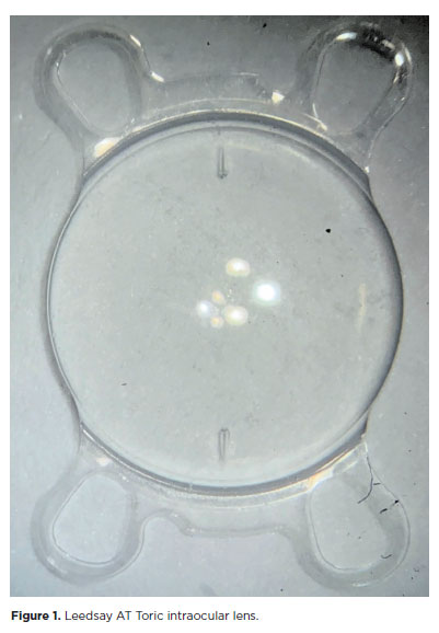
PURPOSE: The purpose of this study was to assess visual outcomes and patient satisfaction following cataract surgery involving the implantation of quad-loop intraocular lenses, including trifocal, bifocal, and toric variants.
METHODS: Information was obtained from both physical and electronic medical records of patients who underwent phacoemulsification cataract surgery with implantation of different intraocular lenses between January 1, 2022, and December 31, 2023. The study included individuals aged over 18 who received bilateral implantation of bifocal, trifocal, or monofocal toric intraocular lenses. Visual acuity was assessed at various postoperative time points using the logMAR scale. Quantitative variables were analyzed using mean and standard deviation.
RESULTS: A total of 92 eyes received premium intraocular lenses: 4 bifocal, 32 trifocal, 52 toric monofocal, and 4 trifocal toric lenses. The average preoperative corrected visual acuity was logMAR 0.478 ± 0.259. On the first postoperative day, the average uncorrected visual acuity was logMAR 0.301 ± 0.207. By day 30, 67.4% of eyes achieved uncorrected distance visual acuity of logMAR 0.2 or better. Patient satisfaction was high, with few reports of glare or halos.
CONCLUSION: Quad-loop intraocular lenses-including trifocal, bifocal, and toric models-demonstrated effective improvement in visual acuity and high levels of patient satisfaction. These lenses represent a suitable option for enhancing visual outcomes after cataract surgery. Additional studies with larger cohorts are recommended to confirm these results.
Keywords: Cataract extraction; Aberrometry/methods; Lenses, intraocular; Lens implantation, intraocular; Prosthesis design
Arq. Bras. Oftalmol. 2024;87 (4 )
:1-7
| DOI: 10.5935/0004-2749.2023-0026
Abstract
PURPOSE: To describe cellular alterations detected by impression cytology of the ocular surface in patients with xeroderma pigmentosum. The secondary objective was to assess the reliability of impression cytology in diagnosing ocular surface squamous neoplasia.
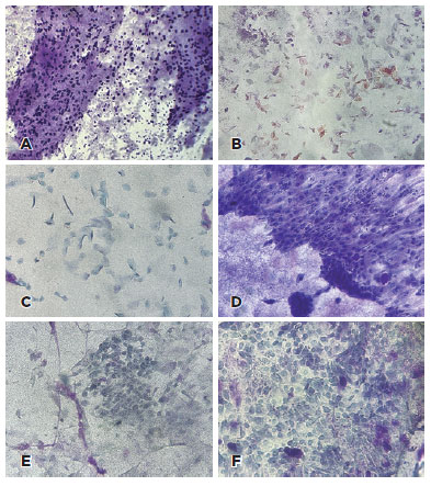
METHODS: Patients with xeroderma pigmentosum underwent a single-day complete ophthalmological examination and impression cytology for ocular surface evaluation using 13 mm diameter mixed cellulose esters membrane filters and combined staining with Periodic Acid Schiff, Hematoxylin and Eosin, and Papanicolaou stains followed by microscopic analysis. The cytological findings were correlated with the clinical diagnosis. The impression cytology findings at baseline and one-year follow-up were correlated with the clinical course (no tumor, treated tumor, residual tumor recurrent tumor, new tumor).
RESULTS: Of the 42 patients examined, impression cytology was performed in 62 eyes of 34 participants (65% females). The mean age of patients was 29.6 ± 17 years (range 7-62). Fifteen eyes had a clinical diagnosis of ocular surface squamous neoplasia. Impression cytology showed goblet cells (47, 75%), inflammatory cells (12, 19%), keratinization (5, 8%), and squamous metaplasia (30, 48%). Impression cytology was positive for atypical cells in 18 patients (12 with and 6 without ocular surface squamous neoplasia). The sensitivity, specificity, positive predictive value, and negative predictive value of impression cytology (at baseline) for diagnosis of ocular surface squamous neoplasia were 80%, 87%, 67%, and 93%, respectively, using clinical diagnosis of ocular surface squamous neoplasia as the reference standard.
CONCLUSION: Impression cytology has a moderate positive predictive value for the diagnosis of ocular surface squamous neoplasia in patients with xeroderma pigmentosum. However, the lack of detection of atypical cells on impression cytology has a high negative predictive value for ocular surface squamous neoplasia. Integration of impression cytology in the long-term management of high-risk patients, such as patients with xeroderma pigmentosum, can avoid unnecessary diagnostic biopsies.
Keywords: Xeroderma pigmentosum; Eye neoplasms; Conjunctiva/cytology; Cornea/cytology; Cytological techniques
Arq. Bras. Oftalmol. 2024;87 (2 )
:1-8
| DOI: 10.5935/0004-2749.2022-0319
Abstract
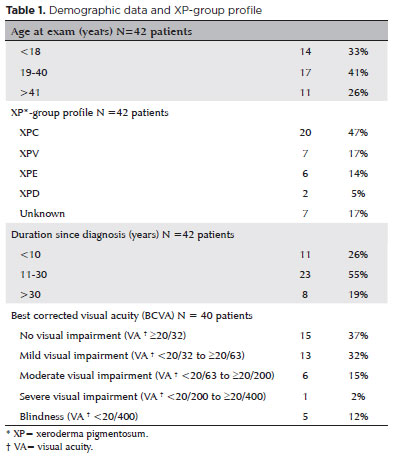
To assess Meibomian gland dysfunction using meibography in patients with xeroderma pigmentosum and correlate with ocular surface changes. This cross-sectional study evaluated patients with xeroderma pigmentosum. All patients underwent a comprehensive and standardized interview. The best-corrected visual acuity of each eye was determined. Detailed ophthalmic examination was conducted, including biomicroscopy examination of the ocular surface, Schirmer test type I, and meibography, and fundus examination was also performed when possible. Meibomian gland dysfunction was assessed by non-contact meibography using Oculus Keratograph® 5M (OCULUS Inc., Arlington, WA, USA). Saliva samples were collected using the Oragene DNA Self-collection kit (DNA Genotek Inc., Ottawa, Canada), and DNA was extracted as recommended by the manufacturer. Factors associated with abnormal meiboscores were assessed using generalized estimating equation models. A total of 42 participants were enrolled, and 27 patients underwent meibography. The meiboscore was abnormal in the upper eyelid in 8 (29.6%) patients and in the lower eyelid in 17 (62.9%). The likelihood of having abnormal meiboscores in the lower eyelid was 16.3 times greater than that in the upper eyelid.In the final multivariate model, age (p=0.001), mutation profile (p=0.006), and presence of ocular surface malignant tumor (OSMT) (p=0.014) remained significant for abnormal meiboscores. For a 1-year increase in age, the likelihood of abnormal meiboscores increased by 12%. Eyes with OSMT were 58.8 times more likely to have abnormal meiboscores than eyes without ocular surface malignant tumor.In the final model, age, xeroderma pigmentosum profile, previous cancer, and clinical alterations on the eyelid correlated with a meiboscore of ≥2.Meibomian gland dysfunction was common in patients with xeroderma pigmentosum, mainly in the lower eyelid. The severity of Meibomian gland dysfunction increases with age and is associated with severe eyelid changes.
Keywords: Meibomian glands/pathology; Meibomian glands/ diagnostic imaging; Photography; Xeroderma pigmentosum; Eyelid diseases/diagnostic imaging; Dry eye syndromes; DNA repair; Humans; Case report
 ABO is licensed under a Creative Commons Attribution-NonComercial 4.0 Internacional.