

Jorge Meireles-Teixeira1; Harley E. A. Bicas2
DOI: 10.1590/S0004-27492005000300004
ABSTRACT
PURPOSE: To evaluate the feasibility of autologous extraocular muscle grafting as a type of muscle expansion surgery. METHODS: The left superior rectus muscle of twenty-nine rabbits was resected and this fragment was attached to the endpoint of the respective right superior rectus (test group). Thereafter, the superior rectus of the left eye was reattached to the sclera (control group). Both groups were examined during different postoperative periods in order to assess their outcomes. RESULTS: The presence of hyperemia was slightly more frequent in the grafted group. Secretion and muscle atrophy were negligible in both groups. Fibrosis was greater in grafted animals. These muscles were weaker than the control muscles, although the force required to split muscular parts was always greater than the physiological one. CONCLUSIONS: This surgical technique was reliable and useful if one intends to achieve muscle expansion without the intrinsic risks of dealing with heterologous/artificial materials.
Keywords: Esotropia; Oculomotor muscles; Transplantation; autologous; Ophthalmologic surgical procedures; Rabbits
RESUMO
OBJETIVO: Avaliar a viabilidade do uso de segmentos de músculos oculares extrínsecos como expansores de tendões musculares. MÉTODOS: Vinte e nove coelhos tiveram seu músculo reto superior esquerdo ressecado e o fragmento de cada um foi transplantado para o reto superior contralateral (grupo-teste). Então, o reto superior esquerdo foi reinserido na esclera (grupo-controle). Os animais foram então examinados em diversos períodos pós-operatórios, até os seus sacrifícios, para que se avaliasse o desenrolar dessa técnica cirúrgica. RESULTADOS: A hiperemia foi maior entre os testes. A secreção e a atrofia muscular foram mínimas nos dois grupos. Houve maior presença de fibrose no grupo-teste, mas não tão expressiva a ponto de inviabilizar os efeitos da cirurgia. Esses músculos também se romperam mais facilmente do que os do grupo-controle, porém, a força de rompimento foi sempre bem maior do que aquela presente numa contração muscular normal. CONCLUSÕES: A técnica de transplante autólogo homotópico de músculos oculares extrínsecos provou ser confiável e eficaz, para o alongamento muscular.
Descritores: Esotropia; Músculos oculomotores; Transplante autólogo; Procedimentos cirúrgicos oftalmológicos; Coelhos
ORIGINAL ARTICLES
Autologous grafting of extraocular muscles: experimental study in rabbits
Transplante autólogo de musculatura ocular extrínseca: estudo experimental em coelhos
Jorge Meireles-TeixeiraI; Harley E. A. BicasII
IDoutorando do setor de Estrabismo da Universidade Federal de São Paulo (UNIFESP). São Paulo (SP)
IIProfessor Titular do Departamento de Oftalmologia, Otorrinolaringologia e Cirurgia da Cabeça e Pescoço da Faculdade de Medicina de Ribeirão Preto da Universidade de São Paulo (USP). Ribeirão Preto (SP)
ABSTRACT
PURPOSE: To evaluate the feasibility of autologous extraocular muscle grafting as a type of muscle expansion surgery.
METHODS: The left superior rectus muscle of twenty-nine rabbits was resected and this fragment was attached to the endpoint of the respective right superior rectus (test group). Thereafter, the superior rectus of the left eye was reattached to the sclera (control group). Both groups were examined during different postoperative periods in order to assess their outcomes.
RESULTS: The presence of hyperemia was slightly more frequent in the grafted group. Secretion and muscle atrophy were negligible in both groups. Fibrosis was greater in grafted animals. These muscles were weaker than the control muscles, although the force required to split muscular parts was always greater than the physiological one.
CONCLUSIONS: This surgical technique was reliable and useful if one intends to achieve muscle expansion without the intrinsic risks of dealing with heterologous/artificial materials.
Keywords: Esotropia/surgery; Oculomotor muscles/transplantation; Transplantation, autologous, Ophthalmologic surgical procedures; Rabbits
RESUMO
OBJETIVO: Avaliar a viabilidade do uso de segmentos de músculos oculares extrínsecos como expansores de tendões musculares.
MÉTODOS: Vinte e nove coelhos tiveram seu músculo reto superior esquerdo ressecado e o fragmento de cada um foi transplantado para o reto superior contralateral (grupo-teste). Então, o reto superior esquerdo foi reinserido na esclera (grupo-controle). Os animais foram então examinados em diversos períodos pós-operatórios, até os seus sacrifícios, para que se avaliasse o desenrolar dessa técnica cirúrgica.
RESULTADOS: A hiperemia foi maior entre os testes. A secreção e a atrofia muscular foram mínimas nos dois grupos. Houve maior presença de fibrose no grupo-teste, mas não tão expressiva a ponto de inviabilizar os efeitos da cirurgia. Esses músculos também se romperam mais facilmente do que os do grupo-controle, porém, a força de rompimento foi sempre bem maior do que aquela presente numa contração muscular normal.
CONCLUSÕES: A técnica de transplante autólogo homotópico de músculos oculares extrínsecos provou ser confiável e eficaz, para o alongamento muscular.
Descritores: Esotropia/cirurgia, Músculos oculomotores/transplante, Transplante autólogo; Procedimentos cirúrgicos oftalmológicos; Coelhos
INTRODUCTION
The surgical treatment of a squint could be defined as a rearrangement of extraocular muscle sites aiming to realign the eyes. Currently, it could be achieved by recession-resection surgeries, which would equalize passive-active force balance(1).
According to Frank-Starling's law for skeletal striated muscles, the more stretched and stressed muscle fibers are, the greater will be their contraction forces, optimizing muscle work. Otherwise, a stretched muscle has a better response to neurological stimuli(2-4).
What actually happens in recession surgical technique is quite the contrary, once the muscle becomes slack. Thus, during a contraction, part of its force will not be converted to mechanical energy (torque). Instead, it will be lost, compensating the created slackness. That is the reason why recession weakens a muscle(5).
The limit to a recession is the equatorial plane or, in other words, the point where the muscle, for the first time, touches the sclera (tangential point), which is usually located 14 mm behind the limbus. Beyond this point, besides muscle weakening, the procedure would also generate translational movements added to the preexisting rotational one, damaging static and dynamic muscle balance. Thus, regardless whether one could achieve eye alignment in primary position, it would also create or enhance incommitancies in secondary and tertiary positions(5-7).
Topical anesthesia and adjustable sutures offer to the surgeon the chance of trying an exaggerated recession and, if it does not fit well, to reduce it. Another way would be to lengthen the muscle instead of doing an exaggerated recession. It could be done by hang-loose sutures (but it does not really expand the muscle once its endpoint would attach to sclera) or using synthetic lengtheners (and deal with the risks of fibrosis and extrusion). To solve this problem the surgeon could use human tissue, especially an autologous one(8-13).
Recently, there have been some papers in Cardiology that refer to the use of autologous tissue to restore damaged infarcted cardiac muscle and their results are promising. In Ophthalmology there are three reports of autologous muscle transplantation aiming at lengthening extraocular muscles. Only in one of them no good results were achieved due to excessive fibrosis. But all studies presented a small number of samples(14-19).
This study aims to analyze the autologous transplantation lengthening technique with a larger sample and using absorbable sutures (Vicryl® 6-0).
METHODS
Twenty-nine rabbits were operated on. A fragment of 6.0 mm was resected from the left superior rectus muscle (SR) and was attached to the endpoint of the right SR. Thereafter, a resection procedure was performed at the left SR (control) and an autologous transplantation surgery was performed on the right SR (test).
During the postoperative (PO) follow-up, hyperemia and secretion were assessed in all 29 rabbits. At the end, fifteen animals were sacrificed on the 45th PO day and underwent mechanical evaluation. The other fourteen remaining rabbits were used for the anatomicopathological examination (4 animals on the 45th PO, 5 on the 60th PO and 5 on the 90th PO day).
After sacrifice, a Stevenson's hook was slid between muscle and sclera in order to assess fibrosis and adhesions.
• Mechanical evaluation: first, the width of muscle (or graft) belly was measured (mm) with a caliper and then, the muscle (or graft) was grabbed with a Stevenson's hook connected to a dynamometer. The muscle/graft was stretched until its disinsertion, rupture or globe luxation. Thereafter the force required to induce any of these phenomena was noted in kilogram-force (kgf).
• Anatomicopathological evaluation: two sections were made of each eye and they were stained with hematoxilin-eosin and Masson's method to microscopically, analyze fibrosis in both groups.
RESULTS
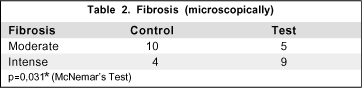
The distribution of hyperemia and secretion was similar between the groups without statistical significance. However fibrosis, as expected, was greater in the test-group in which there was a more intense tissue manipulation (only data of low or intense fibrosis were considered for comparison, the moderate was discharged). Microscopically analyzed fibrosis showed to be greater in the test-group (Tables 1 and 2).

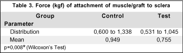
The force of attachment of muscle (or graft) to the sclera was greater in the control-group (Table 3) and there was no correlation between the force required to detach/rupture the muscle and its width, as shown by Spearman's correlation.
• Control-group: p=0,07
• Test-group: p=0,43
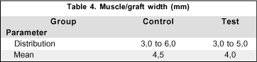
This result was also expected once atrophy was not important and the widths of muscle bellies were similar between the groups (Table 4).
DISCUSSION
Once the same rabbit had one eye used as test and the adelpho used as control, this protocol minimized individual factors that could create bias in the analysis of variables.
Observing data on hyperemia and secretion, one can note that both surgical techniques induced a similar inflammatory reaction. Fibrosis, however, detected either macroscopically or microscopically, was statistically greater in the test-group, due to a more intense surgical trauma. Nevertheless, atrophy was negligible in both groups, even on microscopic evaluation.
One similar study reported total or almost the total change to fibrosis in normal grafted muscle tissue to fibrosis as time passes(17). This event, or at least a tendency to it, was not noted in this research.
Regarding force of attachment to sclera, it was stronger and statistically significant in the control-group. But it should be pointed out that the force of traction supported by muscles (control or test), just before they had been ruptured or detached from the sclera, was much greater than that commonly used during physiological ocular rotations. Hence, clinically, this "weakness" of grafted muscles would not be significant in human beings.
CONCLUSIONS
1- Postoperative outcome (hyperemia and secretion) was similar in both groups even with more manipulation during transplantation surgery.
2- Fibrosis, as expected, was more intense in test animals.
3- Transplantation technique did not trigger atrophy of grafted muscles during a follow-up of 90 days.
4- Undoubtedly, the grafted fragment functioned as a lengthener.
5- Resistance to traction was weaker among test muscles, however it was clinically negligible.
ACKNOWLEDGMENT
To Drs. Maria Cristina Martins and João Pessoa Filho, for their valuable contribution to the anatomicopathological analysis of the specimens.
REFERENCES
1. Lucci L, Goldchmidt M, Souza-Dias CR. Influência dos chamados fatores de supercorreção sobre o resultado da cirurgia para correção de esotropia de grande ângulo. Arq Bras Oftalmol. 1997;60(3):286-9.
2. Guyton AC. Músculo cardíaco; o coração como bomba. In: Guyton AC. Tratado de fisiologia médica. 7ª ed. Rio de Janeiro: Guanabara Koogan; 1989. 129p.
3. Bicas HEA. Consideraciones sobre los factores mecánicos en la acción de los músculos oculares. Acta Estrabologica. 1996;25:161-78.
4. Bicas HEA. Interpretação dos mecanismos de ação dos procedimentos cirúrgicos em estrabismo. In: XII Congreso Del Consejo Latinoamericano de Estrabismo; 1996 mayo; Buenos Aires; 1996. p.297-306.
5. Von Noorden GK. Physiology of the ocular movements. In: Von Noorden GK. Binocular vision and ocular motility: theory and management of strabismus. 5th ed. Missouri: Mosby; 1996. p.53-68.
6. Prieto-Díaz J, Souza-Dias C. Eye motility. In: Prieto-Díaz J, Souza-Dias C. Strabismus. 4th ed. Boston: Butterworth-Heinemann; 2000. p.72-82.
7. Park RS, Park GE. The center of ocular rotation in the horizontal plane. Am J Physiol. 1933;104:545-52.
8. Repka MX, Fishman PJ, Guyton DL. Site of reattachment of the extraocular muscle following hang-back recession. J Ped Ophthal Strabismus. 1990;27(6): 286-90.
9. Reis PPL, Almeida HC. Posicionamento tardio dos retos horizontais, retrocedidos pela técnica da sutura ajustável. Estudo com marcador radiopaco em olhos humanos. Rev Bras Oftalmol. 1996;55(2):25-30.
10. Aggarwal RK, Willshaw HE, Townsend P. New materials for rectus muscle tendon extension in strabismus surgery. Eye. 1993;7(Pt 1):40-2.
11. Dunlap EA. Plastic implants in muscle surgery; plastic materials in the management of extraocular motility restrictions. Arch Ophthalmol. 1968;80:249-57.
12. Morales AG, Polack FM, Arata AF. Silicone implant to extra-ocular muscles. Br J Ophthalmol. 1966;50(5):235-44.
13. Meireles-Teixeira JA, Cunha RP, Mendonça TS. Resultados da correção cirúrgica de esotropias de grande ângulo, em portadores de baixa de acuidade visual unilateral. Arq Bras Oftalmol. 2000;63(5):365-8.
14. Sakai T, Li RK, Weisel RD, Mickle DA, Kim EJ, Tomita S, et al. Autologous heart cell transplantation improves cardiac function after myocardial injury. Ann Thorac Surg. 1999;68(6):2074-80, discussion 2080-1.
15. Menasché P, Hagège AA, Scorsin M, Pouzet B, Desnos M, Duboc D, et al. Myoblast transplantation for heart failure. Lancet 2001;357(9252):279-80.
16. Oakley RM, Ooi OC, Bongso A, Yacoub MH. Myocite transplantation for myocardial repair: a few good cells can mend a broken heart. Ann Thorac Surg. 2001;71(5):1724-33.
17. Hiatt RL. Extraocular muscle transplantation. Trans Am Ophthalmol Soc. 1973;71:426-58.
18. Diamond GR. True transposition procedures. J Ped Ophthal Strabismus. 1990;27(3):153-6.
19. Silva LH, Bicas HEA, Velasco e Cruz AA. Transplante autólogo homotópico de músculo ocular extrínseco. Rev Bras Oftalmol. 1984;43(3):79-84.
 Correspondence to
Jorge Meireles Teixeira
Av. dos Holandeses Nº 9, Q-24
São Luís (MA) CEP 65071-380
E-mail: [email protected]
Recebido para publicação em 20.07.2004
Versão revisada recebida em 03.01.2005
Aprovação em 17.01.2005
Trabalho desenvolvido, como tese de doutorado em Medicina, no Departamento de Oftalmologia da Universidade Federal de São Paulo (UNIFESP).
Nota Editorial: Após concluída a análise do artigo sob sigilo editorial e com a anuência da Dra. Keila Miriam Monteiro de Carvalho sobre a divulgação de seu nome como revisora dele, agradecemos sua participação neste processo.
© 2024 - All rights reserved - Conselho Brasileiro de Oftalmologia
![]()
 English PDF
 Print
Print
 Send this article by email
 How to cite this article
 Submit a comment
 Mendeley
 Scielo
 Pocket
