

Rosana Nogueira Pires da Cunha1; Mauro Campos1; Frederico Luis Dulley2; Bianca Rojas3; Charles Stephen Foster3
DOI: 10.1590/S0004-27492003000100004
ABSTRACT
PURPOSE: The primary ocular targets of chronic graft-versus-host disease (GVHD) are the lacrimal gland (LG) and the conjunctiva, and their involvement results in keratoconjunctivitis sicca (KCS). The purpose of the present study was to evaluate the frequency of signs and symptoms of KCS present in patients submitted to BMT, to identify the cellular phenotype of the conjunctival and lacrimal gland biopsies of these patients by immunohistochemistry and to correlate the findings with the presence of dry eye due to GVHD. METHODS: Forty-seven patients were clinically classified into two groups: Group I, with ocular GVHD, submitted to allogeneic BMT and Group II, without ocular GVHD, submitted to autologous and allogeneic BMT. Thorough eye examination, including clinical lacrimal function tests and biopsies of the conjunctiva and of the LG were performed in the pre- and posttransplantation period. The biopsies were submitted to immunohistochemical analysis using a panel of monoclonal antibodies. RESULTS: Of the 28 (82.4%) patients with chronic GVHD, 13 (46.4%) presented ocular GVHD. Of the six patients without GVHD, one (16.7%) presented ocular GVHD. None of those submitted to autologous BMT presented ocular GVHD and 14 (41.2%) of 34 patients with allogeneic BMT presented ocular GVHD. All patients with ocular GVHD (Group I) had symptoms and the most frequent were burning, foreign body sensation, blurred vision and dryness. The rose bengal test was one of the most sensitive in this study and slit lamp examination was very useful in the evaluation of corneal and conjunctival alterations, since these alterations were always present in patients with dry eye in our study. Neither symptoms and signs of dry eye nor significant immunologic reaction was observed in the conjunctiva and in the LG of patients without ocular GVHD (Group II). There was an increase in the T lymphocyte population, of T helper lymphocyte (Th/i) and T suppressor-cytotoxic lymphocyte (Ts/c) subpopulation in the conjunctiva and in the LG of patients with ocular GVHD after the transplantation. CONCLUSIONS: Patients submitted to allogeneic BMT may develop ocular GHVD characterized by KCS. The immunohistochemical study of the conjunctiva and lacrimal gland biopsies of these patients suggested that these tissues are the target of the T cell mediated immunological reaction.
Keywords: Immunohistochemistry; Conjunctiva; Lacrimal apparatus; Bone marrow transplantation; Graft vs host disease
RESUMO
OBJETIVOS: A glândula lacrimal e a conjuntiva são os tecidos oculares primariamente afetados pela doença enxerto-versus-hospedeiro (DEVH) crônica, e seu envolvimento clínico é caracterizado por ceratoconjuntivite seca (CCS). Os objetivos deste estudo foram os de avaliar a freqüência dos sintomas e sinais de CCS em pacientes submetidos ao transplante de medula óssea (TMO) e identificar a população celular por análise imuno-histoquímica de biópsias da conjuntiva e glândula lacrimal desses pacientes, correlacionando esses achados com o quadro clínico ocular. MÉTODOS: Quarenta e sete pacientes foram classificados em dois grupos: Grupo I, pacientes com DEVH ocular, submetidos ao TMO alogênico e Grupo II, pacientes sem DEVH ocular, submetidos ao TMO alogênico e autólogo. Exame ocular completo, incluindo testes clínicos da função lacrimal, e biópsias de conjuntiva e glândula lacrimal foram realizadas nos períodos pré e pós-transplante. Estudou-se a população celular, por meio de análise imuno-histoquímica das biópsias, utilizando um painel de anticorpos monoclonais. RESULTADOS: Dos 28 (82,4%) pacientes com DEVH crônica, 13 (46,4%) apresentaram DEVH ocular. Dos seis pacientes sem DEVH, um (16,7%) apresentou DEVH ocular. Nenhum paciente submetido a TMO autólogo apresentou DEVH ocular e 14 (41,2%) dos 34 pacientes com TMO alogênico desenvolveram DEVH ocular. Todos os pacientes com DEVH ocular (Grupo I) apresentaram sintomas e os mais freqüentes foram ardor, sensação de corpo estranho, visão borrada e secura ocular. O teste de rosa bengala foi um dos mais sensíveis e o exame biomicroscópico foi considerado muito útil na avaliação das alterações conjuntivais e corneanas, as quais estavam sempre presentes nos pacientes com DEVH ocular deste estudo. Na conjuntiva e na glândula lacrimal dos pacientes sem DEVH ocular não houve reação imunológica significante, concordando com os testes de função lacrimal. Houve aumento da população de linfócitos T, dos linfócitos T auxiliadores (Th/i) e linfócitos T supressores-citotóxicos (Ts/c) após o transplante na conjuntiva e glândula lacrimal de pacientes com DEVH ocular. CONCLUSÕES: Pacientes submetidos ao TMO alogênico podem desenvolver DEVH ocular, caracterizado por ceratoconjuntivite seca. O estudo imuno-histoquímico de biópsias da conjuntiva e glândula lacrimal desses pacientes sugere que esses tecidos são alvo de reação imunológica mediada pelos linfócitos T.
Descritores: Imuno-histoquímica; Conjuntiva; Glândula lacrimal; Transplante de medula óssea; Doença enxerto-hospedeiro
Dry eye evaluation and immunohistochemical study of the conjunctiva and lacrimal gland of patients submitted to bone marrow transplantation
Alterações oculares e imunohistoquímicas na conjuntiva e glândula lacrimal de pacientes submetidos ao transplante de medula óssea
Rosana Nogueira Pires da CunhaI, 1; Mauro CamposI, 1; Frederico Luis DulleyII, 2; Bianca RojasIII, 3; Charles Stephen FosterIII, 3
IDepartment of Ophthalmology, Federal University of São Paulo, UNIFESP
IIBone Marrow Transplantation Sector, Fundação Pró-sangue Hemocentro of São Paulo, São Paulo University, Medicine College
IIIImmunology and Uveitis Service, Department of Ophthalmology, Massachusetts Eye & Ear Infirmary, Harvard Medical School
ABSTRACT
PURPOSE: The primary ocular targets of chronic graft-versus-host disease (GVHD) are the lacrimal gland (LG) and the conjunctiva, and their involvement results in keratoconjunctivitis sicca (KCS). The purpose of the present study was to evaluate the frequency of signs and symptoms of KCS present in patients submitted to BMT, to identify the cellular phenotype of the conjunctival and lacrimal gland biopsies of these patients by immunohistochemistry and to correlate the findings with the presence of dry eye due to GVHD.
METHODS: Forty-seven patients were clinically classified into two groups: Group I, with ocular GVHD, submitted to allogeneic BMT and Group II, without ocular GVHD, submitted to autologous and allogeneic BMT. Thorough eye examination, including clinical lacrimal function tests and biopsies of the conjunctiva and of the LG were performed in the pre- and posttransplantation period. The biopsies were submitted to immunohistochemical analysis using a panel of monoclonal antibodies.
RESULTS: Of the 28 (82.4%) patients with chronic GVHD, 13 (46.4%) presented ocular GVHD. Of the six patients without GVHD, one (16.7%) presented ocular GVHD. None of those submitted to autologous BMT presented ocular GVHD and 14 (41.2%) of 34 patients with allogeneic BMT presented ocular GVHD. All patients with ocular GVHD (Group I) had symptoms and the most frequent were burning, foreign body sensation, blurred vision and dryness. The rose bengal test was one of the most sensitive in this study and slit lamp examination was very useful in the evaluation of corneal and conjunctival alterations, since these alterations were always present in patients with dry eye in our study. Neither symptoms and signs of dry eye nor significant immunologic reaction was observed in the conjunctiva and in the LG of patients without ocular GVHD (Group II). There was an increase in the T lymphocyte population, of T helper lymphocyte (Th/i) and T suppressor-cytotoxic lymphocyte (Ts/c) subpopulation in the conjunctiva and in the LG of patients with ocular GVHD after the transplantation.
CONCLUSIONS: Patients submitted to allogeneic BMT may develop ocular GHVD characterized by KCS. The immunohistochemical study of the conjunctiva and lacrimal gland biopsies of these patients suggested that these tissues are the target of the T cell mediated immunological reaction.
Keyword: Immunohistochemistry; Conjunctiva; Lacrimal apparatus; Bone marrow transplantation/adverse effects; Graft vs host disease
RESUMO
OBJETIVOS: A glândula lacrimal e a conjuntiva são os tecidos oculares primariamente afetados pela doença enxerto-versus-hospedeiro (DEVH) crônica, e seu envolvimento clínico é caracterizado por ceratoconjuntivite seca (CCS). Os objetivos deste estudo foram os de avaliar a freqüência dos sintomas e sinais de CCS em pacientes submetidos ao transplante de medula óssea (TMO) e identificar a população celular por análise imuno-histoquímica de biópsias da conjuntiva e glândula lacrimal desses pacientes, correlacionando esses achados com o quadro clínico ocular.
MÉTODOS: Quarenta e sete pacientes foram classificados em dois grupos: Grupo I, pacientes com DEVH ocular, submetidos ao TMO alogênico e Grupo II, pacientes sem DEVH ocular, submetidos ao TMO alogênico e autólogo. Exame ocular completo, incluindo testes clínicos da função lacrimal, e biópsias de conjuntiva e glândula lacrimal foram realizadas nos períodos pré e pós-transplante. Estudou-se a população celular, por meio de análise imuno-histoquímica das biópsias, utilizando um painel de anticorpos monoclonais.
RESULTADOS: Dos 28 (82,4%) pacientes com DEVH crônica, 13 (46,4%) apresentaram DEVH ocular. Dos seis pacientes sem DEVH, um (16,7%) apresentou DEVH ocular. Nenhum paciente submetido a TMO autólogo apresentou DEVH ocular e 14 (41,2%) dos 34 pacientes com TMO alogênico desenvolveram DEVH ocular. Todos os pacientes com DEVH ocular (Grupo I) apresentaram sintomas e os mais freqüentes foram ardor, sensação de corpo estranho, visão borrada e secura ocular. O teste de rosa bengala foi um dos mais sensíveis e o exame biomicroscópico foi considerado muito útil na avaliação das alterações conjuntivais e corneanas, as quais estavam sempre presentes nos pacientes com DEVH ocular deste estudo. Na conjuntiva e na glândula lacrimal dos pacientes sem DEVH ocular não houve reação imunológica significante, concordando com os testes de função lacrimal. Houve aumento da população de linfócitos T, dos linfócitos T auxiliadores (Th/i) e linfócitos T supressores-citotóxicos (Ts/c) após o transplante na conjuntiva e glândula lacrimal de pacientes com DEVH ocular.
CONCLUSÕES: Pacientes submetidos ao TMO alogênico podem desenvolver DEVH ocular, caracterizado por ceratoconjuntivite seca. O estudo imuno-histoquímico de biópsias da conjuntiva e glândula lacrimal desses pacientes sugere que esses tecidos são alvo de reação imunológica mediada pelos linfócitos T.
Descritores: Imuno-histoquímica; Conjuntiva; Glândula lacrimal; Transplante de medula óssea/efeitos adversos; Doença enxerto-hospedeiro
INTRODUCTION
Bone marrow transplantation (BMT) has been increasingly used in the treatment of severe hematological malignancies and other malignant diseases, aplastic anemia and immunodeficiencies. Patients either receive autologous marrow taken while in remission and stored for reinfusion at a later date, or matched donor marrow either from a relative or an unrelated donor. Prior to infusion of the marrow, the patients receive high-dose chemotherapy alone or in combination with total body irradiation (TBI) to eradicate all host malignant tissue and minimize rejection of the donor marrow(1).
Despite the overall success of this approach, a number of serious and potentially fatal complications may occur following transplantation. The major noninfective complication of allogeneic BMT is graft-versus-host disease (GVHD), in which the lymphocytes of the immunocompetent donor marrow mount an immunological attack on the now immunoincompetent host. This disease affects principally the skin, eyes and gastrointestinal tract and may be fatal(2). The clinicopathologic features resemble the naturally occurring autoimmune connective tissue disorders (Sjögren-like syndrome) and present considerable variability in the mode of onset, the involved organ systems and the rate of progression(3).
Two forms of GVHD have been described: the term acuteGVHD is used to describe a distinctive syndrome of dermatitis, hepatitis and enteritis developing within 100 days of allogeneic BMT. Chronic GVHD is a more pleiotropic syndrome that develops after day 100(4). Patients submitted to autologous BMT do not develop chronic GVHD.
Ocular involvement in GVHD presents as keratoconjunctivitis sicca (KCS) consisting of punctate keratitis, persistent corneal epithelial defects and sterile stromal ulceration(5). The prevalence of ocular GVHD in patients with chronic GVHD is described in the literature as being between 60%(6) and 81.8%(1). There is a correlation between poor prognosis in systemic GVHD and the occurrence of ocular GVHD(6).
Other causes of dry eye after BMT could be attributed to total body irradiation and ocular toxicity of chemotherapy(7). Histological studies in patients with chronic GVHD indicate similarities with Sjögren's syndrome: presence of inflammatory cells in the conjunctiva and cornea, as well as inflammatory and destructive ductile involvement in lacrimal glands(8).
The objectives of this study are:
1) To identify and evaluate the frequency of patients with ocular GVHD in bone marrow transplant recipients, their symptoms and signs and the value of lacrimal function clinical tests for diagnosis of dry eye in these patients.
2) To identify the cellular phenotype of conjunctival and lacrimal gland inflammatory infiltrates by immunohistochemistry in this population and correlate them with clinical findings.
The incidence of GVHD tends to increase with the use of BMT as an alternative to the treatment of neoplasms, and a reasonable number of younger patients, surviving the transplant and its complications could present dry eyes. We understand that the clinical and immunohistochemical ocular alterations are of great value not only for the characterization of lacrimal dysfunction but also for the understanding of the immunopathologic mechanisms of systemic GVHD.
METHODS
Sixty-five patients who underwent BMT between September 1994 and December 1995, were examined at the Department of Ophthalmology, Universidade Federal de São Paulo. Patients were referred to us by the Bone Marrow Transplantation Sector, Fundação Pró-Sangue Hemocentro of São Paulo.
The criteria for patient eligibility were age over 10 years, written consent to participate in the study, no previous ocular diseases and no acute conjunctival or corneal infection on clinical examination. Of the 65 patients fulfilling the inclusion criteria, 15 were subsequently excluded due to problems related to GVHD morbidity, such as hematological and infectious complications and 3 who refused further ocular biopsy. As a result of this selection, 47 patients were submitted to ocular examination, conjunctiva and lacrimal gland biopsies. In 19 patients it was possible to perform two biopsies of the conjunctiva and lacrimal gland, one before and the other after BMT. Patients were divided into groups, according to the presence or absence of ocular GVHD as follows:
Group I:patients with ocular GVHD submitted to allogeneic BMT;
Group II:patients without ocular GVHD submitted to autologous and allogeneic BMT.
All patients underwent a complete ocular examination regarding the presence of ocular GVHD. The tear film was evaluated using the rose bengal staining test, Schirmer test and the tear breakup time (BUT). The rose bengal staining was performed by instillation of one drop of 1% rose bengal (Ophthalmosâ, São Paulo). The intensity of the staining was estimated using a grading system(9). Each eye was divided into three zones and the amount of stain in each of them was recorded according to a scale of 0 to 3. In that way a maximum score of 9 was obtained. A value greater than 3 was considered a positive test. Schirmer test was made using strips from Alcon (Fortworth, TX) which were positioned at the inferior fornix after instillation of one drop of anesthetic 0.5% proxymetacaine (Anestalconâ - Alcon), in each eye during 5 minutes. Values lower than 4 mm of wetting were considered abnormal(10). BUT values were measured after the instillation of 0.25% sodium fluoresceine (Fluoresceinaâ, Frumtost). A test was considered positive when dry spots were seen at the slit lamp less than 10 seconds after blinking(11).
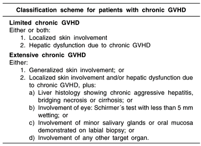
The presence of at least two positive tests associated with the presence of characteristic symptoms were considered diagnostic of keratoconjunctivitis sicca (KCS) and this was the criterion used to define ocular GVHD. The following symptoms were considered related to dry eye: burning, foreign body sensation, blurred vision, ocular pain, mucus discharge, tearing and photophobia. The following findings were considered signs of dry eye: typical changes on slit lamp examination, such as conjunctival hyperemia and punctate keratopathy, and positive results of the lacrimal function tests for dry eye described above.Hematologists from the Fundação Pró-Sangue of São Paulo made the diagnosis of GVHD according to the following criteria(3):
The distribution of the diseases was as follows: non-Hodgkin lymphoma: 8 patients; Hodgkin lymphoma: 2 patients; multiple myeloma: 2 patients; acute myelogenous leukemia: 5 patients; chronic myelogenous leukemia: 21 patients; severe aplastic anemia: 7 patients; breast carcinoma: 1 patient; testicle carcinoma: 1 patient. The conditioning regimen varied according to the type of disease, consisting of busulfan (Myleranâ - Wellcome), cyclophosphamide (Enduxanâ - Abbott) or melfalan (Alkeranâ - Wellcome).
For all diseases, patients undergoing allogeneic BMT received GVHD prophylaxis, consisting of cyclosporine A (Sandimunâ - Sandoz) from one day before BMT to 180 days or 40 weeks after BMT, depending on clinical manifestations of GVHD; methotrexate (Methotrexateâ- Lederle) on days 1, 3 and 6 after BMT and methylprednisolone (Solumedrolâ - Upjohn) or prednisone (Meticortenâ - Schering-Plough), from day 8 to day 72 after BMT depending on the clinical manifestations of GVHD in each patient.
Ocular examination, clinical diagnostic tests for dry eye and biopsies of conjunctiva and lacrimal gland were done in the pre- and posttransplant periods by the first author. The pretransplant procedures were performed from 30 to 10 days before BMT to avoid the effect of drugs used as conditioning regimen. The posttransplant examination and biopsies were performed after a minimum of 100 days after BMT, for examination during the chronic phase of GVHD.
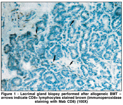
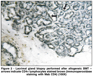
Biopsies were performed with topical 0.5% proxymetacaine anesthetic drops. Specimens of 4 mm were harvested from the superior conjunctiva adjacent to the limbus and from the lacrimal gland at its palpebral portion. Specimens were embedded in OCT compound ("Tissue-freezing Medium", Leica, Reichert & Jung Productsâ, Germany), snap-frozen in liquid nitrogen and stored at -70ºC until they were transported to the Hilles Immunology Laboratory at the Massachusetts Eye and Ear Infirmary, Boston, MA, where they were ultimately processed for immunostaining using the immunoperoxidase technique(12). The primary antibodies (Immunotech, Inc., Westbrook, ME) used were anti-CD3 (mature T cells), anti-CD4 (helper T cells, Th), anti-CD8 (supressor-cytotoxic T cells, Ts/c) and anti-CD 19 (B cells), all diluted 1:50.
Cells displaying a positive brown reaction on the cell surface were counted in three representative power field (x 450) readings with a 10 x 10 mm ocular grid, as shown in Figures 1 and 2. Cell counts were made in a masked fashion using an American Optical Microstar 110 Light microscope (Buffalo, NY) by two independent investigators.
The protocol of this study was approved by the UNIFESP Medical Ethics Committee.
Statistical Analysis - The signal test was used, whenever possible, for the variables determined by the immunohistochemistry examinations, looking for possible differences between before and after Bone Marrow Transplant(13). Level of significance was p £ 0.05 (5%).
RESULTS
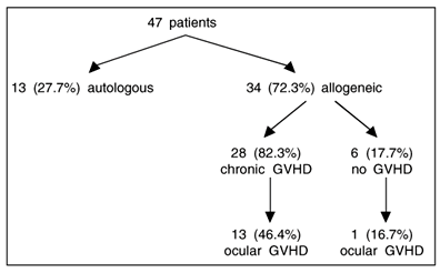
Of the 47 patients undergoing ocular examination, 25 (53.2%) were males and 22 (46.8%) females. The average age was 30 years, and the range was 11 to 57 years. Of the total number of patients, 13 (27.7%) were submitted to autologous BMT and 34 (72.3%) to allogeneic BMT. Of the 34 patients submitted to allogeneic BMT, 28 (82.3%) developed chronic GVHD in one or more organs and 6 (17.7%) did not present GVHD. Of the 28 (82.4%) patients with chronic GVHD, 13 (46.4%) presented ocular GVHD and 15 (53.6%) did not. Of the six patients without GVHD, one (16.7%) presented ocular GVHD and five (83.3%) did not have dry eyes.
None of those 13 patients submitted to autologous BMT presented ocular GVHD and 14 (41.2%) of 34 patients with allogeneic BMT presented ocular GVHD.
All patients with ocular GVHD (Group I) had symptoms and the most frequent were burning, foreign body sensation, blurred vision and dryness (Table 1). Some of these symptoms were also sporadically reported by 5 (15%) patients without ocular GVHD (Group II).
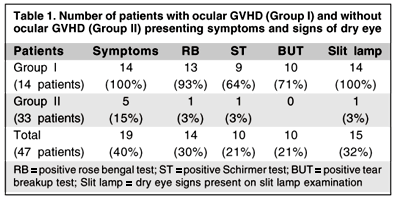
The rose bengal test was one of the most sensitive in this study and the Schirmer test presented a lower sensitivity than the rose bengal test. The breakup time of the lacrimal film (BUT) was highly specific as it was never positive in individuals without ocular GVHD (Table 1). The slit lamp examination was considered the gold standard as all patients of Group I showed conjunctival hyperemia, associated with other findings, such as the existence of filaments, punctate keratitis and tarsal scars.
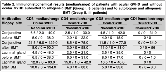
The results of the immunohistochemical analysis are presented in Tables 2 and 3. In the conjunctiva and lacrimal gland of patients presenting dry eye (with ocular GVHD),there was an increase of CD3, CD4 and CD8 cells, respectively indicating an increase of the T, Th/i lymphocyte population and suppressor-cytotoxic T cells after the transplantation.
DISCUSSION
The frequency of ocular GVHD in our series (41.2%) was lower than that reported by other authors, probably due to the criteria we used to diagnose dry eye. We question the utility of the Shirmer test for the diagnosis of chronic GVHD without other manifestations such as symptoms, abnormalities in the lacrimal function or at the slit-lamp examination(14).
One patient of our series, in spite of not presenting systemic GVHD, had dry eye according to our criteria. Ocular manifestations may be the first presentation of GVHD and they may be seen in the absence of systemic manifestations of GVHD. Dry eyes can occur in patients without GVHD(15), probably as a result of total body radiation but other authors(14,16) disagree. In a group of patients without chronic GVHD(14) 10% had a positive Schirmer test, ocular symptoms and an abnormal slit-lamp examination and none of them, as our patient, had received radiation.
Ocular GVHD symptoms can easily be missed while the patient shows already important ocular lesions at the first ophthalmological examination(16). Focusing on the many life-threatening problems present in this group of patients may explain why the ocular complications are seen with such a delay.
It is also possible that patients without systemic GVHD, but with ocular GVHD, could be suffering lymphocytic infiltration also in other organs such as liver or skin, but at the time they were examined they were not presenting clinical manifestations yet. As the ocular GVHD has an autoimmune origin, the immunohistochemical alterations should occur earlier, before clinical manifestations, so probably in the present study the time of ocular examinations and biopsy could influence the results. In these cases, the immunohistochemical examination could be useful for an early diagnosis of ocular GVHD.
The symptoms of patients with ocular GVHD present in this study are in accordance with studies by other authors(7,15), who suggested that the clinical picture was very similar to that present in patients with KCS either idiopathic or secondary to Sjögren's syndrome.
Dry eye in patients submitted to autologous BMT were mentioned(17), attributing the alterations to total body irradiation (TBI) or to the conditioning regimen. None of the patients of this study submitted to autologous BMT developed dry eye. A patient of this group mentioned ocular burning, and another, submitted to TBI a year prior to BMT, complained about ocular pain and tearing. However, their ocular examination did not show all diagnostic characteristics of ocular GVHD. This difference in findings compared to those by other authors(17) can be explained by the time elapsed between the irradiation and ocular examination.
The most used clinical tests of lacrimal function reported in the literature to evaluate patients after BMT are Schirmer test and slit lamp(15). The slit lamp examination was very useful in the evaluation of corneal and conjunctival alterations, since these alterations were always present in patients with dry eye in our study, as they were in other series(17). The rose bengal test, in spite of rarely being mentioned in the literature in the evaluation of patients with ocular GVHD, was one of the most sensitive in this study (Table 1). The Schirmer test, in spite of the fact that it presented a lower sensitivity than the rose bengal test, has the advantage that it can be used by general practitioners and hematologists, as part of propaedeutic evaluation of ocular GVHD, since it is not dependent on slit lamp observation. However, when applied alone, it is not enough for the diagnosis of dry eye, eventually resulting in conflicting information(18).
The development of techniques with monoclonal antibodies against T human cells and subpopulations has made the functional differentiation of these lymphocytes possible and contributed to the knowledge of the immunological composition of the inflammatory infiltrates. Monoclonal antibodies were used to study the lymphocyte subpopulation in the conjunctiva(12). These authors observed the predominance of B cells in the normal conjunctival stroma in relation to T cells, Ts/c cells being predominant over Th/i cells. The same subpopulation was analyzed(19) and different populations from those previously described were found: T cells were predominant over B cells 20 times.
In this research, analyzing the average conjunctival lymphocyte population of patients before being submitted to BMT (Table 2), a greater number of cells was observed with CD3 antigens (T lymphocytes) as opposed to the CD19 antigens (B lymphocytes), according other data(19). We compared our data with those by other authors, in spite of our patients having received aggressive treatment before BMT, because at the moment they were examined and biopsied they were free of malignant diseases on clinical and laboratory evaluation.
In the normal lacrimal gland, the T cells are present in a larger number than B cells, with Ts/c cells predominanting over Th/i cells(20-21). In the present study, on average, in patients before transplantation, the lymphocyte population of the lacrimal gland was also observed to have a tendency of a higher number of T than B cell population (Table 3).
The lymphocyte infiltrates that accumulate in lacrimal tissue of patients with Sjögren's syndrome and promote destruction of its tubular acinous architeture appear to be primarily composed of B cells, disposed in clusters, and they tend to be surrounded by Th/i cells(22-23). The Ts/c cells comprise a small part of the mononuclear infiltrate, the presence of macrophages being rarer. A decreased reflex tearing is associated with lymphocytic infiltration in lacrimal glands in patients with Sjögren syndrome and dry eyes(24).
There is no reported research on immunohistochemistry of the lacrimal gland of patients with GVHD. According to the present study, in contrast to what occurred in patients with Sjögren's syndrome, the T cell population of patients with ocular GVHD exceeded the B cells in the lacrimal gland and the median of Th/i cells was approximately equal to that of Ts/c cells (Table 2). These findings are in agreement with the observation that the effector primary cells in the affected tissues of patients with GVHD are T cells(25).
The proportions of infiltrating lymphocyte subsets in the labial salivary glands of patients with chronic GVHD suffering from xerostomia were measured(26) and it was found that more than 90% of the infiltrating lymphocytes were T cells with a slight predominance of Ts/c over Th/i. In patients with Sjögren's syndrome, Th/i cells were predominant.
An immunohistochemical study of the conjunctiva of six patients with ocular GVHD was performed and it was observed that in the epithelium and in the conjunctival stroma there was a larger number of Th/i cells as opposed to cells found in the conjunctiva of three normal patients(27). It was concluded that the conjunctiva also participates in the T cell mediated immunological reaction in ocular GVHD. Our data are in agreement with the latter(27) and disagree with others(12), who found a larger amount of Ts/c cells than Th/i cells in the conjunctiva, but their conclusion was based on only one patient with GVHD.
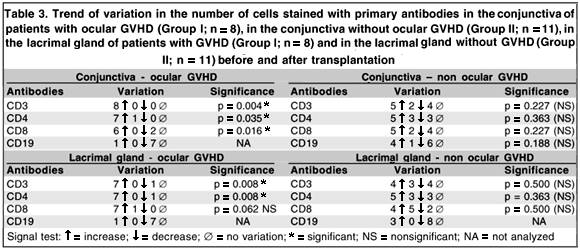
In this research, the data shown in Tables 3 and 4 suggest the existence of a cell mediated immunologic mechanism in the conjunctiva and lacrimal gland of posttransplantation patients. This lymphocytic infiltration may explain the clinical picture of keratoconjunctivitis sicca secondary to GVHD. The destruction of the lacrimal gland tissue by lymphocytic cells was reported(22) and it was also suggested(24) in patients with Sjögren's syndrome.
There are some variables involved in the analysis of this study, such as the size of the sample, consideration of the conjunctival tissue as a whole, and not separately as epithelium and stroma, as done by other authors(12,21). But absolute cellular counts vary from specimen to specimen, with relative amounts of the different populations having to be taken into account. Other variables, such as the use of immunosuppressors, the time elapsed between the transplantation and the biopsy, or even the interpretation of the immunohistochemistry results can have an influence on the results.
Most patients submitted to allogeneic BMT, presenting ocular GVHD, were receiving immunosuppressive medication at the time of the ocular examination and posttransplantation biopsy. It is probable that there was an effect on immunohistochemical results related to the use of cyclosporine A (the medication administered for GVHD prophylaxis), since this drug has a positive selective suppressive effect on T lymphocytes, especially Th/i(28). This drug would reduce the Th/i population in patients submitted to BMT, explaining why, in cases of GVHD, its population equaled that of Ts/c and was not greater, as in Sjögren's syndrome. A study comprising a larger number of patients would be necessary for the confirmation of these results, comparing groups under and without the effect of cyclosporine.
Recent improvements in the systemic management of patients with GVHD have led to the more frequent recognition of the ocular problems. The high prevalence and potentially severe ocular problems in these patients requires close ophthalmic monitoring. It can be assumed that in the future, with the improvement and reduction in costs of immunohistochemical techniques, the biopsy could also be useful promoting early diagnosis of chronic GVHD, always accompanied by ophthalmologic examination in addition to the Schirmer test already performed today.
ACKNOWLEDGMENTS
To Dr Hanna A. Rothschild, medical editor, for the manuscript revision.
To Dr Tong Zhen Zhao, immunologist of Hilles Laboratory, Harvard Medical School, who helped me with the immunohistochemistry technique.
REFERENCES
1. Livesey SJ, Holmes JA, Whittaker JA. Ocular complications of bone marrow transplantation. Eye 1989;3:271-6.
2. Ferreira E, Dulley FL, Morsoletto F, Neto JZ, Pasquini R. Bone marrow transplantation in Brazil. Human Immunol 1985;14:324-32.
3. Shulman HM, Sullivan KM, Weiden PL, McDonald GB, Striker GE, Sale GE, Hackman R, Tsoi MS, Storb R, Thomas ED. Chronic graft-versus-host syndrome in man. A long-term clinicopathologic study of 20 Seattle patients. Am J Med 1980;69:204-17.
4. Sullivan KM. Graft-versus-host disease. In: Stephen J, Forman K, Blume G, Thomas ED, editors. Bone marrow transplantation. Cambridge, MA: Blackwell Scientific Publications; 1994. p.339-62.
5. Tichelli A. Late ocular complications after bone marrow transplantation. Nouv Rev Fr Hematol 1994; 36 suppl 1:579-82.
6. Franklin RM, Kenyon KR, Tutschka PJ, Saral R, Green WR, Santos GW. Ocular manifestations of graft-vs-host disease. Ophthalmology1983;90:4-13.
7. Hirst LW, Jabs DA, Tutschka PJ, Green WR, Santos GW. The eye in bone marrow transplantation. I. Clinical study. Arch Ophthalmol 1983;101:580-4.
8. Mencucci R, Rossi Ferrini C, Bosi A, Volpe R, Guidi S, Salvi G. Ophthalmological aspects in allogeneic bone marrow transplantation: Sjögren-like syndrome in graft-versus-host disease. Eur J Ophthalmol 1997;7:13-8.
9. van Bijsterveld OP. Diagnostic tests in the sicca syndrome. Arch Ophthalmol 1969;82:10-4.
10. Lamberts DW, Foster CS, Perry HD. Schirmer test after topical anesthesia and the tear meniscus height in normal eyes. Arch Ophthalmol 1979;97:1082-5.
11. Gilbard JP. Dry eye disorders. In: Albert DM, Jakobiec F, editors. The principles and practice of ophthalmology: basic sciences.Philadelphia: WB Saunders; 1994. p.257-76.
12. Bhan AK, Fujikawa LS, Foster CS. T-cell subsets and Langerhans cells in normal and diseased conjunctiva. Am J Ophthalmol 1982;94:205-12.
13. Siegel S. Estatística não paramétrica (para as ciências do comportamento).São Paulo: Mekron Books do Brasil; 1975.
14. Abesada-Terk G, Quintero M, Przepiorka D, Shapiro S. Diminished tear production in BMT recipients not receiving radiation. Bone Marrow Transplant 1990;6:151.
15. Calissendorff B, Azazi M, Lonnqvist B. Dry eye syndrome in long-term follow-up of bone marrow transplanted patients. Bone Marrow Transplant 1989;4:675-8.
16. Claes K, Kestelyn P. Ocular manifestations of graft versus host disease following bone marrow transplantation. Bull Soc Belge Ophtalmol 2000;277:21-6.
17. Bray LC, Carey PJ, Proctor SJ, Evans RG, Hamilton PJ. Ocular complications of bone marrow transplantation. Br J Ophthalmol 1991;75:611-4.
18. Goren MB, Goren SB. Diagnostic tests in patients with symptoms of keratoconjunctivitis sicca. Am J Ophthalmol 1988;106:570-4.
19. Sacks EH, Wieczorek R, Jakobiec FA, Knowles DM. Lymphocytic subpopulations in the normal human conjunctiva. A monoclonal antibody study.Ophthalmology 1986;93:1276-83.
20. Wieczorek R, Jakobieck FA, Sacks EH, Knowles DM. The immunoarchitecture of the normal human lacrimal gland. Relevancy for understanding pathologic conditions. Ophthalmology1988;95:100-9.
21. Dua HS, Gomes JA, Jindal VK, Appa SN, Schwarting R, Eagle RC, Donoso LA, Laibson PR. Mucosa specific lymphocytes in the human conjunctiva, corneoscleral limbus and lacrimal gland. Curr Eye Res 1994;13:87-93.
22. Pepose JS, Akata RF, Pflugfelder SC, Voigt W. Mononuclear cell phenotypes and immunoglobulin gene rearrangements in lacrimal gland biopsies from patients with Sjögren's syndrome. Ophthalmology 1990;97:1599-605.
23. Sullivan DA, Sato EH. Immunology of the lacrimal gland. In: Albert DM, Jakobieck FA editors. The principles and practice of ophthalmology: basic sciences. Philadelphia: WB Saunders; 1994. p.479-86.
24. Tsubota K, Xu KP, Fujihara T, Katagiri S, Takeuchi T. Decreased reflex tearing is associated with lymphocytic infiltration in lacrimal glands. J Rheumatol 1996;23:313-20.
25. Foster CS. Pharmacological treatment of immune disorders. In: Albert DM, Jakobiec F editors. The principles and practice of ophthalmology: basic sciences. Philadelphia: WB Saunders; 1994. p.190-217.
26. Hiroki A, Nakamura S, Shinohara M, Gondo H, Ohyama Y, Hayashi S et al. A comparison of glandular involvement between chronic graft-versus-host disease and Sjögren's syndrome. Int J Oral and Maxillofac Surg 1996;25: 298-307.
27. Sainz De La Maza M, Ayliffe W, Foster CS. Immunohistochemical study of conjunctiva from patients with keratoconjunctivitis sicca due to graft-versus-host disease . Invest Ophthalmol & Vis Sci1994;35:1290.
28. Kupiec-Weglinski JW and Tilney NL. Immunopharmacology in the transplantation of vascularized organ allografts. In: Zierhut M, Pleyer U, Thiel HJ. Immunology of corneal transplantation. The Netherlands: Aeolus Press; 1994. p.197-212.
Address to correspondence
Rosana Nogueira Pires da Cunha
Rua Botucatu, 822, São Paulo (SP)
CEP 04023-062
E-mail: [email protected]
Recebido para publicação em 14.12.2001
Aceito para publicação em 11.09.2002
1 Department of Ophthalmology, Federal University of São Paulo, UNIFESP.
2 Bone Marrow Transplantation Sector, Fundação Pró-sangue Hemocentro of São Paulo, São Paulo University, Medicine College.
3 Immunology and Uveitis Service, Department of Ophthalmology, Massachusetts Eye & Ear Infirmary, Harvard Medical School.
© 2024 - All rights reserved - Conselho Brasileiro de Oftalmologia
![]()
 English PDF
 Print
Print
 Send this article by email
 How to cite this article
 Submit a comment
 Mendeley
 Scielo
 Pocket
