
Alberto Diniz Filho1; Sebastião Cronemberger1; Dollores Martins Ferreira1; Rafael Vidal Mérula1; Nassim Calixto1
DOI: 10.1590/S0004-27492010000200011
ABSTRACT
PURPOSE: To investigate, through ultrasound biomicroscopy images, the presence of plateau iris configuration in eyes with narrow-angle from patients with open-angle glaucoma and in eyes with previous acute primary angle-closure and compare the biometric features of eyes with plateau iris configuration with those of normal eyes. METHODS: Ultrasound biomicroscopic images from 196 patients with open-angle glaucoma and narrow-angle and 32 patients with acute primary angle-closure were retrospectively analyzed. The inclusion and specific criteria for the diagnosis of plateau iris configuration was the presence of an anterior positioning of the ciliary processes, supporting the peripheral iris so that it was parallel to the trabecular meshwork; the iris root had a steep rise from its insertion point, followed by a downward angulation from the corneoscleral wall; presence of a central flat iris plane; an absent (complete plateau iris configuration) or partially absent (incomplete plateau iris configuration) ciliary sulcus. The ultrasound biomicroscopic parameters were compared between complete and incomplete plateau iris configuration. The same parameters of both groups were compared with those of normal eyes. The following measurements were performed: anterior chamber depth; angle opening distance at 500 µm from the scleral spur; peripheral iris thickness at 500 µm from the scleral spur; iris-ciliary process distance; trabecular-ciliary process distance and angle recess area. RESULTS: Plateau iris configuration was found in 33 eyes of 20 (10.2%) out of 196 patients with open-angle glaucoma and narrow-angle and in 4 eyes of 2 (6.3%) out of 32 patients with acute primary angleclosure. Seventeen (77.3%) patients with plateau iris configuration were female and 5 (22.7%) male. Twenty-three (62.2%) out of 37 eyes had complete plateau iris configuration, and 14 (37.8%) had incomplete plateau iris configuration. Complete and incomplete plateau iris configuration presented similar biometric features with the exception of the iris-ciliary process distance. All plateau iris configuration eyes showed biometric parameters completely different from those of normal eyes except for peripheral iris thickness at 500 µm from the scleral spur. CONCLUSIONS: Plateau iris configuration was present in 10.2% of patients with open-angle glaucoma and narrow-angle and in 6.3% of patients with acute primary angle-closure. Biometric features were similar in eyes with complete and incomplete plateau iris configuration with the exception of iris-ciliary process distance. Compared to normal eyes, all plateau iris configuration eyes showed biometric parameters completely different except for peripheral iris thickness at 500 µm from the scleral spur.
Keywords: Glaucoma, angle-closure; Anterior chamber; Gonioscopy; Iris diseases
RESUMO
OBJETIVO: Investigar, através de imagens de biomicroscopia ultrassônica, a presença de configuração da íris em platô em olhos com seio camerular estreito em portadores de glaucoma primário de ângulo aberto e em olhos com fechamento angular primário agudo. Avaliar as características biométricas nestes olhos, comparando-os a olhos normais. MÉTODOS: As imagens de biomicroscopia ultrassônica foram analisadas retrospectivamente, sendo que 196 pacientes eram portadores de glaucoma primário de ângulo aberto e 32 pacientes eram portadores de fechamento angular primário agudo. O critério de inclusão para configuração da íris em platô baseado em imagens de biomicroscopia ultrassônica foi definido pela presença de corpo ciliar posicionado anteriormente, íris acentuadamente angulada em seu ponto de inserção seguida de uma angulação descendente a partir da parede corneoescleral, íris central plana e sulco ciliar ausente (configuração da íris em platô completa) ou parcialmente ausente (configuração da íris em platô incompleta). Os parâmetros biométricos medidos pela biomicroscopia ultrassônica foram comparados entre os olhos com configuração da íris em platô completa e incompleta. Os mesmos parâmetros de ambos os grupos foram comparados com os de olhos normais. Foram medidos: profundidade central da câmara anterior; a distância da abertura do ângulo a 500 µm do esporão escleral; a espessura da íris a 500 µm do esporão escleral; a distância íris-processo ciliar, a distância faixa trabecular-processo ciliar e a área de recesso angular. RESULTADOS: A configuração da íris em platô foi encontrada em 33 olhos de 20 pacientes portadores de glaucoma primário de ângulo aberto (10,2% de um total de 196) e 4 olhos de 2 pacientes portadores de fechamento angular primário agudo (6,3% de um total de 32). Dezessete (77,3%) eram do sexo feminino e 5 (22,7%) do sexo masculino. Dos 37 olhos, 23 (62,2%) apresentaram configuração da íris em platô completa e 14 (37,8%) apresentaram configuração da íris em platô incompleta. Olhos com configuração da íris em platô completa e incompleta apresentaram características biométricas muito similares exceto para a distância íris-processo ciliar. Olhos com configuração da íris em platô possuem características biométricas completamente diferentes das de olhos normais exceto a espessura da íris a 500 µm do esporão escleral. CONCLUSÕES: A configuração da íris em platô esteve presente em 10,2% dos pacientes portadores de glaucoma primário de ângulo aberto e em 6,3% dos pacientes portadores de fechamento angular primário agudo. Entre a configuração da íris em platô completa e incompleta, foi encontrada diferença estatisticamente significativa apenas na distância íris-processo ciliar. Quando comparados olhos normais e olhos com configuração da íris em platô, todos os parâmetros apresentaram diferenças altamente significativas, à exceção de espessura da íris a 500 µm do esporão escleral.
Descritores: Glaucoma de ângulo fechado; Câmara anterior; Gonioscopia; Doenças da íris
ORIGINAL ARTICLE
Plateau iris configuration in eyes with narrow-angle: an ultrasound biomicroscopic study
Configuração da íris em platô em olhos com seio camerular estreito: um estudo ultrabiomicroscópico
Alberto Diniz FilhoI; Sebastião CronembergerII; Dollores Martins FerreiraIII; Rafael Vidal MérulaIV; Nassim CalixtoV
IPostgraduate Student, School of Medicine, Universidade Federal de Minas Gerais - UFMG - Belo Horizonte (MG) - Brazil
IIProfessor of Ophthalmology, School of Medicine at UFMG - Belo Horizonte (MG) - Brazil
IIIGraduate Student, School of Medicine at UFMG - Belo Horizonte (MG) - Brazil
IVPostgraduate Student, School of Medicine at UFMG -Belo Horizonte (MG) - Brazil
VProfessor of Ophthalmology, School of Medicine at UFMG - Belo Horizonte (MG) - Brazil
ABSTRACT
PURPOSE: To investigate, through ultrasound biomicroscopy images, the presence of plateau iris configuration in eyes with narrow-angle from patients with open-angle glaucoma and in eyes with previous acute primary angle-closure and compare the biometric features of eyes with plateau iris configuration with those of normal eyes.
METHODS: Ultrasound biomicroscopic images from 196 patients with open-angle glaucoma and narrow-angle and 32 patients with acute primary angle-closure were retrospectively analyzed. The inclusion and specific criteria for the diagnosis of plateau iris configuration was the presence of an anterior positioning of the ciliary processes, supporting the peripheral iris so that it was parallel to the trabecular meshwork; the iris root had a steep rise from its insertion point, followed by a downward angulation from the corneoscleral wall; presence of a central flat iris plane; an absent (complete plateau iris configuration) or partially absent (incomplete plateau iris configuration) ciliary sulcus. The ultrasound biomicroscopic parameters were compared between complete and incomplete plateau iris configuration. The same parameters of both groups were compared with those of normal eyes. The following measurements were performed: anterior chamber depth; angle opening distance at 500 µm from the scleral spur; peripheral iris thickness at 500 µm from the scleral spur; iris-ciliary process distance; trabecular-ciliary process distance and angle recess area.
RESULTS: Plateau iris configuration was found in 33 eyes of 20 (10.2%) out of 196 patients with open-angle glaucoma and narrow-angle and in 4 eyes of 2 (6.3%) out of 32 patients with acute primary angleclosure. Seventeen (77.3%) patients with plateau iris configuration were female and 5 (22.7%) male. Twenty-three (62.2%) out of 37 eyes had complete plateau iris configuration, and 14 (37.8%) had incomplete plateau iris configuration. Complete and incomplete plateau iris configuration presented similar biometric features with the exception of the iris-ciliary process distance. All plateau iris configuration eyes showed biometric parameters completely different from those of normal eyes except for peripheral iris thickness at 500 µm from the scleral spur.
CONCLUSIONS: Plateau iris configuration was present in 10.2% of patients with open-angle glaucoma and narrow-angle and in 6.3% of patients with acute primary angle-closure. Biometric features were similar in eyes with complete and incomplete plateau iris configuration with the exception of iris-ciliary process distance. Compared to normal eyes, all plateau iris configuration eyes showed biometric parameters completely different except for peripheral iris thickness at 500 µm from the scleral spur.
Keywords: Glaucoma, angle-closure; Anterior chamber/ultrasonography; Gonioscopy; Iris diseases
RESUMO
OBJETIVO: Investigar, através de imagens de biomicroscopia ultrassônica, a presença de configuração da íris em platô em olhos com seio camerular estreito em portadores de glaucoma primário de ângulo aberto e em olhos com fechamento angular primário agudo. Avaliar as características biométricas nestes olhos, comparando-os a olhos normais.
MÉTODOS: As imagens de biomicroscopia ultrassônica foram analisadas retrospectivamente, sendo que 196 pacientes eram portadores de glaucoma primário de ângulo aberto e 32 pacientes eram portadores de fechamento angular primário agudo. O critério de inclusão para configuração da íris em platô baseado em imagens de biomicroscopia ultrassônica foi definido pela presença de corpo ciliar posicionado anteriormente, íris acentuadamente angulada em seu ponto de inserção seguida de uma angulação descendente a partir da parede corneoescleral, íris central plana e sulco ciliar ausente (configuração da íris em platô completa) ou parcialmente ausente (configuração da íris em platô incompleta). Os parâmetros biométricos medidos pela biomicroscopia ultrassônica foram comparados entre os olhos com configuração da íris em platô completa e incompleta. Os mesmos parâmetros de ambos os grupos foram comparados com os de olhos normais. Foram medidos: profundidade central da câmara anterior; a distância da abertura do ângulo a 500 µm do esporão escleral; a espessura da íris a 500 µm do esporão escleral; a distância íris-processo ciliar, a distância faixa trabecular-processo ciliar e a área de recesso angular.
RESULTADOS: A configuração da íris em platô foi encontrada em 33 olhos de 20 pacientes portadores de glaucoma primário de ângulo aberto (10,2% de um total de 196) e 4 olhos de 2 pacientes portadores de fechamento angular primário agudo (6,3% de um total de 32). Dezessete (77,3%) eram do sexo feminino e 5 (22,7%) do sexo masculino. Dos 37 olhos, 23 (62,2%) apresentaram configuração da íris em platô completa e 14 (37,8%) apresentaram configuração da íris em platô incompleta. Olhos com configuração da íris em platô completa e incompleta apresentaram características biométricas muito similares exceto para a distância íris-processo ciliar. Olhos com configuração da íris em platô possuem características biométricas completamente diferentes das de olhos normais exceto a espessura da íris a 500 µm do esporão escleral.
CONCLUSÕES: A configuração da íris em platô esteve presente em 10,2% dos pacientes portadores de glaucoma primário de ângulo aberto e em 6,3% dos pacientes portadores de fechamento angular primário agudo. Entre a configuração da íris em platô completa e incompleta, foi encontrada diferença estatisticamente significativa apenas na distância íris-processo ciliar. Quando comparados olhos normais e olhos com configuração da íris em platô, todos os parâmetros apresentaram diferenças altamente significativas, à exceção de espessura da íris a 500 µm do esporão escleral.
Descritores: Glaucoma de ângulo fechado; Câmara anterior/ ultrassonografia; Gonioscopia; Doenças da íris
INTRODUCTION
Classically, the plateau iris configuration (PIC) was defined as presurgical changes of an eye with normal central anterior chamber depth (ACD), flat iris by conventional biomicroscopy, but with an extremely narrow or closed angle on gonioscopic examination. On the other hand, the plateau iris syndrome (PIS) was defined as an acute glaucoma crisis in one eye with normal central anterior chamber depth in spite of a patent iridotomy on direct examination, presenting angle-closure confirmed by gonioscopic examination after mydriasis(1-3).
The ultrasound biomicroscopy (UBM) enabled one to anatomically recognize the so-called PIC and explained the PIS mechanism that occurs in many eyes. Some UBM studies showed that the ciliary processes were in an anterior position when compared to their position in normal subjects and in patients with angle-closure glaucoma caused by pupillary block. Therefore, a significant angle narrowing was found, even though the central depth of the anterior chamber was almost normal(4).
The purpose of this study is to investigate, through UBM images, the presence of PIC in eyes with narrow-angle from patients with open-angle glaucoma (OAG) and from eyes with acute primary angle-closure (APAC). Biometric features of PIC eyes and normal eyes were also compared.
METHODS
Ultrasound biomicroscopy images of 196 consecutive OAG subjects and 32 consecutive APAC subjects in one or both eyes submitted or not to laser peripheral iridotomy (LPI) from September, 1995 to August, 2006, were retrospectively analyzed.
The inclusion and specific criteria for the diagnosis of PIC was the presence of an anterior positioning of the ciliary processes, supporting the peripheral iris so that it was parallel to the trabecular meshwork; the iris root had a steep rise from its point of insertion, followed by a downward angulation from the corneoscleral wall; presence of a central flat iris plane; an absent (complete PIC) or partially absent (incomplete PIC) ciliary sulcus(5), through the analysis of UBM imaging in the four quadrants.
The following criteria were used to define cases of previous APAC: 1) presence of at least two of the following symptoms: ocular or periocular pain, nausea and/or vomiting, antecedent history of intermittent blurring of vision with haloes; and 2) presenting intraocular pressure (IOP) higher than 28 mmHg (as measured by Goldmann applanation tonometry) and the presence of at least three of the following signs: conjunctival injection, corneal epithelial edema, mid-dilated unreactive pupil, and shallow anterior chamber; and 3) the presence of an occluded angle in the affected eye, verified by gonioscopy.
The criteria used to define OAG were: 1) intraocular pressure (IOP) higher than 18 mmHg; 2) presence of glaucomatous optic neuropathy defined by at least two of the following items: cup/disc ratio (C/D) asymmetry between fellow eyes of greater than 0.2, rim thinning, notching, cup/disc ratio (C/D) more than 0.7, optic disc haemorrhage or retinal nerve fiber layer (RNFL) defect; 3) glaucomatous visual field defects were detected by automated perimetry evaluated by means of Interzeag Octopus 1-2-3 (the visual field test was considered abnormal if two of the following three criteria were met on at least two consecutive visual fields: (a) an abnormal Bebie curve (deviation more than 0.5); (b) 3 contiguous nonedge points (allowing the two nasal step edge points) on a Octopus program G1X visual field with p<0.05 on the probability plot, with at least 1 point at p<0.01; and (c) a corrected loss variance p<0.05; and 4) open-angle evaluated by gonioscopy [clinically narrow-angles as those angles regarded by the examiner as being grade III (posterior portion of trabecular meshwork hidden) using the Scheie classification based upon the most posterior structure visible in the angle, without goniosynechia].
Gonioscopy in primary gaze position was performed by the members of the Glaucoma Service and confirmed by one of the authors (SC), using a Goldmann three-mirror goniolens (Haag-Streit AG, Köniz, Switzerland), in the undilated state under room light illumination (approximately 240 lux), and dark (approximately 0.1 lux) (the room was darkened in 5 minutes), both conditions previously measured with an illumination meter (Minolta T10 Illumination Meter, Konica Minolta Sensing Inc., Osaka, Japan), being uniforms the conditions for all examination. The angle was fully gonioscopically evaluated (360º), and the classification was based on the visibility of the posterior trabecular meshwork (180º).
The following exclusion criteria were used: presence of iridociliary cysts; nuclear sclerosis defined as Lens Opacities Classification System (LOCS) II more than grade 2 (NC2, NO2); secondary glaucoma (traumatic, uveitic, aphakic, neovascular, pseudophakic, phacomorphic or phacoanaphylatic); peripheral anterior synechia; corneal opacity in both eyes; previous surgical procedure except LPI; and closed angle under dark gonioscopic examination. Blind eyes were also excluded.
The UBM examination was performed by a single examiner (SC) with the commercial instrument (UBM model 840, Zeiss-Humphrey Instruments Inc., San Leandro, California, USA) and the 50-MHz transducer. After surface anaesthesia with 0.5% proparacaine, a plastic eyecup containing 2% methylcellulose and physiologic saline was applied to the eyeball between the eyelids. The scanning was performed by placing the probe at the limbus and ciliary body region, always perpendicular to the surface of the eyeball, and multiple cross-sectional scans passing trough the ciliary process were obtained per eye with the patient in a supine position. The UBM examination was done in the undilated state under room light illumination (approximately 240 lux), and dark (approximately 0.1 lux), being uniforms the conditions for all examination.
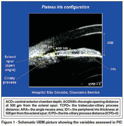
Ultrasound biomicroscopic images of the central anterior chamber, and superior, inferior, nasal and temporal quadrants of each eye were evaluated, and the following parameters were measured (Figure 1): central anterior chamber depth (ACD); the angle opening distance at 500 µm from the scleral spur (AOD500); the peripheral iris thickness at 500 µm from the scleral spur (ID1); the iris-ciliary process distance (ICPD); the trabecular-ciliary process distance (TCPD) and the angle recess area (ARA) in mm2. All the parameters, with the exception of the ACD, were measured in the temporal quadrant. The UBM Pro 2000 software program (Paradigm Medical Industries, Salt Lake City, UT, USA) was used to automatically measure the ARA after spotting the scleral spur(6). We compared the ACD, AOD500, ID1 and TCDP measurements between complete and incomplete PIC eyes. We also compared the same biometric parameters of complete and incomplete PIC eyes with those of 89 normal eyes who were studied by one of the authors (SC)(7). All normal patients presented normal ophthalmological examination and none of them had PIC on UBM examination.

We compared the ICPD measurements between the complete and incomplete PIC eyes with those of 42 normal eyes from another study which this parameter was measured(8).
Finally, we compared the ARA measurements of the eyes with PIC with those of 30 normal eyes from another study(9).
A SPSS 13.0 program was used for the statistical analysis. The significance level of 1% (p<0.01) was adopted. This study was approved by the Ethics and Research Committee of the Federal University of Minas Gerais through Certificate 226/05.
RESULTS
One hundred ninety-six patients were previously classified as having OAG. One hundred and seventy-one (75%) out of 228 patients were female and 57 (25%) were male. PIC was found in 33 eyes of 20 patients out of 196 OAG patients (10.2%) (seven eyes of these patients were excluded) and in four eyes of two patients out of 32 APAC patients (6.3%).
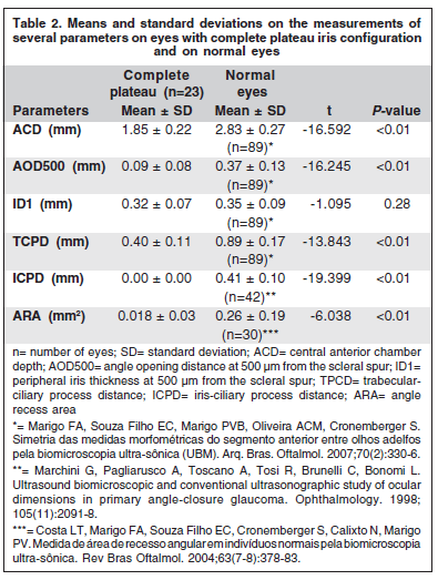
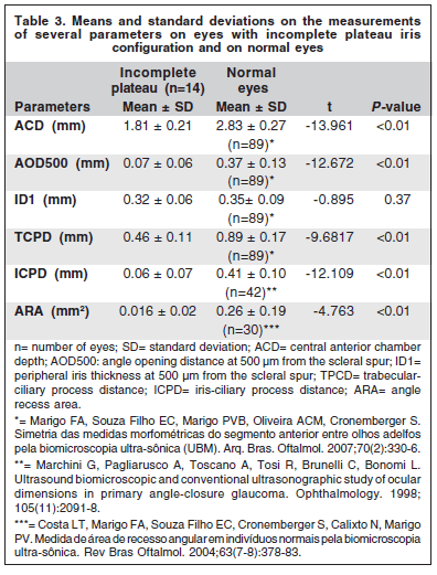
Twenty-three eyes (62.2%) of 15 patients had complete PIC, and 14 eyes of 7 patients (37.8%) had incomplete PIC. Seventeen (77.3%) out of 22 patients with PIC were female, and 5 (22.7%) were male. The patients' age ranged from 45 to 78, with an average of 60.1 years. Table 1 shows the comparisons between the biometric parameters of eyes with complete and incomplete PIC. The biometric features were similar in patients with complete and incomplete PIC with the exception of ICPD. The results of the comparisons among eyes with complete and incomplete PIC and normal eyes are seen in tables 2 and 3. As expected, all PIC eyes presented biometric parameters statistically different from those of normal eyes except for ID1. In tables 2 and 3, attention is drawn to the fact that eyes with complete and incomplete PIC showed shallower (1 mm average) ACD than normal eyes. The ICPD equaled zero in complete PIC and showed a minimum value in the incomplete PIC.


DISCUSSION
The gonioscopy can lead to a diagnosis of PIC. However, UBM plays a fundamental role by confirm the PIC diagnosis. Furthermore, UBM enables the measurement of many parameters of the ocular anterior segment of PIC patients and their comparison to those of normal eyes or OAG eyes. Therefore, it is recommended that the eyes suspected of PIC, PIS and APAC by conventional biomicroscopy or gonioscopy should be assessed by UBM.
In this paper, we observed that 17 (77.3%) out of 22 patients with PIC were female, and 5 (22.7%) were male, which is in agreement with previous studies that showed that PIC is more prevalent in women than in men(10-11). The patients with PIS were mostly women (74.3%). Some authors also found that PIS was more frequent in young women with angle-closure glaucoma(10-11).
In the present study, the patients' age ranged from 45 to 78, with an average of 60.1 years old, which is much higher when compared to another study: 34.9 years old on average(10).
PIC is an ocular bilateral anomaly. In four patients with bilateral complete PIC, we considered only one eye. The contralateral eye was excluded because it had undergone a trabeculectomy.
One more recent study found a smaller PIS prevalence of 22% in younger patients with recurrent angle-closure(11). In both studies the eyes presented acute crisis of glaucoma after LPI. Four eyes of two patients with APAC had previously undergone a LPI in the present study.
Some years ago we have established through UBM the normal values of several measurements of the structures of the anterior segment of normal patients(7). None of the normal eyes had PIC on UBM examination.
In the present study, the complete PIC was more prevalent than the incomplete PIC, which is in agreement with another paper(10).
PIS was responsible for the highest frequency of angleclosure in young patients found in 35 (52.2%) out of the 67 patients with angle-closure glaucoma(10).
In this paper, PIC was found in 10.2% of the patients with OAG and narrow-angle and in 6.3% of the patients with APAC.
A cohort cross-sectional study was published determining the prevalence of plateau iris in primary angle-closure suspects (PACSs) using UBM(12). Two hundred five subjects were enrolled; UBM images of 167 patients were available for analysis. Plateau iris was found in 54 (32.3%) out of 167 PACS eyes after LPI. Plateau iris was most commonly observed in the superior and inferior quadrants(12). This percentage was higher than that found in this paper, perhaps because of the differences envolving the methodogy and the population studied.
In this study, no statistical significant differences were found between the ID1 of complete and incomplete PIC, or in relation to the ID1 of normal eyes (Tables 1, 2 and 3)(7-9). In fact, the ID1 measurement was performed at 500 µm from the scleral spur, which in most eyes with PIC does not correspond to the position of the plateau iris. Moreover, table 1 shows that, with the exception of ID1, all the other parameters presented statistically significant lower values than those found in normal eyes(7-9).
In accordance with some authors(13), in this study, the average ACD was proven to be much inferior in eyes with complete and incomplete PIC than in normal ones (Tables 1, 2 and 3). However, this result is in disagreement with other studies(3).
The average age of the patients studied was 60.1 years old, very similar to the one found in the study by Mandell et al.(13), in which the PIC patients averaged 57.5 years old, and different from the patients assessed in the study of Wand et al.(3), in which the average age was 39.6 years old.
Another important finding concerns the ICPD which equaled zero in complete PIC and presented a low average value in incomplete PIC, with statistically significant differences. The ICPD average in eyes with incomplete PIC and the absence of ICPD (ICPD = zero) in complete PIC have different values from the ones found on normal eyes, in which the ciliary sulcus is present (Tables 1, 2 and 3)(8). In practical terms, complete and incomplete plateau iris theoretically have the same chance to develop PIS, depending on the level of the iris relative to the Schwalbe's line and the structures of the angle wall(14). The ARA measurements on patients with PIC were inferior to those found on normal eyes (Tables 1, 2 and 3)(9).
The importance of the diagnosis of PIC by UBM resides in the fact that preventive measures may be adopted to avoid an acute crisis of glaucoma or its repetition (PIS) despite the presence of a patent iridotomy. Therefore, the UBM is of capital importance for the indication of an appropriate treatment.
This study has many limitations. The most of limitations of the current study are due to the fact that it is retrospective. The glaucoma definition used in the present study is not identical to the definition adopted in other studies. The prevalence rates might be affected by selection bias, including the need to clearly clarify study population characteristics, particularly when this may affect study results and the absence of adjustment for demographic characteristics.
Despite all these limitations, the present study based on an expressive sample certainly provides important information about the role of PIC.
CONCLUSIONS
This study showed a PIC percentage of 10.2% in narrowangle from OAG patients and of 6.3% in APAC. No statistically significant difference was found between the morphometric findings of complete and incomplete PIC, except for ICPD. The biometric parameters of complete and incomplete PIC are completely different of those of normal eyes, except for ID1. The ACD of eyes with complete and incomplete PIC is significantly shallower than that of normal eyes.
REFERENCES
1. Tornquist R. Angle-closure glaucoma in an eye with a plateau type of iris. Acta Ophthalmol (Copenh). 1958;36(3):419-23.
2. Shaffer RN. Primary glaucomas. Gonioscopy, ophthalmoscopy and perimetry. Trans Am Acad Ophthalmol Otolaryngol. 1960;64:112-27.
3. Wand M, Grant WM, Simmons RJ, Hutchinson BT. Plateau iris syndrome. Trans Sect Ophthalmol Am Acad Ophthalmol Otolaryngol. 1977;83(1):122-30.
4. Tran HV, Liebmann JM, Ritch R. Iridociliary apposition in plateau iris syndrome persists after cataract extraction. Am J Ophthalmol. 2003;135(1):40-3. Comment in: Am J Ophthalmol. 2003;136(2):395; author reply 395-6.
5. Pavlin CJ, Ritch R, Foster FS. Ultrasound biomicroscopy in plateau iris syndrome. Am J Ophthalmol. 1992;113(4):390-5.
6. Ishikawa H, Esaki K, Liebmann JM, Uji Y, Ritch R. Ultrasound biomicroscopy dark room provocative testing: a quantitative method for estimating anterior chamber angle width. Jpn J Ophthalmol. 1999;43(6):526-34.
7. Marigo FA, Souza-Filho EC, Marigo PVB, Oliveira ACM, Cronemberger S. Simetria das medidas morfométricas do segmento anterior entre olhos adelfos pela biomicroscopia ultra-sônica (UBM). Arq Bras Oftalmol. 2007; 70(2):330-6.
8. Marchini G, Pagliarusco A, Toscano A, Tosi R, Brunelli C, Bonomi L. Ultrasound biomicroscopic and conventional ultrasonographic study of ocular dimensions in primary angle-closure glaucoma. Ophthalmology. 1998;105 (11):2091-8.
9. Costa LT, Marigo FA, Souza Filho EC, Cronemberger S, Calixto N, Marigo PVB. Medida da área de recesso angular em indivíduos normais pela biomicroscopia ultra-sônica. Rev Bras Oftalmol. 2004;63(7/8):378-83.
10. Ritch R, Chang BM, Liebmann JM. Angle-closure in younger patients. Ophthalmology. 2003;110(10):1880-9.
11. Stieger R, Kniestedt C, Sutter F, Bachmann LM, Stuermer J. Prevalence of plateau iris syndrome in young patients with recurrent angle-closure. Clin Experiment Ophthalmol. 2007;35(5):409-13.
12. Kumar RS, Baskaran M, Chew PT, Friedman DS, Handa S, Lavanya R, et al. Prevalence of plateau iris in primary angle-closure suspects an ultrasound biomicroscopy study. Ophthalmology. 2008;115(3):430-4.
13. Mandell MA, Pavlin CJ, Weisbrod DJ, Simpson ER. Anterior chamber depth in plateau iris syndrome and pupillary block as measured by ultrasound biomicroscopy. Am J Ophthalmol. 2003;136(5):900-3.
14. Ritch R, Lowe RF. Angle-closure glaucoma: clinical types. In: Ritch R, Shields MB, Krupin T, editors. The glaucomas. 2nd ed. St Louis: Mosby; 1996. p.821-40.
 Correspondence address:
Alberto Diniz Filho
Avenida Afonso Pena, 270 - Curvelo (MG)
CEP 35790-000
E-mail: [email protected]
Recebido para publicação em 10.12.2008
Última versão recebida em 31.01.2010
Aprovação em 15.03.2010
From the Department of Ophthalmology and Otorrinolaringology, Universidade Federal de Minas Gerais -UFMG - Belo Horizonte (MG) - Brazil.
Nota Editorial: Depois de concluída a análise do artigo sob sigilo editorial e com a anuência do Dr. Lisandro Massanori Sakata sobre a divulgação de seu nome como revisor, agradecemos sua participação neste processo.