Arq. Bras. Oftalmol. 2021;84 (4 )
:316-323
| DOI: 10.5935/0004-2749.20210045
Abstract
OBJETIVO: O objetivo deste estudo foi analisar a segurança do implante de lente intraocular primária em um grande número de olhos em crianças <24 meses.
MÉTODOS: Foram revisados os prontuários de pacientes com idade entre 5-24 meses, submetidos a implante primário de lente intraocular no saco capsular. Uma lente intraocular acrílica de três peças dobrável foi implantada pelo mesmo cirurgião usando uma única técnica cirúrgica. Pacientes que tiveram <1 ano de acompanhamento após a cirurgia foram excluídos. Os principais resultados incluíram medidas de acuidade visual, mudança miópica, complicações pós operatórias e cirurgias adicionais.
RESULTADOS: Foram analisados 68 pacientes (93 olhos). A média de idade dos pacientes no momento da cirurgia foi de 15,06 ± 6,19 (5 a 24) meses, e o equivalente esférico 1 mês após a cirurgia foi de 3,62 ± 2,32 D. Após 5,67 ± 3,10 anos, o equivalente esférico foi de -0,09 ± 3,22 D, e a acuidade visual corrigida à distância foi de 0,33 ± 0,33 e 0,64 ± 0,43 logMAR em casos bilaterais e casos unilaterais, respectivamente (p=0,000). A maior mudança míopica foi observado em bebês submetidos à cirurgia aos 5 e 6 meses de idade. As complicações mais frequentes incluíram opacificação do eixo visual e corectopia. Glaucoma e descolamento de retina não foram relatados.
CONCLUSÃO: O implante primário de lente intraocular no saco capsular em crianças de 5-24 meses é seguro e está associado à baixas taxas de eventos adversos e cirurgias adicional.
Keywords: Catarata pediátrica; Lente intraocular; Implante primário LIO; Mudança miópica; Catarata congênita
Arq. Bras. Oftalmol. 2021;84 (3 )
:214-219
| DOI: 10.5935/0004-2749.20210029
Abstract
OBJETIVO: Avaliar a influência da dinâmica pupilar na curva de desfoco de olhos implantados com lente intraoculares multifocais difrativas.
MÉTODOS: Estudo prospectivo e randomizado realizado na Faculdade de Medicina de Ribeirão Preto - Universidade de São Paulo - Departamento de Oftalmologia. Trinta e oito pacientes foram aleatoriamente designados para receber bilateralmente lentes intraoculares SN6AD1 (n=20) (mfIOL) ou SN60WF (n=18) (aIOL). Além da acuidade visual para longe e perto, corrigida e não corrigida, e curva de desfoco, foi ainda realizada pupilometria dinâmica. A área sob a curva de desfoco foi calculada usando um modelo polinomial empírico.
RESULTADOS: Um total de 16 e 17 pacientes (n=32 e 34 olhos) completaram 1 ano de seguimento nos grupos mfIOL e aIOL, respectivamente. Não houve diferenças significativas entre grupos para as acuidades visuais seja para longe ou perto. As curvas de desfoco do grupo mfIOL mostraram um pico duplo; enquanto o SN60WF mostrou apenas um pico, típico para uma lente intraoculares monofocal. A média da área sob a curva de desfoco do grupo aIOL foi (4,66 ± 1,51 logMAR.dp), e essa é estatisticamente significante diferente da métrica do grupo mfIOL (1,99 ± 1,31 logMAR.dp). A pupila na contração máxima após a exposição a um flash de 30 cd/m2 por 1 segundo foi significativamente correlacionada com uma melhor área de foco no grupo mfIOL (r=0,54; p=0,0017), essa relação não foi observada para o grupo aIOL.
CONCLUSÃO: Estes dados indicam que quanto menor a pupila durante contração, melhor é a área sob a curva de desfoco e, portanto, o desempenho visual dos olhos implantados com essa mfIOL. Esta correlação não foi encontrada para lentes intraoculares monofocais.
Keywords: Lentes intraoculares multifocais; Pupila/fisiologia, Catarata; Facoemulsificacão
Arq. Bras. Oftalmol. 2025;88 (6 )
:1-5
| DOI: 10.5935/0004-2749.2025-0085
Abstract
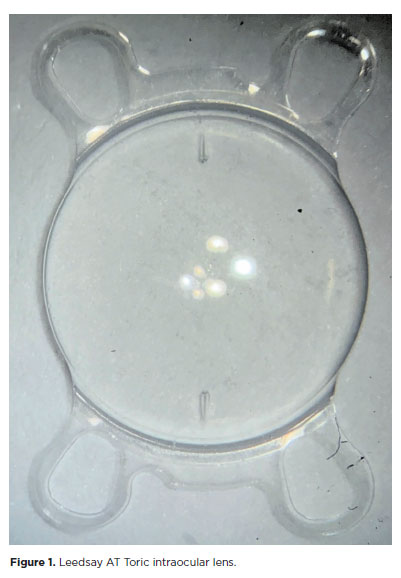
PURPOSE: The purpose of this study was to assess visual outcomes and patient satisfaction following cataract surgery involving the implantation of quad-loop intraocular lenses, including trifocal, bifocal, and toric variants.
METHODS: Information was obtained from both physical and electronic medical records of patients who underwent phacoemulsification cataract surgery with implantation of different intraocular lenses between January 1, 2022, and December 31, 2023. The study included individuals aged over 18 who received bilateral implantation of bifocal, trifocal, or monofocal toric intraocular lenses. Visual acuity was assessed at various postoperative time points using the logMAR scale. Quantitative variables were analyzed using mean and standard deviation.
RESULTS: A total of 92 eyes received premium intraocular lenses: 4 bifocal, 32 trifocal, 52 toric monofocal, and 4 trifocal toric lenses. The average preoperative corrected visual acuity was logMAR 0.478 ± 0.259. On the first postoperative day, the average uncorrected visual acuity was logMAR 0.301 ± 0.207. By day 30, 67.4% of eyes achieved uncorrected distance visual acuity of logMAR 0.2 or better. Patient satisfaction was high, with few reports of glare or halos.
CONCLUSION: Quad-loop intraocular lenses-including trifocal, bifocal, and toric models-demonstrated effective improvement in visual acuity and high levels of patient satisfaction. These lenses represent a suitable option for enhancing visual outcomes after cataract surgery. Additional studies with larger cohorts are recommended to confirm these results.
Keywords: Cataract extraction; Aberrometry/methods; Lenses, intraocular; Lens implantation, intraocular; Prosthesis design
Arq. Bras. Oftalmol. 2026;89 (2 )
:1-8
| DOI: 10.5935/0004-2749.2025-0175
Abstract
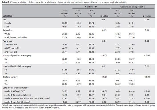
PURPOSE: Endophthalmitis is one of the most important adverse events after cataract surgery, as it can lead to total vision loss. This study aimed to describe the occurrence of endophthalmitis after phacoemulsification with intraocular lens implantation in patients treated in a community setting in Porto Velho, Rondônia, Brazil.
METHODS: This retrospective cohort study was conducted using a database of 649 medical records of patients who underwent surgery and were followed for three months. Poisson regression analysis was used to estimate relative risks and 95% confidence intervals (95% CIs).
RESULTS: The incidence of confirmed endophthalmitis was 11.94% (95% CI, 9.50-14.76), while the incidence of confirmed and probable cases was 20.50% (95% CI, 17.52-23.73). For confirmed cases, bilateral surgery and the use of lens model 3 were identified as risk factors for endophthalmitis, whereas age over 70 yr and preoperative antibiotic use were protective factors. For confirmed and probable cases, brown and yellow skin color, bilateral surgery, and the use of lens model 3 were also identified as risk factors. Gram-negative bacteria were the predominant etiological agents, and corneal edema was the main clinical manifestation. The mean duration of treatment was eight days, and 27.12% of patients used antibiotics.
CONCLUSION: The incidence observed was substantially higher than that reported in the literature, with a predominance of Gram-negative agents and an association with bilateral surgeries and the Eyeol intraocular lens model. These findings reinforce the need for continuous epidemiological surveillance and the implementation of specific biosafety and infection control protocols during cataract surgery campaigns.
Keywords: Endophthalmitis; Disease outbreaks; Phacoemulsification; Lens implantation, intraocular; Lenses, intraocular; Cataract; Risk factors; Anti-bacterial agents
Arq. Bras. Oftalmol. 2025;88 (4 )
:1-6
| DOI: 10.5935/0004-2749.2024-0083
Abstract
PURPOSE: We developed an artificial intelligence program for calculating intraocular lenses and analyzed its accuracy rate via ultrasonic biometry. This endeavor is aimed at enhancing precision and efficacy in the selection of intraocular lenses, particularly in cases where optical biometry is unavailable.
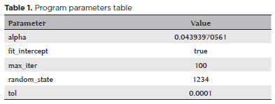
METHODS: Data was collected from the Hospital de Clínicas de Porto Alegre, which included cases of phacoemulsification with intraocular lens implantation, in which the lens selection was based on ultrasonic biometry. The program, implemented in Python, Java, and PHP, employs the ridge regression method. Two design options were developed: a basic model, which uses only keratometry variables (K1 and K2), axial size and final target refraction in the spherical equivalent, and an advanced model, which incorporates preoperative refraction and the patient's age. The Universal Barrett II formula was used to compare both models.
RESULTS: The sample consisted of 486 eyes from 313 patients, with 350 eyes used for program training and 136 for program validation. The spherical equivalent hit rates, with a variation of ±0.5 D, were 86% and 87.5% for the basic and advanced models, respectively, with no statistically significant difference between them. With the Barret Universal II formula, the success rate was 69%, which was significantly different from the values of the two aforementioned models (p<0.0001). The system was better for medium and long eyes but worse for short eyes (<=22.00 mm).
CONCLUSION: The developed artificial intelligence program was superior to the Barrett formula in terms of performance, in the general context and within the subgroup of patients with longer eyes. This innovation can considerably contribute to the selection of intraocular lenses, particularly in cases where optical biometry is unavailable.
Keywords: Biometry; Intraocular lens; Cataract; Artificial intelligence
Arq. Bras. Oftalmol. 2026;89 (3 )
:1-4
| DOI: 10.5935/0004-2749.2025-0263
Abstract
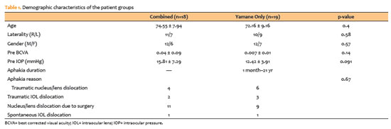
PURPOSE: To compare patients who underwent scleral fixation using the Yamane technique with and without simultaneous pars plana vitrectomy.
METHODS: A total of 37 patients were included in the study. Eighteen underwent simultaneous pars plana vitrectomy. The Yamane technique alone was performed only in patients with aphakia who had previously undergone pars plana vitrectomy for various reasons. Final lens position, best corrected visual acuity spherical equivalent, complication rates, and optical coherence tomography findings were recorded.
RESULTS: The duration of aphakia before intraocular lens implantation ranged from 1 month to 21 yr. Postoperative best corrected visual acuity improved in both groups, with no statistically significant difference (with pars plana vitrectomy: 0.42 ± 0.34; without pars plana vitrectomy: 0.32 ± 0.26; p=0.33). The spherical equivalent was also not significantly different between groups (with pars plana vitrectomy: 0.29 ± 1.08; without pars plana vitrectomy: 0.65 ± 2.23; p=0.53). There were no significant differences between the groups in complication rates, postoperative intraocular lens position or optical coherence tomography findings.
CONCLUSION: There was no difference in terms of safety or efficacy between the two approaches. Surgical decisions may be based on the surgeon’s experience and the patient’s systemic and ocular condition.
Keywords: Lens implantation, intraocular; Tomography, optical coherence; Vitrectomy; Intraocular lenses; Visual acuity; Aphakia; Yamane technique
Arq. Bras. Oftalmol. 2026;89 (4 )
:1-5
| DOI: 10.5935/0004-2749.2026-0010
Abstract
PURPOSE: To evaluate changes in scotopic pupil diameter before and after cataract surgery performed by phacoemulsification with intraocular lens implantation.
METHODS: This prospective longitudinal observational study included patients who underwent cataract surgery. Scotopic pupil diameter was measured preoperatively and 30-40 days postoperatively using an automated keratometer after a standardized dark-adaptation period under controlled ambient illumination. Each eye was considered an independent unit of observation. Because some participants contributed both eyes, intraindividual correlation was accounted for using a linear mixed-effects model with random patient intercepts. Time of assessment (preoperative versus postoperative), age, sex, and eye laterality were included as fixed effects.
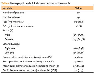
RESULTS: A total of 354 eyes from 251 patients were analyzed. The mean patient age was 69.3±7.2 yr. Mean scotopic pupil diameter decreased from 5.3±0.9mm preoperatively to 4.8±0.8mm postoperatively, representing a mean reduction of 0.5mm (9.4%). In the linear mixed-effects model, cataract surgery was associated with a significant reduction in pupil diameter, with an adjusted mean difference of 0.45mm (95% confidence interval [95% CI], 0.39-0.51; p<0.001). Age (p=0.061), sex (p=0.920), and eye laterality (p=0.152) were not significantly associated with the magnitude of pupil diameter change.
CONCLUSION: Phacoemulsification with intraocular lens implantation was associated with a significant reduction in scotopic pupil diameter, independent of age, sex, and eye laterality. This finding should be considered during preoperative planning, particularly when selecting intraocular lenses whose optical performance depends on postoperative pupil size.
Keywords: Cataract; Pupil; Phacoemulsification; Lens implantation, intraocular; Lenses, intraocular; Pseudophakia
Arq. Bras. Oftalmol. 2025;88 (5 )
:1-7
| DOI: 10.5935/0004-2749.2024-0368
Abstract
PURPOSE: To compare endothelial corneal cell changes following cataract surgery performed by phacoemulsification with intraocular lens implantation, conducted by surgeons with varying levels of experience.
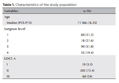
METHODS: Two hundred and eighty-three eyes diagnosed with cataract were included. Lens opacity was classified into three categories (I, II, and III). Surgeons were categorized into four experience levels (1, 2, 3, and 4), based on years of practice and lifetime surgeries performed. Corneal endothelial characteristics were assessed using non-contact specular microscopy, with measurements taken before surgery and 30-60 days post-surgery.
RESULTS: Pre- and postoperative endothelial analysis showed no significant differences between surgeon levels regarding visual acuity achieved, corneal thickness, and endothelial hexagonality. However, the central endothelial cell density index showed a significantly greater reduction among level 1 surgeons (p=0.026). Grade II cataracts exhibited significant variations in the central endothelial cell density (p=0.011) and average cell size, with level 1 surgeons showing the largest increases (p=0.024).
CONCLUSIONS: The analysis revealed significant differences in visual acuity and endothelial indices between surgeon experience levels, with less experienced surgeons showing greater variations and poorer performance. Clinical protocols should consider these data to establish safer training protocols.
Keywords: Cataract extraction; Phacoemulsification; Endothelium; corneal; Lens implantation, intraocular; Visual acuity; Internship and residency; Surgeons
Arq. Bras. Oftalmol. 2025;88 (5 )
:1-6
| DOI: 10.5935/0004-2749.2024-0015
Abstract
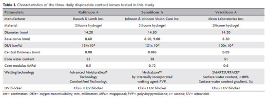
PURPOSE: This study aimed to compare the effects of three different daily disposable contact lens materials on contrast sensitivity.
METHODS: The participants were aged 18–45 years, with spherical equivalent refraction between -0.50 D and -6.00 D, astigmatism below 0.75 D, and best contact lens-corrected visual acuity of 0.0 logMAR or better. Each patient was fitted binocularly with three daily disposable contact lenses made of different materials on three separate examination days. These materials were kalifilcon A, senofilcon A, and verofilcon A. The contrast sensitivity of each patient was recorded at spatial frequencies of 3, 6, 12, and 18 cycles per degree (cpd) under photopic (85 cd/m2) and mesopic (3 cd/m2) conditions.
RESULTS: The current study comprised 72 eyes of 34 female and two male patients. The mean age of the participants was 25.63 (± 0.80) years. Under photopic conditions, the participants’ contrast sensitivity was significantly better with senofilcon A than with kalifilcon A at a frequency of 12 cpd (p=0.008). Under mesopic conditions, participants’ contrast sensitivity was significantly higher with kalifilcon A than verofilcon A at 3 cpd (p=0.001), and with senofilcon A than verofilcon A at 12 cpd (p=0.004). The pre-lens non-invasive break-up times did not differ significantly between the three daily disposable contact lenses (p>0.05).
CONCLUSION: In both photopic and mesopic lighting conditions, the participants in this study exhibited differences in contrast sensitivity when wearing three different daily disposable contact lens types, despite similar visual acuity and pre-lens tear film stability results in their clinical evaluations. These findings demonstrate the potential for subjective visual complaints arising from variations in the contrast sensitivity achieved by different daily disposable contact lenses.
Keywords: Contact lenses; Contrast sensitivity; Astigmatism; Lighting; Visual acuity


 ABO is licensed under a Creative Commons Attribution-NonComercial 4.0 Internacional.


02-tab01tb.jpg)
03-fig01.jpg)
02-fig01.jpg)
01-fig01.jpg)
13-fig01tb.jpg)