Arq. Bras. Oftalmol. 2023;86 (5 )
:1-5
| DOI: 10.5935/0004-2749.20230063
Abstract
Objetivo: A injeção peribulbar de triancinolona é um tratamento alternativo para doenças oculares da tireoide; no entanto, a segurança desse procedimento continua controversa. O objetivo deste artigo é descrever os efeitos adversos locais e sistêmicos de injeções peribulbares de triancinolona em pacientes com doença ocular da tireoide.
Métodos: Estudo retrospectivo de uma série de casos. Foram analisados os prontuários médicos dos pacientes com doença ocular da tireoide tratados com injeções de triancinolona peribulbar em uma única instituição acadêmica entre 2007 e 2019. Foram documentadas as complicações locais e sistêmicas.
Resultados: Um total de 123 pacientes foram tratados. Apenas 11 (8,9%) pacientes apresentaram complicações locais, sendo a mais frequente a presença de equimoses palpebrais superficiais (7,3%), enquanto 2 (1,6%) pacientes apresentaram complicações sistêmicas (hiperglicemia e inibição da suprarrenal após a interrupção do tratamento). Todas estas complicações foram transitórias e nenhum paciente apresentou sequelas de longo prazo.
Conclusões: As injeções peribulbares de triancinolona nas doenças oculares da tireoide têm uma taxa muito baixa de complicações, tanto locais quanto sistêmicas. São necessários estudos prospectivos para aprofundar este tópico.
Keywords: Órbita/diagnóstico por imagem; Imageamento por ressonância magnética; Oftalmopatia de Graves; Triancinolona/efeitos adversos; Injeções.
Arq. Bras. Oftalmol. 2021;84 (2 )
:149-157
| DOI: 10.5935/0004-2749.20210023
Abstract
OBJETIVO: Descrever alterações microvasculares na mácula em diabéticos do tipo 2 sem retinopatia diabética e pacientes saudáveis, e correlacionar achados com perfil clínico nos diabéticos.
MÉTODOS: Foram incluídos 60 olhos de 30 diabéticos e 30 pacientes saudáveis. Diabéticos realizaram fundoscopia, retinografia® (CR2; Canon Inc., New York, New York, USA) e angiografia fluoresceínica® (TRC-50DXC; Topcon Inc., Tokyo, Japan) para descartar a presença de retinopatia. Os 60 pacientes realizaram a angiografia por tomografia de coerência óptica® (RTVue XR, Avanti, Optovue, Fremont, CA, USA) (área macular: 6 x 6 mm2) e foram analisados densidade vascular total, foveal e parafoveal no plexo capilar superficial e plexo capilar profundo, espessura foveal, espessura parafoveal, área da zona avascular da fóvea no plexo capilar superficial e área de fluxo da coriocapilar. Resultados da angiografia por tomografia de coerência óptica foram comparados entre os 2 grupos e correlacionados com acuidade visual, tempo de diabetes, controle glicêmico, perfil lipídico e função renal nos diabéticos.
RESULTADOS: Observou-se aumento mínimo da área de fluxo da coriocapilar nos diabéticos, média das áreas foi de 22,3 ± 4,6 mm² no grupo controle e 22,6 ± 3,9 mm² em diabéticos (p=0,017). Não foi observada diferença estatisticamente significante entre outras variáveis da angiografia por tomografia de coerência óptica analisadas nos dois grupos. Hemoglobina glicosilada e glicemia de jejum apresentaram correlação negativa estatisticamente significante com densidade vascular foveal de ambos os plexos e a glicemia de jejum se correlacionou positivamente com área de fluxo da coriocapilar (p=0,034). Outros dados clínicos avaliados não apresentaram correlação com achados da angiografia por tomografia de coerência óptica.
CONCLUSÃO: Resultados sugerem que a angiografia por tomografia de coerência óptica pode não ser a melhor ferramenta na detecção de alterações pré-clínicas em diabéticos, não substituindo o exame clínico, e corroboram a ideia de que o controle glicêmico deve ser o principal parâmetro clínico a ser considerado na triagem da retinopatia. Estudos com amostras maiores são necessários para confirmar os achados.
Keywords: Angiografia; Diabetes mellitus; Retinopatia diabética; Diagnóatico por imagem; Tomografia de coerência óptica
Arq. Bras. Oftalmol. 2022;85 (2 )
:166-173
| DOI: 10.5935/0004-2749.20220034
Abstract
Objetivos: Mensurar a perfusão do complexo retina/coróide com ressonância magnética em olhos com fechamento angular primário agudo (FAPA).
Métodos: Três sequências de ressonância magnética, duas anatômicas e uma de perfusão com gadolínio, foram adquiridas em pacientes com fechamento angular primário agudo. Regiões de interesse foram desenhadas na sequência de perfusão e sobrepostas à sequência anatômica. O volume de sangue relativo nos 2 primeiros segundos foi considerado como referência, e sua variação nos 28 segundos subsequentes foi analisada.
Resultados: Cinco olhos de 5 pacientes com fechamento angular primário agudo foram incluídos (3 com crise unilateral e 2 com crise bilateral). Três olhos contralaterais e 2 olhos de 2 pacientes saudáveis, pareados por sexo e idade, foram incluídos no grupo controle. Pacientes com fechamento angular primário agudo incluíam 4 (80%) mulheres, com idade média de 65,8 ± 12,37 anos, pressão intraocular média de 56,2 ± 14,67 mmHg, pressão arterial média de 113,4 ± 8,17 mmHg e pressão de perfusão ocular de 57,2 ± 13,46mmHg. No grupo controle, pressão intraocular média foi de 15,6 ± 2,61 mmHg (p=0,0625), pressão arterial média de 107,4 ± 6,57 mmHg (p=1,00) e pressão de perfusão ocular de 91,8 ± 6,72 mmHg (p=0,0625). O volume de sangue relativo do complexo retina/coróide foi de -0,127 ± 0,048 nos olhos em fechamento angular primário agudo e -0,213 ± 0,116 nos olhos controles (p=0,3125).
Conclusões: A sequência de ressonância magnética com gadolínio não demonstrou diferença na perfusão de retina/coroide em olhos com fechamento angular primário agudo.
Keywords: Glaucoma de ângulo fechado; Imagem por ressonância magnética; Gadolínio; Retina; Perfusão
Arq. Bras. Oftalmol. 2026;89 (3 )
:1-9
| DOI: 10.5935/0004-2749.2025-0259
Abstract
PURPOSE: To evaluate the reliability and comparability of a Scheimpflug-based tomographer relative to a Placido-based topographer and specular microscopy in healthy eyes.
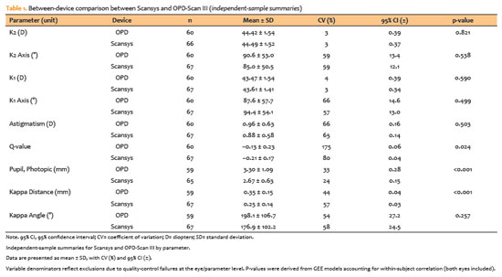
METHODS: This cross-sectional study included 40 patients (80 eyes). Each eye underwent randomized imaging with a Scheimpflug-based tomographer, a Placido-based topographer, and Tomey EM-4000 specular microscopy. Three acquisitions per device were obtained. For interdevice comparisons, the best-quality scan per eye/device was selected, whereas all three scans were used for intradevice repeatability analyses. Unreliable scans were repeated (up to five attempts) and excluded if acceptable quality was not achieved, resulting in variable denominators. Between-device comparisons were performed using generalized estimating equations
with participant-level clustering and robust standard errors and were supplemented by Bland–Altman analysis.
RESULTS: The effective sample size varied by parameter (independent summaries: 59–67 eyes; paired comparisons: 48–51 eyes). In paired-eye analyses, the Scheimpflug-based tomographer measured slightly higher keratometry values than the Placido-based topographer (K1: 43.95 vs. 43.78 D, p=0.003; K2: 44.91 vs 44.73 D, p=0.002), more negative Q-values (p=0.001), smaller photopic pupil diameter (p<0.001), and shorter kappa distance (p<0.001). Mean absolute differences were 0.32 D for K1 and 0.30 D for K2, with high dispersion for angular metrics (kappa angle coefficient of variation: 195%).
CONCLUSIONS: The Scheimpflug-based tomographer provides reproducible corneal measurements in healthy eyes. However, systematic differences relative to the Placido-based topographer—particularly for keratometry, asphericity, and pupil and kappa metrics—suggest limited interchangeability. Consistent device use is recommended when these parameters inform clinical decision-making.
Keywords: Scheimpflug tomography; Placido topography; Specular microscopy; keratometry; Corneal imaging; Refractive surgical procedures; Lenses, intraocular
Arq. Bras. Oftalmol. 2025;88 (2 )
:1-5
| DOI: 10.5935/0004-2749.2024-0113
Abstract
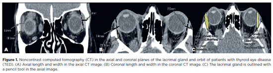
This study aimed to evaluate the morphometric and volumetric dimensions of the lacrimal gland in patients with inactive thyroid eye disease and compare them with the values reported in the literature. This case series evaluated consecutive patients with inactive thyroid eye disease treated at a tertiary eye hospital from 2015 to 2020. The patients' baseline demographics and clinical characteristics were obtained. The axial and coronal length, width, and volume of the lacrimal gland were measured on computed tomography scan images, and the results were statistically analyzed. A total of 21 patients (42 orbits) with inactive thyroid eye disease were evaluated. Their mean age was 49.0 ± 14.6 years, and 12 (57.1%) of them were men. The main complaint was dryness, and the majority of the patients had good vision and mild proptosis. The mean axial length and width of the lacrimal gland were 19.3 ± 3.9 mm and 7.5 ± 2.1 mm, respectively; coronal length and width, 20.4 ± 4.5 mm and 7.5 ± 2.1 mm, respectively; and lacrimal gland volume, 0.825 ± 0.326 mm3. Age, sex, or laterality were not found to be determinants of lacrimal gland enlargement. Patients with thyroid eye disease have enlarged lacrimal gland even in the nonactive phase of the disease multifactorial aspects influence the lacrimal gland in thyroid eye disease, making it difficult to establish a clear correlation with predisposing factors. Further studies are warranted to better understand the association between thyroid eye disease and the lacrimal gland.
Keywords: Graves' ophthalmology; Graves' disease; Lacrimal apparatus; Lacrimal apparatus diseases; X-ray computed tomography
Arq. Bras. Oftalmol. 2024;87 (2 )
:1-6
| DOI: 10.5935/0004-2749.2021-0435
Abstract
PURPOSE: This study aimed to analyze the association between magnetic resonance imaging apparent diffusion coefficient map value and histopathological differentiation in patients who underwent eye enucleation due to retinoblastomas.
METHODS: An observational chart review study of patients with retinoblastoma that had histopathology of the lesion and orbit magnetic resonance imaging with apparent diffusion coefficient analysis at Hospital de Clínicas de Porto Alegre between November 2013 and November 2016 was performed. The histopathology was reviewed after enucleation. To analyze the difference in apparent diffusion coefficient values between the two major histopathological prognostic groups, Student's t-test was used for the two groups. All statistical analyses were performed using SPSS version 19.0 for Microsoft Windows (SPSS, Inc., Chicago, IL, USA). Our institutional review board approved this retrospective study without obtaining informed consent.
RESULTS: Thirteen children were evaluated, and only eight underwent eye enucleation and were included in the analysis. The others were treated with photocoagulation, embolization, radiotherapy, and chemotherapy and were excluded due to the lack of histopathological results. When compared with histopathology, magnetic resonance imaging demonstrated 100% accuracy in retinoblastoma diagnosis. Optic nerve invasion detection on magnetic resonance imaging showed a 66.6% sensitivity and 80.0% specificity. Positive and negative predictive values were 66.6% and 80.0%, respectively, with an accuracy of 75%. In addition, the mean apparent diffusion coefficient of the eight eyes was 0.615 × 103 mm2/s. The mean apparent diffusion coefficient value of poorly or undifferentiated retinoblastoma and differentiated tumors were 0.520 × 103 mm2/s and 0.774 × 103 mm2/s, respectively.
CONCLUSION: This study revealed that magnetic resonance imaging is useful in the diagnosis of retinoblastoma and detection of optic nerve infiltration, with a sensitivity of 66.6% and specificity of 80%. Our results also showed lower apparent diffusion coefficient values in poorly differentiated retinoblastomas with a mean of 0.520 ×
103 mm2/s, whereas in well and moderately differentiated, the mean was 0.774 × 103 mm2/s.
Keywords: Retinoblastoma; Prognosis; Retinal neoplasms; Orbit; Diffusion magnetic resonance imaging
 ABO is licensed under a Creative Commons Attribution-NonComercial 4.0 Internacional.
05-tab01.jpg)
10-tab01tb.jpg)
05-fig01.jpg)
10-fig01.jpg)
11-fig01tb.jpg)
11-fig01.jpg)
05-fig01tb.jpg)
03-fig01.jpg)
01-fig01.jpg)
14-fig01.jpg)
05-fig01.jpg)