Arq. Bras. Oftalmol. 2023;86 (6 )
:1-7
| DOI: 10.5935/0004-2749.2021-0383
Abstract
Objetivo: Avaliar as taxas de sucesso anatômico e funcional da ceratoplastia penetrante associada à cirurgia vitreorretiniana assistida por ceratoprótese temporária.
Métodos: Foram incluídos neste estudo retrospectivo 15 olhos de 14 pacientes. Registraram-se as características demográficas, as patologias pré-operatórias dos segmentos anteriores e posteriores, as complicações perioperatórias, a condição pós-operatória do implante e a fixação e as complicações da retina. Foram avaliadas as taxas de sucesso anatômico e funcional.
Resultados: O período médio de acompanhamento foi de 29,8 ± 19,1(6-60) meses. A patologia corneana pós-operatória mais comum foi o abscesso do implante (7 olhos, 46,7%) e o diagnóstico mais comum no segmento posterior foi a endoftalmite (7 olhos, 46,7%). Cinco casos (33,3%) mostraram acuidade visual entre 0,001 e 0,08. Foi diagnosticada endoftalmite pré-operatória em todos os 5 casos com insucesso anatômico.
Conclusão: A cirurgia vitreorretiniana assistida por ceratoprótese temporária associada à ceratoplastia penetrante é um método eficaz de tratamento de patologias agudas e subagudas concomitantes nos segmentos anterior e posterior. Porém, os resultados podem variar de caso a caso. A endoftalmite pré-operatória é um fator de pior prognóstico de sucesso de longo prazo.
Keywords: Ceratoplastia penetrante; Cirurgia vitreorretiniana; Vitrectomia; Segmento anterior do olho; Período pré-operatório; Endoftalmite.
Arq. Bras. Oftalmol. 2022;85 (3 )
:249-254
| DOI: 10.5935/0004-2749.20220036
Abstract
Objetivo: Criar modelos, em catarata pediátrica, para estimar valores futuros de ceratometria e comprimento axial, com base na ceratometria e no comprimento axial medidos na cirurgia, para previsão do poder da lente intraocular para emetropia em idades futuras.
Métodos: Olhos com catarata bilateral, ceratometria e comprimento axial medidos na cirurgia e pelo menos um exame pós-operatório com medidas de ceratometria e comprimento axial foram considerados para este estudo. Os modelos para estimar futuras ceratometrias e comprimentos axiais foram criados considerando (1) ceratometria e comprimento axial medidos na cirurgia, (2) a inclinação média da regressão logarítmica da ceratometria e comprimento axial criada para cada olho e (3) a idade na cirurgia. A lente intraocular para emetropia em idades futuras pode ser estimada usando esses valores em fórmulas de terceira geração. Os erros de estimativa da ceratometria, comprimento axial e poder da lente intraocular, usando os modelos, também foram calculados.
Resultados: 57 olhos de 29 pacientes preencheram os critérios de inclusão. A idade média na cirurgia e acompanhamento foram de 36,96 ± 32,04 meses e 2,39 ± 1,46 anos, respectivamente. A inclinação média da regressão logarítmica criada para cada olho foi de -3.286 para ceratometria e + 3.189 para o comprimento axial. Os erros médios de estimativa absoluta para ceratometria e comprimento axial foram respectivamente: 0,61 ± 0,54 D e 0,49 ± 0,55 mm, e para o poder da lente intraocular usando as fórmulas SRK-T, Hoffer-Q e Holladay I foram: 2,04 ± 1,73 D, 2,49 ± 2,10 D e 2,26 ± 1,87 D, respectivamente.
Conclusões: Os modelos apresentados podem ser utilizados para estimar o poder da lente intraocular que levaria a emetropia em idades futuras e orientar a escolha do poder da lente intraocular a ser implantada na catarata pediátrica.
Keywords: Catarata; Biometria/métodos; Emetropia; Comprimento axial do olho; Lentes intraoculares; Criança
Arq. Bras. Oftalmol. 2025;88 (6 )
:1-5
| DOI: 10.5935/0004-2749.2025-0085
Abstract
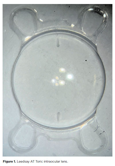
PURPOSE: The purpose of this study was to assess visual outcomes and patient satisfaction following cataract surgery involving the implantation of quad-loop intraocular lenses, including trifocal, bifocal, and toric variants.
METHODS: Information was obtained from both physical and electronic medical records of patients who underwent phacoemulsification cataract surgery with implantation of different intraocular lenses between January 1, 2022, and December 31, 2023. The study included individuals aged over 18 who received bilateral implantation of bifocal, trifocal, or monofocal toric intraocular lenses. Visual acuity was assessed at various postoperative time points using the logMAR scale. Quantitative variables were analyzed using mean and standard deviation.
RESULTS: A total of 92 eyes received premium intraocular lenses: 4 bifocal, 32 trifocal, 52 toric monofocal, and 4 trifocal toric lenses. The average preoperative corrected visual acuity was logMAR 0.478 ± 0.259. On the first postoperative day, the average uncorrected visual acuity was logMAR 0.301 ± 0.207. By day 30, 67.4% of eyes achieved uncorrected distance visual acuity of logMAR 0.2 or better. Patient satisfaction was high, with few reports of glare or halos.
CONCLUSION: Quad-loop intraocular lenses-including trifocal, bifocal, and toric models-demonstrated effective improvement in visual acuity and high levels of patient satisfaction. These lenses represent a suitable option for enhancing visual outcomes after cataract surgery. Additional studies with larger cohorts are recommended to confirm these results.
Keywords: Cataract extraction; Aberrometry/methods; Lenses, intraocular; Lens implantation, intraocular; Prosthesis design
Arq. Bras. Oftalmol. 2025;88 (4 )
:1-6
| DOI: 10.5935/0004-2749.2024-0083
Abstract
PURPOSE: We developed an artificial intelligence program for calculating intraocular lenses and analyzed its accuracy rate via ultrasonic biometry. This endeavor is aimed at enhancing precision and efficacy in the selection of intraocular lenses, particularly in cases where optical biometry is unavailable.
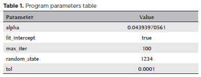
METHODS: Data was collected from the Hospital de Clínicas de Porto Alegre, which included cases of phacoemulsification with intraocular lens implantation, in which the lens selection was based on ultrasonic biometry. The program, implemented in Python, Java, and PHP, employs the ridge regression method. Two design options were developed: a basic model, which uses only keratometry variables (K1 and K2), axial size and final target refraction in the spherical equivalent, and an advanced model, which incorporates preoperative refraction and the patient's age. The Universal Barrett II formula was used to compare both models.
RESULTS: The sample consisted of 486 eyes from 313 patients, with 350 eyes used for program training and 136 for program validation. The spherical equivalent hit rates, with a variation of ±0.5 D, were 86% and 87.5% for the basic and advanced models, respectively, with no statistically significant difference between them. With the Barret Universal II formula, the success rate was 69%, which was significantly different from the values of the two aforementioned models (p<0.0001). The system was better for medium and long eyes but worse for short eyes (<=22.00 mm).
CONCLUSION: The developed artificial intelligence program was superior to the Barrett formula in terms of performance, in the general context and within the subgroup of patients with longer eyes. This innovation can considerably contribute to the selection of intraocular lenses, particularly in cases where optical biometry is unavailable.
Keywords: Biometry; Intraocular lens; Cataract; Artificial intelligence
Arq. Bras. Oftalmol. 2025;88 (6 )
:1-8
| DOI: 10.5935/0004-2749.2024-0394
Abstract
The advantages and disadvantages of using perioperative subconjunctival steroid injections in dropless cataract surgery continue to be debated. A systematic review of PubMed, EMBASE, and the Cochrane Central database identified five studies—two randomized controlled trials and three non-randomized studies—encompassing 70,751 eyes. Among these, 12,319 eyes (17.4%) received subconjunctival steroid injections, while 58,432 eyes (82.6%) were managed with topical steroids. The Cochrane Collaboration’s RoB 2 tool was applied for bias assessments in randomized controlled trials, and heterogeneity was assessed using the I² statistics. No statistically significant differences were found between the two groups regarding macular edema (p=0.249), visual acuity (p=0.73), or laser flare count (p=0.45). Both subconjunctival injections and topical steroids demonstrated comparable efficacy and safety in controlling postoperative inflammation after cataract surgery. Additional research is warranted to validate these conclusions.
Keywords: Cataract extraction; Phacoemulsification; Lens implantation, intraocular; Postoperative care; Intravitreal injections; Anti-inflammatory agents, non-steroidal/administration & dosage; Glucocorticoids; Triamcinolone acetonide; Research design; Randomiz
Arq. Bras. Oftalmol. 2024;87 (5 )
:0-0
| DOI: 10.5935/0004-2749.2022-0063
Abstract
Objetivo: Comparar os parâmetros de câmara anterior obtidos através da tomografia de coerência óptica de segmento anterior antes e após a iridectomia periférica a laser.
Métodos: Quatorze pacientes com fechamento angular primário e seis com glaucoma primário de ângulo fechado foram prospectivamente avaliados neste estudo. Gonioscopia e tomografia de coerência óptica de segmento anterior com DRI OCT Triton®foram realizadas antes e após a iridectomia periférica a laser. Os seguintes parâmetros de tomografia de coerência óptica de segmento anterior, baseados na localização do esporão escleral, foram avaliados: ângulo de abertura angular a 250 µm, 500 µm e 750 µm, área do espaço entre a íris e o trabeculado a 500 µm, ângulo entre a íris e o trabeculado, extensão do contato entre a íris e o trabeculado e curvatura da íris.
Resultados: A tomografia de coerência óptica de segmento anterior identificou 61% dos indivíduos com dois ou mais quadrantes fechados. A gonioscopia identificou mais quadrantes com ângulo fechado do que tomografia de coerência óptica de segmento anterior antes da iridectomia periférica a laser. Quanto aos parâmetros angulares, apenas ângulo de abertura angular a 250 µm no quadrante nasal não aumentou significativamente após a iridectomia
periférica a laser. A curvatura da íris e a extensão do contato entre a íris e o trabeculado apresentaram redução significativa induzida pelo procedimento a laser. Mesmo nos olhos em que a gonioscopia não identificou aumento da amplitude angular após iridectomia periférica a laser (n=7), ângulo de abertura angular a 750 µm aumentou (nasal: 0,15 ± 0,10 mm para 0,27
± 0,16 mm, p=0,01; temporal: 0,14 ± 0,11 mm para 0,25 ± 0,12 mm, p=0,001), e ICURVE diminuiu (nasal: 0,25 ±
0,04 mm vs. 0,11 ± 0,07 mm, p=0,02; temporal: 0,25 ± 0,07 mm vs. 0,14 ± 0,08 mm, p=0,007).
Conclusão: As alterações na câmara anterior induzidas pelo iridectomia periférica a laser puderam ser avaliadas quantitativamente e documentadas pelo DRI OCT Triton®.
Keywords: Gonioscopia; Tomografia de coerência óptica; Segmento anterior do olho; Glaucoma de ângulo fechado; Iridectomia; Terapia a laser; Lasers
Arq. Bras. Oftalmol. 2024;87 (4 )
:1-6
| DOI: 10.5935/0004-2749.2022-0035
Abstract
Objetivo: Comparar as diferenças entre a chord aparente µ e o chord real µ.
Métodos: Estudo prospectivo, comparativo, não randomizado e não intervencionista. Os exames de imagem (Pentacam e HD Analyzer) foram realizados na mesma sala e nas mesmas condições escotópicas. Os critérios de inclusão foram idade de 21 a 71 anos; compreensão do termo de consentimento; miopia até 4D e astigmatismo topográfico anterior até 1D. Os critérios de exclusão foram usuários de lentes de contato; pacientes com doenças oculares prévias ou cirurgias; opacidades da córnea; a presença de alterações tomográficas da córnea ou suspeita de ceratocone.
Resultados: Em nosso estudo foram analisados 116 olhos de 58 pacientes. A média de idade foi de 30,69 anos (± 7,85). Análises de correlação foram desenvolvidas e o coeficiente de correlação de Pearson (0,647) indica uma relação linear positiva moderada entre as variáveis. A média do chord µ real foi 226,21± 128,53 µm e a média do chord µ média foi 278,66 ± 123,90 µm, com diferença média de 52,45 µm (p=0,01).
A análise do diâmetro pupilar médio apresentou: 5,76mm no HD Analyzer e 3,31mm no Pentacam.
Conclusões: Entendemos a existência de uma diferença significativa entre os métodos e assim a medida de ambos os dispositivos com base em princípios diferentes devemos respeitar suas peculiaridades. Como encontramos correlação entre as duas medidas, acreditamos que ambas podem ser utilizadas na prática diária.
Keywords: Imagem óptica; Percepção visual; Pupila; Segmento anterior do olho; Córnea; Técnicas de diagnóstico oftalmológico
Arq. Bras. Oftalmol. 2024;87 (3 )
:1-7
| DOI: 10.5935/0004-2749.2021-0493
Abstract
Objetivo: Avaliar a qualidade de vida e o nível de estresse relacionada à função visual após a cirurgia de catarata pediátrica em um hospital público brasileiro.
Métodos: Estudo prospectivo em crianças de seis a 14 anos submetidas à cirurgia de catarata. A Escala de Stresse Infantil e o Questionário de Função Visual em Crianças foram usados para avaliar o nível de estresse e a qualidade de vida, respectivamente. Ambos os instrumentos foram aplicados por duas psicólogas antes e após a cirurgia. O exame oftalmológico foi realizado por dois oftalmologistas. Os dados coletados no pré e pós-operatório foram comparado.
Resultados: Vinte e três crianças (32 olhos) foram incluídas no estudo, nove delas apresentavam catarata bilateral. A média de idade na cirurgia foi de 9,65±2,26 (6 a 14) anos. Um mês após a cirurgia, o equivalente esférico foi de -0,90 ± 1,66D e a acuidade visual corrigida a distância foi de 0,13 ± 0,10 (0-0,3) LogMAR em casos bilaterais e 0,50 ± 0,39 (0-1,3) LogMAR em casos unilaterais (p<0.01). De acordo com a Escala de Stresse Infantil, 77,7% dos casos de catarata bilaterais, e 57,1% dos casos unilaterais mantiveram o nível de estresse e 34,7% das crianças melhoraram o nível de estresse. A análise do Questionário de Função Visual em Crianças foi baseada em pontuações para saúde geral, saúde geral da visão, competência, personalidade e tratamento. Após a cirurgia de catarata, 78,2% dos pacientes melhoraram ou mantiveram o escore do Questionário de Função Visual em Crianças na saúde geral, 82,6% na saúde geral da visão, 95,6% na competência, 56,5% na personalidade e 78,2% no tratamento.
Conclusão: A cirurgia de catarata pediátrica melhora a função visual e a qualidade de vida em pacientes submetidos a procedimento cirúrgico, sem aumentar o nível de estresse.
Keywords: Catarata; Extração de catarata; Experiências adversas da infância Qualidade de vida; Criança
Arq. Bras. Oftalmol. 2025;88 (5 )
:1-8
| DOI: 10.5935/0004-2749.2024-0328
Abstract
PURPOSE: Posterior capsule rupture is defined as an intraoperative posterior capsule tear resulting in vitreous loss. This study aimed to analyze the clinical characteristics, preoperative risk factors, intraoperative management strategies, and postoperative complications associated with posterior capsule rupture during phacoemulsification surgery.
METHODS: This was a retrospective observational cohort study of the medical records for 25,224 phacoemulsification surgeries performed at our tertiary eye care center between 2017 and 2022. We collected and collated the demographic characteristics and clinical findings of the patients in our cohort. Intraoperative management strategies and postoperative outcomes over a 1-year followup period were also recorded.
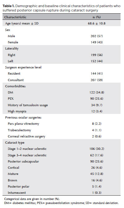
RESULTS: Posterior capsule rupture occurred in 351 eyes (351 patients), giving an overall posterior capsule rupture rate of 1.3%. The mean patient age was 68.6 ± 10.8 years. Pseudoexfoliation syndrome, mature cataracts, brown cataracts, and surgery performed by a resident were identified as risk factors for posterior capsule rupture (p<0.05 for each; the risk ratios were 2.70, 2.15, 2.44, 1.34, respectively). The most common intraoperative complications were dislocated lens fragments in the vitreous (8%) and iris damage (7.1%). The mean best-corrected visual acuity improved from 1.31 ± 0.84 (logMAR) postoperatively to 0.51 ± 0.56 at the end of the 1-year follow-up period (p<0.001). Corneal edema (55.6%) and elevated intraocular pressure (33.3%) were the most common early postoperative complications. Persistently elevated intraocular pressure (11.1%) and cystoid macular edema (5.1%) were the most common late postoperative complications.
CONCLUSION: Posterior capsule rupture is a common complication of phacoemulsification surgery that requires prolonged postoperative follow-up and a multidisciplinary approach. Despite the increased incidence of complications when rupture occurs, appropriate intraoperative and postoperative management can lead to satisfactory visual outcomes.
Keywords: Cataract extraction; Phacoemulsification; Posterior capsule rupture; Corneal edema; Risk factors; Postoperative complications; Intraoperative complications
Arq. Bras. Oftalmol. 2025;88 (5 )
:1-7
| DOI: 10.5935/0004-2749.2024-0368
Abstract
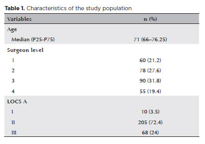
PURPOSE: To compare endothelial corneal cell changes following cataract surgery performed by phacoemulsification with intraocular lens implantation, conducted by surgeons with varying levels of experience.
METHODS: Two hundred and eighty-three eyes diagnosed with cataract were included. Lens opacity was classified into three categories (I, II, and III). Surgeons were categorized into four experience levels (1, 2, 3, and 4), based on years of practice and lifetime surgeries performed. Corneal endothelial characteristics were assessed using non-contact specular microscopy, with measurements taken before surgery and 30-60 days post-surgery.
RESULTS: Pre- and postoperative endothelial analysis showed no significant differences between surgeon levels regarding visual acuity achieved, corneal thickness, and endothelial hexagonality. However, the central endothelial cell density index showed a significantly greater reduction among level 1 surgeons (p=0.026). Grade II cataracts exhibited significant variations in the central endothelial cell density (p=0.011) and average cell size, with level 1 surgeons showing the largest increases (p=0.024).
CONCLUSIONS: The analysis revealed significant differences in visual acuity and endothelial indices between surgeon experience levels, with less experienced surgeons showing greater variations and poorer performance. Clinical protocols should consider these data to establish safer training protocols.
Keywords: Cataract extraction; Phacoemulsification; Endothelium; corneal; Lens implantation, intraocular; Visual acuity; Internship and residency; Surgeons
Arq. Bras. Oftalmol. 2024;87 (2 )
:1-6
| DOI: 10.5935/0004-2749.2023-2022-0341
Abstract
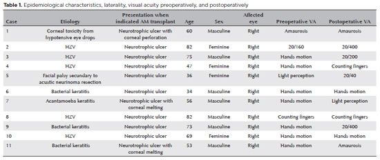
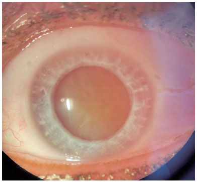
PURPOSE: To evaluate the clinical results of cryopreserved amniotic membrane transplantation as a treatment option for refractory neurotrophic corneal ulcers.
METHODS: This prospective study included 11 eyes of 11 patients who underwent amniotic membrane transplantation for the treatment of refractory neurotrophic corneal ulcers at Hospital de Clínicas da Universidade Federal do Paraná, in the city of Curitiba, from May 2015 to July 2021. Patients underwent different surgical techniques in which the amniotic membrane was applied with the epithelium facing upward to promote corneal re-epithelialization.
RESULTS: The median age of the patients was 60 years (range, 34-82 years), and 64% were men. The predominant etiology of corneal ulcers was herpes zoster (45% of cases). Approximately one-third of the patients (27%) were chronically using hypotensive eye drops, and more than half (54%) had previously undergone penetrating corneal transplantation. At the time of amniotic membrane transplantation, 18% of the eyes had corneal melting, 9% had corneal perforation, and the others had corneal ulceration without other associated complications (73%). The time between clinical diagnosis and surgical treatment ranged from 9 days to 2 years. The corrected visual acuity was worse than 20/400 in 90% of the patients preoperatively, with improvement in 36% after 3 months of the procedure, worsening in 18% and remaining stable in 36%. Of the patients, 81% complained of preoperative pain, and 66% of them reported total symptom relief after the surgical procedure. In one month, 54.6% of the patients presented a closure of epithelial defect, and half of the total group evolved with corneal thinning. The failure rate was 45.5% of the cases.
CONCLUSION: Cryopreserved amniotic membrane transplantation can be considered a good alternative for treating refractory neurotrophic corneal ulcers, as it resulted in significant improvement in pain (66%) and complete epithelial closure (60%) in many patients at 1 month postoperatively. Notably, the high failure rate highlights the need for further studies to identify patient- and ulcer-related factors that may influence the outcomes of this procedure.
Keywords: Amnion/transplantation; Corneal ulcer; Anterior eye segment; Keratitis
 ABO is licensed under a Creative Commons Attribution-NonComercial 4.0 Internacional.
10-fig01tb.jpg)
07-tab01tb.jpg)
11-fig01tb.jpg)
07-fig01tb.jpg)
14-tab01tb.jpg)
13-fig01.jpg)
06-fig01.jpg)