Showing of 1 until 15 from 112 result(s)
Search for: Contact lenses; Scleral lenses, fitting; Keratoconus; Keratotomy, radial; Refractive surgical procedures; Rehabilitation; Learning curve
Abstract
Introdução: Alterações topográficas podem ocorrer secundariamente ao uso de uso de lentes de contato duras ou gelatinosas. O termo "corneal warpage" é utilizado para denominar as deformidades corneanas causadas pelas lentes. O quadro topográfico da fase inicial do ceratocone pode ser muito parecido com o de um paciente com este quadro. Objetivo: Mostrar um paciente portador de ceratocone, usuário de lentes de contato rígida gás-permeável (RGP) que desenvolveu quadro de "corneal warpage", diagnosticado e acompanhado por topografias e exames clínicos seriados. Relato de caso: O paciente é um engenheiro de 40 anos com diagnóstico de ceratocone bilateral há 12 anos, usando lentes RGP desde então. No primeiro exame em 5/95, a refração de OD foi impossível de se determinar e a ceratometria era maior que 60,00 D. Foi feita topografia, que se mostrou compatível com ceratocone, tendo sido adaptada uma lente Soper, com visão de 20/70. Após um ano, uma topografia de controle mostrou aumento da curvatura central e aplanamento da periferia inferior. O uso da lente foi suspenso e após 6 meses houve regressão das mudanças, tendo sido adaptadas novas lentes com melhor padrão e visão de 20/40. Discussão: O caso apresentado evidencia a ocorrência de deformidade corneana em um portador de ceratocone usuário de lente RGP. O autor discute a fisiopatologia e o diagnóstico clínico e topográfico do "corneal warpage", lembrando que a exemplo de pacientes normais, os pacientes com ceratocone podem apresentar estas alterações, que devem ser diferenciadas da própria evolução da doença.
Keywords: Ceratocone; Lente de contato; Deformidade corneana
08-fig01.jpg)
Abstract
Objetivo: Este estudo tem como objetivo avaliar a eficácia de lentes de contato híbridas de nova geração nos resultados visuais e na qualidade de vida relacionada à visão em pacientes com ceratocone com intolerância ou insucesso dos métodos de correção habituais, tais como lentes flexíveis de silicone-hidrogel ou rígidas permeáveis a gases.
Métodos: Foram incluídos neste estudo transversal prospectivo 42 olhos de 28 pacientes com ceratocone. Uma lente Airflex (Swisslens) foi aplicada nos olhos de acordo com as instruções do fabricante. Um exame oftalmológico, incluindo refração manifesta, melhor acuidade visual corrigida para longe, biomicroscopia com lâmpada de fenda e a aplicação do National Eye Institute Visual Function Questionnaire-25 (NEI-VFQ-25), foi realizado no início do estudo e na visita de 6 meses.
Resultados: Foi possível obter um ajuste adequado em 39 olhos (92,9%) de 26 pacientes. Foram excluídos do estudo 6 olhos de 3 pacientes devido à cessação do uso de lentes. A idade média dos usuários bem-sucedidos era de 20,3 ± 4,9 anos. A média da melhor acuidade visual corrigida para longe foi melhorada estatisticamente de 0,62 ± 0,30 para 0,11 ± 0,06 logMAR com as lentes de contato híbridas Airflex (p<0,001). A pontuação média geral composta no questionário NEI-VFQ-25 aumentou de forma estatisticamente significativa com a lente de contato híbrida Airflex na visita de 6 meses, em comparação com a pontuação inicial (de 77,1 ± 16,3 para 90,9 ± 7,3, p=0,036). As lentes de contato híbridas Airflex apresentaram pontuações melhores com significância estatística em todos os itens das sub-escalas do NEI-VFQ-25 (todos com p<0,05). Nenhum efeito adverso significativo foi observado.
Conclusões: Lentes de contato híbridas de nova geração podem ser usadas como uma alternativa eficaz para a correção do astigmatismo irregular em pacientes com ceratocone com intolerância ou insucesso dos métodos habituais. Com essas lentes, pode-se alcançar uma melhora significativa na qualidade de vida relacionada à visão em pacientes com ceratocone.
Keywords: Lentes de contato; Ceratocone; Refração ocular; Acuidade visual; Qualidade de vida; Inquéritos e questionários
Abstract
PURPOSE: This study aimed to identify the strategies adopted by Brazilian ophthalmologists to control myopia in clinical practice.
METHODS: This was a prospective cross-sectional study. Data were collected using an online questionnaire.
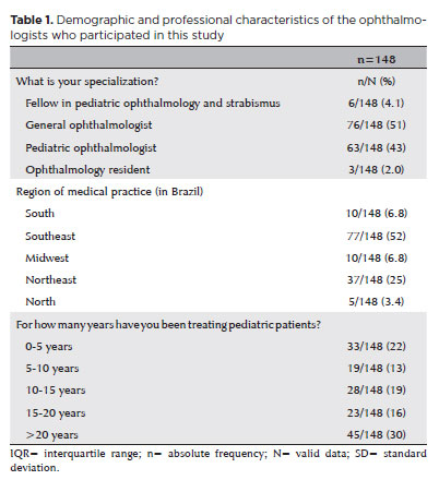
RESULTS: Responses from 148 participants were collected between March and May 2024. The majority of respondents were general ophthalmologists (51%) and pediatric ophthalmologists (43%). They came from all regions of Brazil, but more than half (52%) were from the Southeast region. Most participants (30%) had over 20 years of clinical practice experience. A significant proportion (89.2%) treated progressive myopia. The most requested complementary exams were optical biometry (83.78%) and corneal topography or tomography (69.59%). Behavioral measures were considered the most effective myopia treatment strategies by 41.2% of the respondents, followed by optical (33.8%) and pharmacological interventions (25%). Most recommended spending more time outdoors (94.59%) and reducing screen time (93.92%). Spectacle lenses for myopia (83.11%) and 0.025% atropine eye drops (54.73%) were the most prescribed treatments after the recommendation of environmental and behavioral changes.
CONCLUSION: This study presents a novel analysis of the clinical strategies for myopia control among Brazilian ophthalmologists. Understanding current clinical practices and identifying possible improvements are essential steps toward developing evidence-based guidelines and professional education aimed at improving patient care.
Keywords: Myopia/epidemiology; Refractive errors; Contact lenses; Myopia/drug therapy; Atropine/therapeutic use; Ophthalmologists; Practice patterns, physicians’; Surveys and questionnaires; Brazil/epidemiology
15-tab01.jpg)
Abstract
Objetivo: O objetivo deste estudo foi analisar dados epidemiológicos de pacientes e resultados laboratoriais para todas as amostras de córnea coletadas de pacientes atendidos no Departamento de Oftalmologia do Hospital São Paulo, Brasil, durante um período de 30 anos e correlacionar com o uso de lentes de contato.
Métodos: Amostras de córnea de pacientes com diagnóstico clínico de ceratite microbiana (de janeiro de 1987 a dezembro de 2016) foram incluídas neste estudo. Resultados laboratoriais para culturas positivas para bactérias, fungos e Acanthamoeba spp. foram analisados retrospectivamente. Para verificar se o número de pacientes com ceratite microbiana associada à lente de contato, fator de risco para infecção microbiana, mudou ao longo do tempo, a análise foi dividida em três décadas: 1987-1996, 1997-2006 e 2007-2016. As informações incluindo o sexo do paciente, idade e tipo de organismo isolado foram comparadas entre os períodos. A análise estatística foi realizada no software SAS/STAT 9.3 e SPSS (v20.0).
Resultados: Amostras de córnea de 10.562 pacientes com ceratite microbiana foram incluídas no estudo, das quais 1.848 foram relacionadas ao uso de lentes de contato. Os resultados revelaram que a frequência de ceratite microbiana associada à lente de contato aumentou nas últimas duas décadas analisadas. No geral, os homens compreendiam uma proporção maior do grupo ceratite microbiana não associada à lente de contato (CMNLC) (60,3%) e as mulheres eram mais frequentes no grupo ceratite microbiana associada à lente de contato (59,5%). Pacientes com idade entre 19 e 40 anos foram mais frequentemente observados no grupo ceratite microbiana associada à lente de contato em todos os períodos. Staphylococcus spp. foi a bactéria Gram-positiva mais frequentes, enquanto Pseudomonas spp. foi a bactéria Gram-negativa nos grupos ceratite microbiana. Entre os fungos ceratite microbiana, os fungos filamentosos foram os fungos mais frequentes durante todo o período do estudo, com Fusarium spp. sendo o mais frequente no grupo ceratite microbiana não associada à lente de contato. Acanthamoeba spp. e Pseudomonas spp. amostras positivas foram significativamente correlacionadas com ceratite microbiana associada à lente de contato.
Conclusões: A maior prevalência de ceratite microbiana associada à lente de contato no nosso estudo foi observada em mulheres e adultos jovens com idade entre 19 e 40 anos. Staphylococcus spp. e Fusarium spp. foram as bactérias e fungos predominantes isolados nas amostras da córnea. Pseudomonas spp. e Acanthamoeba spp. foram significativamente correlacionados a ceratite microbiana associada à lente de contato neste estudo.
Keywords: Lentes de contato/efeitos adversos; Infecções oculares bacterianas/microbiologia; Ceratite por Acanthamoeba; Úlcera de córnea
Abstract
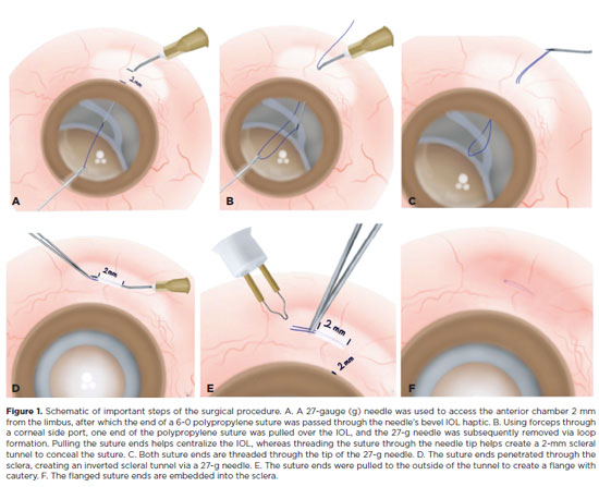
PURPOSE: The aim of this study is to describe a minimally invasive and atraumatic technique for managing the polypropylene suture-assisted scleral fixation of intraocular lens–capsular bag complex or artificial iris–intraocular lens complex for repositioning late luxated or subluxated intraocular lens–capsular bags and artificial iris–intraocular lens complexes.
METHODS: In this retrospective and observational study, we evaluated 11 patients, including 10 patients with capsular bag–intraocular lens complex subluxation or luxation into the vitreous cavity and 1 patient with an aniridia–intraocular lens complex. A single senior surgeon performed the procedures. After anesthesia, a 4 × 4 mm conjunctival peritomy was created, and a 6-0 polypropylene suture was passed through the sclera marked 2.0 mm posterior to the limbus. The suture ends were cauterized into a flange under 0.5 mm and inserted inversely into a scleral tunnel, concealed within a 2-mm scleral tunnel to ensure secure intraocular lens positioning.
RESULTS: We analyzed 11 patients with dislocated or dropped capsular bag–intraocular lens complexes. The patients' median age was 67 (range 44–78) years, with a median follow-up of 10 (range: 4–16) months. There were 8 (72%) men and 3 (27%) women. Conjunctival peritomy was performed in 4 (36%) patients. Predominantly, preoperative diagnoses indicated 7 (63%) patients with dislocated capsular bag–intraocular lens complexes. The capsular bag–intraocular lens complexes were centralized in all patients, and optical coherence tomography confirmed accurate suture positioning within the sclera. No suture-related complications were observed throughout the follow-up period, and no vision-threatening complications were reported during the postoperative follow-up.
CONCLUSIONS: Our technique provides a simple, effective solution for treating decentralized or dislocated capsular bag–intraocular lens complexes, eliminating the need for complex interventions such as large corneal wounds, scleral flaps, intraocular lens exchange, and intraocular lens externalization.
Keywords: Scleral fixation; Intraocular lens dislocation; Ophthalmologic surgical procedures; Sutures; Intraocular lens; Lens subluxation
Abstract
PURPOSE: Using advanced imaging techniques, this study aimed to evaluate corneal stability, epithelial remodeling, and tear film changes over a one-year period in first-time soft-contact lens wearers.
METHODS: A retrospective study was conducted on 100 eyes of 50 first-time daily soft-contact lens users aged 21–65 years with no prior rigid gas-permeable lens wear. The Sirius Scheimpflug imaging system was used to assess corneal topography, epithelial thickness, and non-invasive tear break-up time at baseline, 3, 6, and 12 months. Corneal warpage was evaluated using symmetry indices and Baiocchi Calossi Versaci indices. We performed statistical analysis using repeated-measures analyses of variance with Greenhouse-Geisser correction.
RESULTS: The mean baseline central corneal thickness was 537.83 (±7.92) µm, with no significant thinning after one year. The average simulated keratometry values remained stable, indicating no progressive corneal steepening or flattening. There were no significant changes in warpage indices over time, suggesting corneal shape preservation. Higher-order aberrations (coma, trefoil, and spherical aberrations) and non-invasive tear break-up time remained unchanged throughout the study period.
CONCLUSIONS: Modern silicone hydrogel soft-contact lenses do not induce significant corneal warpage, epithelial remodeling, or optical aberrations over a one-year period. We found that corneal morphology and tear film stability were preserved, supporting the safety of soft-contact lens use. These findings provide clinically relevant insights into the long-term impact of contact lens wear. They may facilitate improved lens fitting strategies and preoperative refractive surgery assessments.
Keywords: Contact lenses, hydrophilic; Cornea/surgery; Corneal diseases; Corneal topography; Adaptation, ocular/physiology; Endothelium, corneal/pathology; Refractive errors; Tears/metabolism.
Abstract
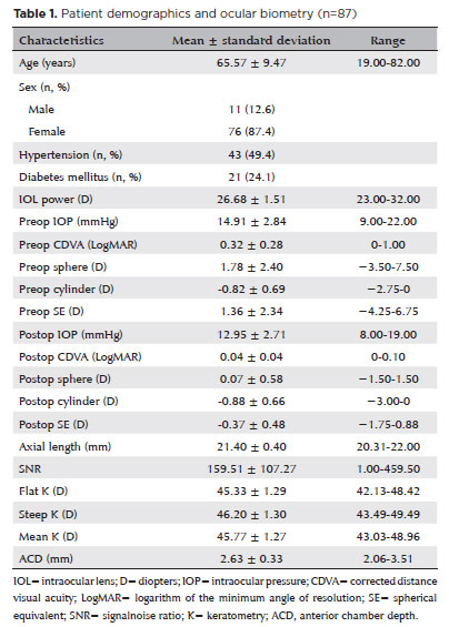
PURPOSE: To compare the refractive prediction error of Hill-radial basis function 3.0 with those of 3 conventional formulas and 11 combination methods in eyes with short axial lengths.
METHODS: The refractive prediction error was calculated using 4 formulas (Hoffer Q, SRK-T, Haigis, and Hill-RBF) and 11 combination methods (average of two or more methods). The absolute error was determined, and the proportion of eyes within 0.25-diopter (D) increments of absolute error was analyzed. Furthermore, the intraclass correlation coefficients of each method were computed to evaluate the agreement between target refractive error and postoperative spherical equivalent.
RESULTS: This study included 87 eyes. Based on the refractive prediction error findings, Hoffer Q formula exhibited the highest myopic errors, followed by SRK-T, Hill-RBF, and Haigis. Among all the methods, the Haigis and Hill-RBF combination yielded a mean refractive prediction error closest to zero. The SRK-T and Hill-RBF combination showed the lowest mean absolute error, whereas the Hoffer Q, SRK-T, and Haigis combination had the lowest median absolute error. Hill-radial basis function exhibited the highest intraclass correlation coefficient, whereas SRK-T showed the lowest. Haigis and Hill-RBF, as well as the combination of both, demonstrated the lowest proportion of refractive surprises (absolute error >1.00 D). Among the individual formulas, Hill-RBF had the highest success rate (absolute error ≤0.50 D). Moreover, among all the methods, the SRK-T and Hill-RBF combination exhibited the highest success rate.
CONCLUSIONS: Hill-radial basis function showed accuracy comparable to or surpassing that of conventional formulas in eyes with short axial lengths. The use and integration of various formulas in cataract surgery for eyes with short axial lengths may help reduce the incidence of refractive surprises.
Keywords: Cataract; Lenses, intraocular; Axial length, eye; Refractive errors; Artificial intelligence
Abstract
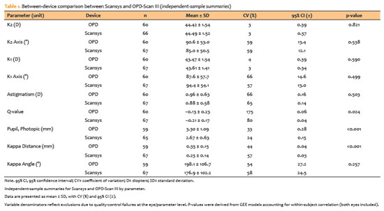
PURPOSE: To evaluate the reliability and comparability of a Scheimpflug-based tomographer relative to a Placido-based topographer and specular microscopy in healthy eyes.
METHODS: This cross-sectional study included 40 patients (80 eyes). Each eye underwent randomized imaging with a Scheimpflug-based tomographer, a Placido-based topographer, and Tomey EM-4000 specular microscopy. Three acquisitions per device were obtained. For interdevice comparisons, the best-quality scan per eye/device was selected, whereas all three scans were used for intradevice repeatability analyses. Unreliable scans were repeated (up to five attempts) and excluded if acceptable quality was not achieved, resulting in variable denominators. Between-device comparisons were performed using generalized estimating equations
with participant-level clustering and robust standard errors and were supplemented by Bland–Altman analysis.
RESULTS: The effective sample size varied by parameter (independent summaries: 59–67 eyes; paired comparisons: 48–51 eyes). In paired-eye analyses, the Scheimpflug-based tomographer measured slightly higher keratometry values than the Placido-based topographer (K1: 43.95 vs. 43.78 D, p=0.003; K2: 44.91 vs 44.73 D, p=0.002), more negative Q-values (p=0.001), smaller photopic pupil diameter (p<0.001), and shorter kappa distance (p<0.001). Mean absolute differences were 0.32 D for K1 and 0.30 D for K2, with high dispersion for angular metrics (kappa angle coefficient of variation: 195%).
CONCLUSIONS: The Scheimpflug-based tomographer provides reproducible corneal measurements in healthy eyes. However, systematic differences relative to the Placido-based topographer—particularly for keratometry, asphericity, and pupil and kappa metrics—suggest limited interchangeability. Consistent device use is recommended when these parameters inform clinical decision-making.
Keywords: Scheimpflug tomography; Placido topography; Specular microscopy; keratometry; Corneal imaging; Refractive surgical procedures; Lenses, intraocular
09-tab01.jpg)
Abstract
Objetivo: Explorar os efeitos terapêuticos das lentes de ortoceratologia combinados com colírio atropina 0,01% em miopia juvenil.
Métodos: Um total de 340 pacientes com miopia juvenil (340 olhos) tratados entre 2018 e Dezembro de 2020 foram divididos em Grupo Controle (170 casos com 170 olhos, lentes de ortoceratologia) e Grupo Observação (170 casos com 170 olhos, lentes de ortoceratologia combinadas com colírio atropina 0,01%). A acuidade visual melhor corrigida para longe, acuidade visual melhor corrigida para perto, dioptria, comprimento axial, amplitude de acomodação, diâmetro da pupila brilhante, diâmetro da pupila escura, espessura da camada lipídica da película lacrimal e tempo de ruptura do rasgo foram medidos antes do tratamento e 1 ano depois. A incidência de reações adversas foi observada.
Resultados: Antes do tratamento, o grau esférico equivalente foi significativamente melhorado em 0,22 (0,06, 0,55) D e 0,40 (0,15, 0,72) D respectivamente no Grupo Observação e no Grupo Controle após o tratamento (p<0,01). Após tratamento, o comprimento axial foi significativamente aumentado em (0,15 ± 0,12) mm e (0,24 ± 0,11) mm respectivamente nos Grupos Observação e controle (p<0,01), enquanto, no grupo de observação, a amplitude de acomodação diminuiu significativamente e foi inferior a do Grupo Controle, e o diâmetro da pupila brilhante e o diâmetro da pupila escura aumentaram significativamente e foram maiores do que os do Grupo Controle (p<0,01). A espessura da camada lipídica da película lacrimal e o tempo de ruptura do rasgo diminuíram significativamente nos dois grupos (p<0,01) após o tratamento.
Conclusões: As lentes de ortoceratologia combinadas com colírio atropina 0,01% podem melhorar significativamente o efeito controle em miopia juvenil com elevada segurança.
Keywords: Atropina; Miopia; Procedimentos ortoceratológicos; Comprimento axial do olho; Topografia da córnea; Acuidade visual; Lentes de contato.
Abstract
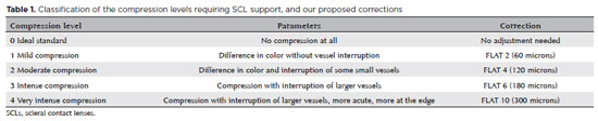
PURPOSE: This study aimed to modify scleral contact lenses to achieve a desired compression standard and to evaluate the effectiveness and reliability of the adjustments.
METHODS: In this nonrandomized, noncomparative, and partially masked study Scleral contact lens fittings were analyzed in 20 eyes of 12 patients (50% women, 50% men) diagnosed with keratoconus. Participants were selected based on their need for scleral contact lenses (SCLs), which was determined in complete ophthalmological examinations. Patients were tested with Zenlens scleral contact lenses (Bausch & Lomb, Vaughan, Ontario, Canada). We evaluated compression in the lens support area after one hour of use, excluding cases of peripheral lifting. Photos of the adaptations were sent to five experts for analysis of the quadrants (nasal, temporal, superior, and inferior). We used Fisher's exact test for statistical analysis.
RESULTS: The proposed adjustment was highly effective (93.5% correct) in lens delivery (BL=0), with the interrater agreement between doctors ranging from 68.8% to 80.9%.
CONCLUSION: The clinical parameters proposed for scleral contact lenses adjustment proved useful and reproducible, enabling their practical application to scleral lens adaptation.
Keywords: Contact lenses; Lifting; Keratoconus; Rehabilitation
Abstract
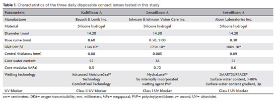
PURPOSE: This study aimed to compare the effects of three different daily disposable contact lens materials on contrast sensitivity.
METHODS: The participants were aged 18–45 years, with spherical equivalent refraction between -0.50 D and -6.00 D, astigmatism below 0.75 D, and best contact lens-corrected visual acuity of 0.0 logMAR or better. Each patient was fitted binocularly with three daily disposable contact lenses made of different materials on three separate examination days. These materials were kalifilcon A, senofilcon A, and verofilcon A. The contrast sensitivity of each patient was recorded at spatial frequencies of 3, 6, 12, and 18 cycles per degree (cpd) under photopic (85 cd/m2) and mesopic (3 cd/m2) conditions.
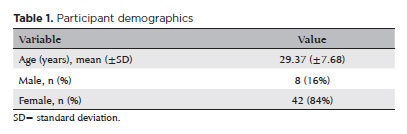
RESULTS: The current study comprised 72 eyes of 34 female and two male patients. The mean age of the participants was 25.63 (± 0.80) years. Under photopic conditions, the participants’ contrast sensitivity was significantly better with senofilcon A than with kalifilcon A at a frequency of 12 cpd (p=0.008). Under mesopic conditions, participants’ contrast sensitivity was significantly higher with kalifilcon A than verofilcon A at 3 cpd (p=0.001), and with senofilcon A than verofilcon A at 12 cpd (p=0.004). The pre-lens non-invasive break-up times did not differ significantly between the three daily disposable contact lenses (p>0.05).
CONCLUSION: In both photopic and mesopic lighting conditions, the participants in this study exhibited differences in contrast sensitivity when wearing three different daily disposable contact lens types, despite similar visual acuity and pre-lens tear film stability results in their clinical evaluations. These findings demonstrate the potential for subjective visual complaints arising from variations in the contrast sensitivity achieved by different daily disposable contact lenses.
Keywords: Contact lenses; Contrast sensitivity; Astigmatism; Lighting; Visual acuity
02-fig01.jpg)
Abstract
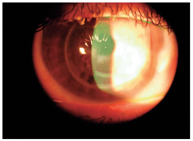
Uma paciente de 38 anos desenvolveu afacia e aniridia secundárias a um trauma, levando à perda da visão. Para melhorar sua visão, um complexo de íris e lente intraocular (Reper®) foi fixado à esclera com a técnica de flange duplo de Canabrava. Houve um aumento satisfatório na acuidade visual do paciente e nenhuma complicação foi observada durante o acompanhamento de 6 meses. A técnica de Canabrava simplifica e melhora a fixação do complexo de íris e lente intraocular na esclera. É uma opção segura que não requer retalhos ou pontos esclerais.
Keywords: Afacia/etiologia; Aniridia; Implante de lente intraocular; Lentes intraoculares; Esclera/cirurgia; Acuidade visual; Humanos; Relatos de casos.
04-fig01.jpg)
Abstract
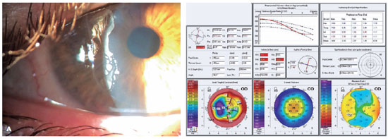
O ceratocone é uma doença progressiva que se manifesta como uma elevação semelhante a um cone da córnea central ou paracentral inferior e é associada a uma redução irregular da espessura do estroma. Há uma diminuição gradual da acuidade visual devido à assimetria da córnea, ao astigmatismo irregular e a um aumento das aberrações ópticas, o que prejudica a qualidade de vida. Foram desenvolvidos vários procedimentos para tentar interromper ou mesmo reverter a evolução da doença. Um deles é o chamado procedimento de Bader, que inclui um padrão de incisões em volta da circunferência da córnea e na base do cone protuberante. Essas incisões penetram até 70%-90% da profundidade da córnea e têm o objetivo de achatar a topografia e diminuir a assimetria da córnea e o astigmatismo irregular. Embora essa técnica seja muito promissora, segundo um estudo anterior, aqui se apresenta o caso de um paciente no qual esses objetivos não foram atingidos. Esse paciente recebeu lentes de contato para restaurar e manter sua visão, enquanto sua ectasia corneana e a redução da espessura progrediram ao longo da década seguinte.
Keywords: Ceratocone; Astigmatismo; Córnea; Topografia da córnea; Procedimentos cirúrgicos oftalmológicos; Lentes de contato; Dilatação patológica; Acuidade visua; Qualidade de vida.
Abstract
We present a case of a patient complaining of monocular diplopia due to a decentered ablation after LASIK. The patient underwent a wavefront-guided retreatment, which resulted in an epithelial ingrowth complication. Additionally, the patient developed cataract, with cataract surgery requiring reliable biometric measurements. Therefore, we opted for corneal treatment and corneal surface regularization. Although we attempted to lift the flap and wash the interface initially, the procedure proved unsuccessful, thereby necessitating immediate flap amputation. Once the corneal surface was regularized in the seventh postoperative month, transepithelial photorefractive keratectomy was successfully performed to homogenize the ocular surface, thereby significantly improving the patient's corrected visual acuity and resolving monocular diplopia. The surface and corneal curvature stabilized by the fifth month after the procedure. Phacoemulsification was then performed along with the implantation of a toric monofocal lens, which was selected using an appropriate formula, resulting in an excellent uncorrected visual acuity.
Keywords: Refractive surgical procedures; Surgical flap/surgery; Keratomileusis laser In situ/methods; Biometry; Corneal topography; Lasers, Excimer/adverse effects; Dipoplia/etiologia; Visual acuity; Humans; Case reports
Abstract
O ceratocone é uma doença progressiva que se manifesta como uma elevação semelhante a um cone da córnea central ou paracentral inferior e é associada a uma redução irregular da espessura do estroma. Há uma diminuição gradual da acuidade visual devido à assimetria da córnea, ao astigmatismo irregular e a um aumento das aberrações ópticas, o que prejudica a qualidade de vida. Foram desenvolvidos vários procedimentos para tentar interromper ou mesmo reverter a evolução da doença. Um deles é o chamado procedimento de Bader, que inclui um padrão de incisões em volta da circunferência da córnea e na base do cone protuberante. Essas incisões penetram até 70%-90% da profundidade da córnea e têm o objetivo de achatar a topografia e diminuir a assimetria da córnea e o astigmatismo irregular. Embora essa técnica seja muito promissora, segundo um estudo anterior, aqui se apresenta o caso de um paciente no qual esses objetivos não foram atingidos. Esse paciente recebeu lentes de contato para restaurar e manter sua visão, enquanto sua ectasia corneana e a redução da espessura progrediram ao longo da década seguinte.
Keywords: Ceratocone; Astigmatismo; Córnea; Topografia da córnea; Procedimentos cirúrgicos oftalmológicos; Lentes de contato; Dilatação patológica; Acuidade visua; Qualidade de vida
 ABO is licensed under a Creative Commons Attribution-NonComercial 4.0 Internacional.
About
Issues
Editorial Board
Submission
Arquivos Brasileiros de Oftalmologia
Official publication of Brazilian Council of Ophthalmology - Conselho Brasileiro de Oftalmologia (CBO)
Rua Casa do Ator, 1.117 - 2nd floor - Zip Code: 04546-004
São Paulo - SP, Brazil
TEL: +55 11 3266-4000
E-mail: [email protected]