

Showing of 1 until 14 from 34 result(s)
Search for: Sclera; Transplantation heterologous; Dogs; Animal
12-tab01tb.jpg)
Abstract
Objetivo: Comparar o teste de meniscometria em tira com o teste lacrimal de Schirmer 1 e o tempo de ruptura do filme lacrimal, em cães com olhos normais e cães com diagnóstico de ceratoconjuntivite seca.
Métodos: Cento e cinquenta e seis olhos de 78 cães, 88 olhos normais e 68 olhos com diagnóstico de ceratoconjuntivite seca. Os testes foram realizados na seguinte sequência: o teste lacrimal de Schirmer 1 foi utilizado para alocar os cães no grupo normal ou no grupo ceratoconjuntivite seca, seguido pelo teste de meniscometria em tira e tempo de ruptura do filme lacrimal.
Resultados: As médias e desvios-padrão para os olhos normais foram: teste lacrimal de Schirmer 1 = 22,75 ± 3,88 mm / min; teste de meniscometria em tira = 10,01 ± 2,35 mm / 5 seg; tempo de ruptura do filme lacrimal = 25,82 ± 5,47 seg; e para os olhos do grupo ceratoconjuntivite seca foram: teste lacrimal de Schirmer 1 = 6,10 ± 4,44 mm / min; teste de meniscometria em tira = 3,03 ± 2,62 mm / 5 seg; tempo de ruptura do filme lacrimal = 10,78 ± 4,23 seg. O teste de correlação de Spearman no grupo ceratoconjuntivite seca foi muito alto, com diferença significativa entre teste de meniscometria em tira e teste lacrimal de Schirmer 1 (r=0,848, p<0,001), moderada e significativa entre teste de meniscometria em tira e tempo de ruptura do filme lacrimal (r=0,773, p<0,001). O cut-off para teste de meniscometria em tira para ceratoconjuntivite seca foi identificado em 7,0 mm / 5 seg, valores abaixo de 10 mm / 5 seg podem ser considerados suspeitos para KCS.
Conclusões: Este estudo forneceu valores de teste de meniscometria em tira em olhos normais e com ceratoconjuntivite seca em cães, revelando alta sensibilidade e especificidade em comparação com o teste lacrimal de Schirmer 1. No futuro, o teste de meniscometria em tira pode ser outro teste quantitativo importante e pode complementar o teste lacrimal de Schirmer padrão ouro para o diagnóstico de ceratoconjuntivite seca em cães, um excelente modelo animal para o estudo de ceratoconjuntivite seca.
Keywords: Ceratoconjuntivite seca; Teste lacrimal de Schirmer; Teste de meniscometria de tira; Tempo de ruptura do filme lacrimal; Cães.
Abstract
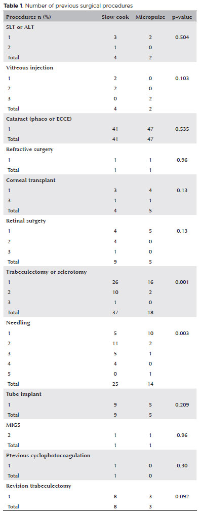
PURPOSE: This study aimed to compare the safety and effectiveness of intraocular pressure reduction between micropulse transscleral cyclophotocoagulation and “slow cook” transscleral cyclophotocoagulation in patients with refractory primary open-angle glaucoma.
METHODS: We included patients with primary open angle glaucoma with at least 12 months of follow-up. We collected and analyzed data on the preoperative characteristics and postoperative outcomes. The primary outcomes were a reduction of ≥20% of the baseline value (criterion A) and/or intraocular pressure between 6 and 21 mmHg (criterion B).
RESULTS: We included 128 eyes with primary open-angle glaucoma. The preoperative mean intraocular pressure was 25.53 ± 6.40 and 35.02 ± 12.57 mmHg in the micropulse- and “slow cook” transscleral cyclophotocoagulation groups, respectively (p<0.001). The mean intraocular pressure was reduced significantly to 14.33 ± 3.40 and 15.37 ± 5.85 mmHg in the micropulse- and “slow cook” transscleral cyclophotocoagulation groups at the last follow-up, respectively (p=0.110). The mean intraocular pressure reduction at 12 months was 11.20 ± 11.46 and 19.65 ± 13.22 mmHg in the micropulse- and “slow cook” transscleral cyclophotocoagulation groups, respectively (p<0.001). The median preoperative logMAR visual acuity was 0.52 ± 0.69 and 1.75 ± 1.04 in the micropulse- and “slow cook” transscleral cyclophotocoagulation groups, respectively (p<0.001). The mean visual acuity variation was -0.10 ± 0.35 and -0.074 ± 0.16 in the micropulse- and “slow cook” transscleral cyclophotocoagulation, respectively (p=0.510). Preoperatively, the mean eye drops were 3.44 ± 1.38 and 2.89 ± 0.68 drugs in the micropulse- and “slow cook” transscleral cyclophotocoagulation groups, respectively (p=0.017), but those were 2.06 ± 1.42 and 1.02 ± 1.46 at the end of the study in the slow cook” and micropulse transscleral cyclophotocoagulation groups, respectively (p<0.001). The success of criterion A was not significant between both groups. Compared with 11 eyes (17.74%) in the slow cook” transscleral cyclophotocoagulation group, 19 eyes (28.78%) in the micropulse transscleral cyclophotocoagulation group showed complete success (p=0.171). For criterion B, 28 (42.42%) and 2 eyes (3.22%) showed complete success after micropulse- and slow cook” transscleral cyclophotocoagulation, respectively (p<0.001).
CONCLUSION: Both techniques reduced intraocular pressure effectively.
Keywords: Sclera/surgery; Glaucoma, open-angle/surgery; Ciliary body/surgery; Intraocular pressure; Laser coagulation/methods; Lasers, semiconductor; Comparative study; Effectiveness
Abstract
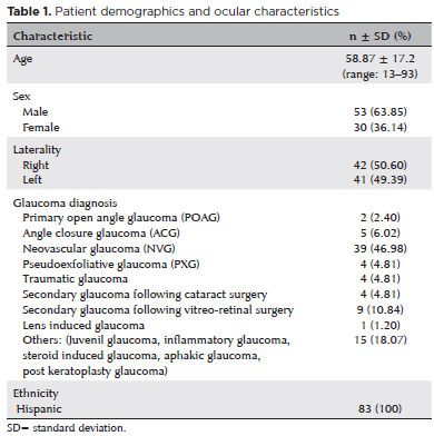
PURPOSE: Glaucoma is one of the leading causes of irreversible blindness worldwide. When topical hypotensive agents or laser trabeculoplasty fail to adequately control the disease, escalation of therapy becomes necessary, with transscleral cyclophotocoagulation being one of the available options. Several variations of transscleral cyclophotocoagulation exist, including traditional continuous wave, MicroPulse, and slow-coagulation techniques. We propose a novel variation – custom slow-coagulation transscleral cyclophotocoagulation – which combines elements of both continuous wave and slow-coagulation approaches. This study aimed to evaluate the outcomes of this technique in patients with refractory glaucoma.
METHODS: This retrospective, interventional study included 104 eyes of 83 patients with refractory glaucoma who underwent custom slow-coagulation transscleral cyclophotocoagulation. Changes in intraocular pressure, visual acuity, the number of glaucoma medications, and postoperative complications were analyzed. A paired t test was used to compare changes in intraocular pressure and visual acuity, while the Wilcoxon signed-rank test was applied to categorical variables. Success rates following custom slow-coagulation transscleral cyclophotocoagulation were estimated using Kaplan–Meier survival analysis.
RESULTS: Mean intraocular pressure decreased significantly from 38.9 ± 15.8 mmHg at baseline to 16.3 ± 9.9 mmHg at Month 12 (p<0.001). The mean number of glaucoma medications also decreased significantly from 3.6 ± 0.6 to 1.8 ± 1.4 (p<0.001). No significant reduction in mean visual acuity was observed during follow-up.
CONCLUSIONS: Custom slow-coagulation transscleral cyclophotocoagulation effectively reduced baseline intraocular pressure and the number of glaucoma medications, with a low rate of complications and no decline in visual acuity over a 12-month follow-up period. This novel technique demonstrated a high safety profile in a Hispanic population and represents a low-cost, minimally invasive procedure with rapid recovery and promising efficacy in intraocular pressure control.
Keywords: Glaucoma/surgery; Sclera; Filtering surgery; Laser coagulation/methods; Lasers, semiconductor/therapeutic use; Intraocular pressure; Blindness/prevention & control; Vision, low/epidemiology; Visual acuity
13-tab01.jpg)
Abstract
Objetivo: Avaliar o perfil clínico e epidemiológico dos transplantes de córnea realizados em um centro de referência oftalmológica de Recife no estado de Pernambuco, localizado no nordeste do Brasil.
Métodos: Esse estudo transversal coletou através de prontuários médicos dados clínicos e epidemiológicos de pacientes submetidos a ceratoplastia na Fundação Altino Ventura, de janeiro a dezembro de 2017.
Resultados: Um total de 356 procedimentos foram realizados em 327 pacientes dos quais 165 (50.5%) eram mulheres. A média de idade na cirurgia foi de 50.9 ± 22.6 anos (variação, 10 - 89 anos). A maioria dos pacientes (n=152 [46.5%]) era da capital e região metropolitana. A média de tempo de espera na fila para o transplante de córnea foi de 52.4 ± 58.9 dias (variação, 0 - 460 dias). As principais indicações de transplante foram ceratite infecciosa (n=88 [24.7%]), ceratocone (n=80 [22.5%]) e falência de transplante prévio (n=75 [21.1%]). Transplante penetrante foi a técnica mais realizada (n=213 [59.9%]) e foi mais comum em homens (n=132 [76.7%]), enquanto os transplantes lamelares posteriores (n=143 [41.1%]) foram mais realizados nas mulheres (p<0.001).
Conclusão: Ceratites infecciosas foram a causa mais comum de transplante, com prevalência similar em adultos economicamente ativos de ambos os sexos. Transplante penetrantes foram os prevalentes em homens e os transplantes lamelares em mulheres.
Keywords: Doença da córnea/epidemiologia; Transplante de córnea; Ceratoplastia penetrante; Brasil/epidemiologia
06-tab01tb.jpg)
Abstract
MÉTODOS: Córneas humanas de treinamento disponibilizadas foram randomizadas em quatro grupos: Pachy-100 (profundidade de incisão = espessura corneana central - margem de segurança de 100 µm), Pachy-50 (margem de segurança de 50 µm), Pachy-0 (sem margem de segurança) e Pachy+50 (profundidade de incisão = espessura corneana central + 50 µm). Todas as lamelas foram dissecadas através um método padronizado e já publicado (Pachy-DSEK). As espessuras das lamelas (centro, zona de 3,0mm e zona de 6,0mm) foram medidas com tomografia de coerência óptica. A razão de espessura centro-periferia foi calculada aos 3,0 e 6,0 mm de diâmetro.
RESULTADOS: Perfuração endotelial ocorreu apenas no grupo Pachy+50 (n=3, 30%). A espessura central da lamela nos grupos Pachy-100, Pachy-50, Pachy-0 e Pachy+50 foi de 185 ± 42 µm, 122 ± 29 µm, 114 ± 29 µm, e 58 ± 31 µm, respectivamente (p<0,001). As razões C/P aos 3,0 e 6,0 mm foram de 0,97 ± 0,06 e 0,92 ± 0,14, respectivamente. Os parâmetros de características do doador não se correlacionaram com os resultados de espessura de lamela. A profundidade planejada de incisão se correlacionou com a maioria dos parâmetros de espessura de lamela (p<0,001). A espessura de lamela se correlacionou negativamente com a profundidade planejada da incisão (p<0.001, r=-0,580). O melhor resultado foi observado no grupo Pachy-0, em que 75% das lamelas mediram abaixo de 130 µm e não houve perfuração endotelial.
CONCLUSÃO: Através de um método padronizado de dissecção, a maioria das lamelas endoteliais apresentou uma configuração planar. O planejamento de profundidade de incisão igual à espessura corneana central resultou em alta porcentagem de lamelas ultrafinas sem ocorrência de perfuração.
Keywords: Transplante de córnea; Ceratoplastia lamelar; Endotélio corneano; Dissecção; Tomografia de coerência óptica

Abstract
PURPOSE: To evaluate the impact of the COVID-19 pandemic and characterize the serological profile of discarded corneal donations in the coverage area of the Banco de Olhos de Londrina, through reverse transcription-polymerase chain reaction testing for COVID-19 and serological screening of cornea donors excluded because of positive test results.
METHODS: This observational retrospective study included 776 cornea donors who’s serological and reverse transcription-polymerase chain reaction test results were processed at the Hospital of Universidade Estadual de Londrina between May 2020 and 2022. The number of corneal donations and tissue utilization rates throughout the years of operation of the Banco de Olhos de Londrina were also analyzed.
RESULTS: The mean donor age was 53.14 years; 332 donors (43%) were female, and 444 (57%) were male. Positive results were identified in 15.76% of donors for hepatitis B core antibody antibodies, 0.65% for hepatitis B surface antigen, 1.03% for hepatitis C antibodies, and 0.52% for human immunodeficiency virus and human T-lymphotropic vírus. Positive reverse transcription-polymerase chain reaction results for SARS-CoV-2 were observed in 2.7% of cases. Older adults were 2.6 times more likely to test positive for SARS-CoV-2 (95% CI, 1.06-6.34) and 3.0 times more likely to test positive for hepatitis B core antibody (95% CI, 1.95-4.41) than younger individuals. A 75.2% reduction in corneal donations was observed in 2020 compared with 2019, accompanied by a 5% increase in tissue utilization, possibly associated with the effectiveness of donor screening during the pandemic.
CONCLUSION: The COVID-19 pandemic had a profound impact on the number of corneal transplants worldwide, in Brazil, and at the Banco de Olhos de Londrina because of the substantial decline in donations during this period. Hepatitis B was the leading cause of corneal tissue discard due to positive serology in both this study and previous reports, highlighting the importance of prevention programs and improved vaccination coverage. Strict legislation, comprehensive serological screening, and appropriate processing of donated tissue remain essential to eliminate potential sources of infection and ensure transplantation safety.
Keywords: Cornea; Corneal transplantation; COVID-19; Eye banks; Serology
Abstract
PURPOSE: This study aimed to report the use, efficacy, and safety of intracameral voriconazole as an adjuvant treatment for deep fungal keratitis.
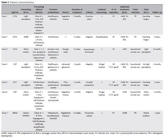
METHODS: This was a prospective case series of seven eyes with fungal keratitis with anterior chamber involvement or a corneal ulcer refractory to conventional topical treatment. In addition to topical treatment with 0.15% amphotericin B eye drops, voriconazole 50 μg/ 0.1 mL
was administered to the anterior chamber of each affected eye up to four times within 72 h. The primary outcome measures were healing (fungal eradication) and the need for therapeutic keratoplasty. Best-corrected visual acuity was a secondary outcome measure.
RESULTS: Three cases were confirmed by confocal microscopy, and four were diagnosed from positive culture tests. At presentation, one patient had a best-corrected visual acuity of 20/80, while all others had hand motion or worse. Four cases received one intracameral injection, two cases received three, and one case received four injections. There were no complications after any of the intracameral voriconazole injections. Four patients had imminent corneal perforations and were treated with cyanoacrylate adhesive and bandage contact lenses. Four patients recovered from the infection, and three underwent therapeutic keratoplasty. The final best-corrected visual acuity was improved in two cases but all patients had a final visual acuity of counting fingers or worse.
CONCLUSION: As an adjuvant treatment for deep fungal keratitis, intracameral voriconazole injection is a feasible option. Although fungal eradication was achieved in all patients, three required therapeutic keratoplasty and all patients had unsatisfactory visual acuity outcomes.
Keywords: Antifungal agents; Fungi; Corneal transplantation; Keratitis; Eye infections, fungal; Voriconazole
Abstract
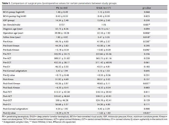
PURPOSES: This study aims to assess and compare the postoperative visual and topographic outcomes, complications, and graft survival rates following deep anterior lamellar keratoplasty and penetrating keratoplasty in patients with macular corneal dystrophy.
METHODS: In this study we enrolled 59 patients (23 male; and 36 female) with macular corneal dystrophy comprising 81 eyes. Out of these, 64 eyes underwent penetrating keratoplasty, while 17 eyes underwent deep anterior lamellar keratoplasty. The two groups were analyzed and compared based on best-corrected visual acuity, corneal tomography parameters, pachymetry, complication rates, and graft survival rates.
RESULTS: After 12 months, 70.6% of the patients who underwent deep anterior lamellar keratoplasty (DALK) and 75% of those who had penetrating keratoplasty (PK) achieved a best-corrected visual acuity of 20/40 or better (p=0.712). Following surgery, DALK group showed lower front Kmean (p=0.037), and Q values (p<0.01) compared to the PK group. Postoperative interface opacity was observed in seven eyes (41.2%) in the DALK group. Other topography values and other complications (graft rejection, graft failure, cataract, glaucoma, microbial keratitis, optic atrophy) did not show significant differences between the two groups. The need for regrafting was 9.4% and 11.8% in the PK and DALK groups, respectively (p=0.769). Graft survival rates were 87.5% and 88.2% for PK and DALK; respectively (p=0.88 by Log-rank test).
CONCLUSION: Both PK and DALK are equally effective in treating macular corneal dystrophy, showing similar visual, topographic, and survival outcomes. Although interface opacity occurs more frequently after DALK the visual results were comparable in both groups. Therefore, DALK emerges as a viable surgical choice for patients with macular corneal dystrophy without Descemet membrane involvement is absent.
Keywords: Macular corneal dystrophy; Corneal dystrophies; Hereditary; Keratoplasty; Penetrating; Corneal transplantation
Abstract
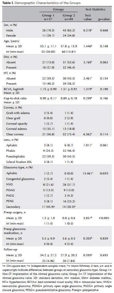
PURPOSE: As superotemporal implantation of the Ahmed glaucoma valve is not always feasible in cases of refractory glaucoma, this study examined the characteristics and surgical outcomes of cases in which the valve was implanted in a nonsuperotemporal quadrant using a modified long scleral tunnel technique.
METHODS: This retrospective case-control study included 37 eyes with nonsuperotemporal quadrant-Ahmed glaucoma valve implantation in Group 1 and 69 eyes with superotemporal Ahmed glaucoma valve implantation in Group 2. The demographic characteristics of these groups, surgical outcomes, including complications, further surgical interventions, and surgical success rates were compared. Surgical success was defined as an intraocular pressure not exceeding 21 mmHg, accompanied by a minimum reduction of 20% in intraocular pressure from the baseline without any additional intraocular pressure-lowering procedures, and the absence of light perception loss or phthisis bulbi.
RESULTS: Group 1 had significantly higher numbers of eyes with secondary glaucoma and preoperative surgical procedures than Group 2 (p<0.05). Both groups had mean preoperative intraocular pressure values, and mean intraocular pressure values at the last visit of 34.2 and 27.9 months, 35.5 ± 1.5 and 35.8 ± 1.2 mmHg, and 14.5 ± 5 and 14.9 mmHg, respectively. Although both groups had 70.2% and 75.8% as their five-year cumulative probability of success, respectively, the rates of complications, revisional surgery, and additional surgical procedures did not differ significantly (p>0.05).
CONCLUSION: The modified long scleral tunnel technique for Ahmed glaucoma valve implantation in nonsuperotemporal quadrants achieves intraocular pressure control and complication rates comparable to superotemporal implantation.
Keywords: Glaucoma/surgery; Sclera/surgery; Glaucoma drainage implant; Intraocular pressure; Tenon capsule
Abstract
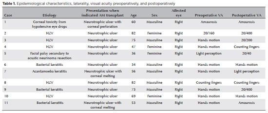
PURPOSE: To evaluate the clinical results of cryopreserved amniotic membrane transplantation as a treatment option for refractory neurotrophic corneal ulcers.
METHODS: This prospective study included 11 eyes of 11 patients who underwent amniotic membrane transplantation for the treatment of refractory neurotrophic corneal ulcers at Hospital de Clínicas da Universidade Federal do Paraná, in the city of Curitiba, from May 2015 to July 2021. Patients underwent different surgical techniques in which the amniotic membrane was applied with the epithelium facing upward to promote corneal re-epithelialization.
RESULTS: The median age of the patients was 60 years (range, 34-82 years), and 64% were men. The predominant etiology of corneal ulcers was herpes zoster (45% of cases). Approximately one-third of the patients (27%) were chronically using hypotensive eye drops, and more than half (54%) had previously undergone penetrating corneal transplantation. At the time of amniotic membrane transplantation, 18% of the eyes had corneal melting, 9% had corneal perforation, and the others had corneal ulceration without other associated complications (73%). The time between clinical diagnosis and surgical treatment ranged from 9 days to 2 years. The corrected visual acuity was worse than 20/400 in 90% of the patients preoperatively, with improvement in 36% after 3 months of the procedure, worsening in 18% and remaining stable in 36%. Of the patients, 81% complained of preoperative pain, and 66% of them reported total symptom relief after the surgical procedure. In one month, 54.6% of the patients presented a closure of epithelial defect, and half of the total group evolved with corneal thinning. The failure rate was 45.5% of the cases.
CONCLUSION: Cryopreserved amniotic membrane transplantation can be considered a good alternative for treating refractory neurotrophic corneal ulcers, as it resulted in significant improvement in pain (66%) and complete epithelial closure (60%) in many patients at 1 month postoperatively. Notably, the high failure rate highlights the need for further studies to identify patient- and ulcer-related factors that may influence the outcomes of this procedure.
Keywords: Amnion/transplantation; Corneal ulcer; Anterior eye segment; Keratitis
13-fig01tb.jpg)
Abstract
Relato de uma técnica que combina o implante de uma lente intraocular com fixação intraescleral sem sutura e uma ceratoplastia endotelial da membrana de Descemet em paciente com ceratopatia bolhosa pseudofácica anterior. Foram criados dois túneis esclerais. Foram feitas incisões na córnea e a lente intraocular dobrável foi cortada e removida da câmara anterior. Foi então efetuada uma vitrectomia anterior e uma lente intraocular dobrável de 3 peças foi implantada na câmara anterior. Um dos hápticos da lente intraocular foi pinçado com um fórceps e puxado para fora do túnel escleral. A extremidade do háptico foi cauterizada. Manobras semelhantes foram feitas no outro háptico. Foi preparado um tecido de doador com 8 mm de diâmetro e o tecido endotelial da área receptora foi removido do centro da córnea. O tecido preparado do doador foi injetado na câmara anterior. Após abertura e posicionamento adequados do tecido do doador, foi injetada uma bolha de ar abaixo do tecido. Não foi observada nenhuma complicação pós-operatória durante um mês de acompanhamento.
Keywords: Lâmina limitante posterior; Ceratoplastia penetrante; Implante de lente intraocular; Lentes intraoculares; Esclera/cirurgia; Humanos; Relatos de casos
15-fig01.jpg)
Abstract
O objetivo deste estudo é discutir um caso de ceratite tardia por Klebsiella oxytoca, após ceratoplastia lamelar anterior profunda, bem como seu tratamento. Uma paciente de 21 anos apresentou vermelhidão e efluxo no olho esquerdo 5 meses após cirurgia de ceratoplastia lamelar anterior profunda sem complicações. Ao exame, havia uma única sutura solta na região nasal superior e uma área de infiltração com defeito epitelial no enxerto e na junção com o leito receptor na área da sutura solta. Iniciou-se empiricamente um tratamento tópico com vancomicina e ceftazidima fortificada de hora em hora, porém com resposta insuficiente. Após o crescimento de K. oxytoca a partir de cultura de swab e sutura retirados da paciente, a vancomicina fortificada foi substituída por imipenem fortificado. Observou-se que a área de infiltração regrediu rapidamente e que o defeito epitelial foi fechado com o tratamento com imipenem fortificado. O imipenem fortificado pode ser considerado um tratamento alternativo, especialmente nos casos sem resposta ao tratamento e detecção de crescimento na cultura.
Keywords: Transplante e córnea; Ceratoplastia lamelar; Klebsiella oxytoca; Ceratite; Imipenem
09-fig01.jpg)
Abstract
Em decorrência de complicações, da biocompatibilidade e da escassez de tecido doador para transplantes de córnea natural, foram elaboradas córneas artificiais que são potenciais para reabilitar funções ópticas. Nessa perspectiva, objetivou-se a análise da eficácia da reabilitação visual entre os implantes: Boston tipo I, Boston tipo II, Aurolab, osteo-odonto-ceratoprótese e ceratoprótese de Osso Tibial. De modo geral, a princípio observou-se uma tendência de melhoria da Best-corrected visual acuity em todos os tipos de lentes, mas considerável queda durante acompanhamento a longo prazo. O dispositivo com melhor reabilitação visual em pacientes com superfícies oculares úmidas é a Boston tipo I, seguida pela Aurolab, que é economicamente viável em países emergentes. Ao considerar pacientes com olhos secos, o implante de Boston tipo II apresenta maior reabilitação visual. Por fim, em virtude de não apresentarem dados equiparáveis, as lentes osteo-odonto-ceratoprótese e de osso tibial não puderam ser analisadas.
Keywords: Transplante de córnea; Próteses visuais; Córnea; Reabilitação; Acuidade visual
Abstract
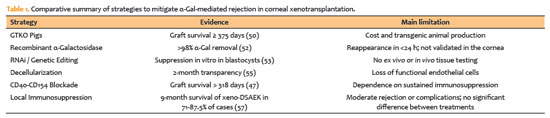
Corneal blindness affects more than 12 million individuals worldwide; however, the availability of human donor corneas meets only a small fraction of the demand. Porcine corneal xenotransplantation represents a promising alternative because of the cornea's immune privilege and the anatomical similarities between human and porcine corneas. However, the galactose-α1,3-galactose antigen expressed on porcine cells induces hyperacute rejection through the activation of human anti-Gal antibodies, complement pathways, and innate immune cell infiltration. This review summarizes the mechanisms underlying corneal immune privilege and contrasts them with galactose-α1,3-galactose-mediated rejection pathways. In addition, it evaluates five major mitigation strategies: GGTA1-knockout pigs, enzymatic α-galactosidase treatment, RNA interference, decellularization, and immunosuppressive approaches, including CD40-CD154 blockade and topical tacrolimus. Preclinical studies have demonstrated that GTKO corneas can maintain transparency for longer than 1 year, whereas decellularized matrices preserve corneal architecture while exhibiting low antigenicity. These findings support the future clinical translation of porcine corneal xenotransplantation.
Keywords: Corneal transplantation; Xenotransplantation; Graft rejection; Galactose; Immunosuppressive agents.
 ABO is licensed under a Creative Commons Attribution-NonComercial 4.0 Internacional.
About
Issues
Editorial Board
Submission

Arquivos Brasileiros de Oftalmologia
Official publication of Brazilian Council of Ophthalmology - Conselho Brasileiro de Oftalmologia (CBO)
Rua Casa do Ator, 1.117 - 2nd floor - Zip Code: 04546-004
São Paulo - SP, Brazil
TEL: +55 11 3266-4000
E-mail: [email protected]
