Arq. Bras. Oftalmol. 2023;86 (5 )
:1-5
| DOI: 10.5935/0004-2749.20230063
Abstract
Objetivo: A injeção peribulbar de triancinolona é um tratamento alternativo para doenças oculares da tireoide; no entanto, a segurança desse procedimento continua controversa. O objetivo deste artigo é descrever os efeitos adversos locais e sistêmicos de injeções peribulbares de triancinolona em pacientes com doença ocular da tireoide.
Métodos: Estudo retrospectivo de uma série de casos. Foram analisados os prontuários médicos dos pacientes com doença ocular da tireoide tratados com injeções de triancinolona peribulbar em uma única instituição acadêmica entre 2007 e 2019. Foram documentadas as complicações locais e sistêmicas.
Resultados: Um total de 123 pacientes foram tratados. Apenas 11 (8,9%) pacientes apresentaram complicações locais, sendo a mais frequente a presença de equimoses palpebrais superficiais (7,3%), enquanto 2 (1,6%) pacientes apresentaram complicações sistêmicas (hiperglicemia e inibição da suprarrenal após a interrupção do tratamento). Todas estas complicações foram transitórias e nenhum paciente apresentou sequelas de longo prazo.
Conclusões: As injeções peribulbares de triancinolona nas doenças oculares da tireoide têm uma taxa muito baixa de complicações, tanto locais quanto sistêmicas. São necessários estudos prospectivos para aprofundar este tópico.
Keywords: Órbita/diagnóstico por imagem; Imageamento por ressonância magnética; Oftalmopatia de Graves; Triancinolona/efeitos adversos; Injeções.
Arq. Bras. Oftalmol. 2023;86 (4 )
:359-364
| DOI: 10.5935/0004-2749.20230056
Abstract
Objetivo: Comparar as características radiológicas e clínicas do adenoma pleomórfico primário e do carcinoma adenoide cístico da glândula lacrimal.
Métodos: Este estudo revisou retrospectivamente os achados de imagem e os prontuários médicos de casos de adenoma pleomórfico e carcinoma adenoide cístico da glândula lacrimal.
Resultados: Foram avaliados 11 pacientes com adenoma pleomórfico e 16 pacientes com carcinoma adenoide cístico. Não houve diferenças estatisticamente significativas em relação à idade e sexo. Proptose foi o sintoma de apresentação mais comum em ambos os grupos. Os carcinomas adenoides císticos foram mais propensos que os adenomas pleomórficos a apresentarem massas palpáveis, diplopia, dor e perda sensorial, mas essa diferença entre os grupos não foi estatisticamente significativa. Não houve diferenças estatísticas em termos de homogeneidade e indentação do globo ocular entre os dois tipos de tumores em imagens de tomografia computadorizada (p>0,05). Também à tomografia computadorizada, a invasão óssea, a calcificação do tumor e o sinal em cunha foram mais frequentes nos carcinomas adenoides císticos, enquanto a remodelação óssea foi mais frequente nos adenomas pleomórficos, com significância estatística para todas essas manifestações (p<0,05). À ressonância magnética, os adenomas pleomórficos foram significativamente mais propensos a terem margens bem definidas, contornos lobulados, realce heterogêneo pelo contraste e hiperintensidade na ressonância magnética ponderada em T2 (p<0,05).
Conclusão: Ao se diferenciar o adenoma pleomórfico e o carcinoma adenoide cístico da glândula lacrimal, é muito importante avaliar as características radiológicas juntamente com as características clínicas. Os contornos lobulados podem ser uma característica radiológica significativamente distinta em favor do adenoma pleomórfico.
Keywords: Aparelho lacrimal/patologia; Adenoma pleomorfo; Carcinoma adenoide cístico; Tomografia computadorizada por raios x; Imagem por ressonância magnética.
Arq. Bras. Oftalmol. 2022;85 (2 )
:166-173
| DOI: 10.5935/0004-2749.20220034
Abstract
Objetivos: Mensurar a perfusão do complexo retina/coróide com ressonância magnética em olhos com fechamento angular primário agudo (FAPA).
Métodos: Três sequências de ressonância magnética, duas anatômicas e uma de perfusão com gadolínio, foram adquiridas em pacientes com fechamento angular primário agudo. Regiões de interesse foram desenhadas na sequência de perfusão e sobrepostas à sequência anatômica. O volume de sangue relativo nos 2 primeiros segundos foi considerado como referência, e sua variação nos 28 segundos subsequentes foi analisada.
Resultados: Cinco olhos de 5 pacientes com fechamento angular primário agudo foram incluídos (3 com crise unilateral e 2 com crise bilateral). Três olhos contralaterais e 2 olhos de 2 pacientes saudáveis, pareados por sexo e idade, foram incluídos no grupo controle. Pacientes com fechamento angular primário agudo incluíam 4 (80%) mulheres, com idade média de 65,8 ± 12,37 anos, pressão intraocular média de 56,2 ± 14,67 mmHg, pressão arterial média de 113,4 ± 8,17 mmHg e pressão de perfusão ocular de 57,2 ± 13,46mmHg. No grupo controle, pressão intraocular média foi de 15,6 ± 2,61 mmHg (p=0,0625), pressão arterial média de 107,4 ± 6,57 mmHg (p=1,00) e pressão de perfusão ocular de 91,8 ± 6,72 mmHg (p=0,0625). O volume de sangue relativo do complexo retina/coróide foi de -0,127 ± 0,048 nos olhos em fechamento angular primário agudo e -0,213 ± 0,116 nos olhos controles (p=0,3125).
Conclusões: A sequência de ressonância magnética com gadolínio não demonstrou diferença na perfusão de retina/coroide em olhos com fechamento angular primário agudo.
Keywords: Glaucoma de ângulo fechado; Imagem por ressonância magnética; Gadolínio; Retina; Perfusão
Arq. Bras. Oftalmol. 2025;88 (2 )
:1-5
| DOI: 10.5935/0004-2749.2024-0113
Abstract
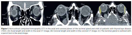
This study aimed to evaluate the morphometric and volumetric dimensions of the lacrimal gland in patients with inactive thyroid eye disease and compare them with the values reported in the literature. This case series evaluated consecutive patients with inactive thyroid eye disease treated at a tertiary eye hospital from 2015 to 2020. The patients' baseline demographics and clinical characteristics were obtained. The axial and coronal length, width, and volume of the lacrimal gland were measured on computed tomography scan images, and the results were statistically analyzed. A total of 21 patients (42 orbits) with inactive thyroid eye disease were evaluated. Their mean age was 49.0 ± 14.6 years, and 12 (57.1%) of them were men. The main complaint was dryness, and the majority of the patients had good vision and mild proptosis. The mean axial length and width of the lacrimal gland were 19.3 ± 3.9 mm and 7.5 ± 2.1 mm, respectively; coronal length and width, 20.4 ± 4.5 mm and 7.5 ± 2.1 mm, respectively; and lacrimal gland volume, 0.825 ± 0.326 mm3. Age, sex, or laterality were not found to be determinants of lacrimal gland enlargement. Patients with thyroid eye disease have enlarged lacrimal gland even in the nonactive phase of the disease multifactorial aspects influence the lacrimal gland in thyroid eye disease, making it difficult to establish a clear correlation with predisposing factors. Further studies are warranted to better understand the association between thyroid eye disease and the lacrimal gland.
Keywords: Graves' ophthalmology; Graves' disease; Lacrimal apparatus; Lacrimal apparatus diseases; X-ray computed tomography
Arq. Bras. Oftalmol. 2024;87 (2 )
:1-6
| DOI: 10.5935/0004-2749.2021-0435
Abstract
PURPOSE: This study aimed to analyze the association between magnetic resonance imaging apparent diffusion coefficient map value and histopathological differentiation in patients who underwent eye enucleation due to retinoblastomas.
METHODS: An observational chart review study of patients with retinoblastoma that had histopathology of the lesion and orbit magnetic resonance imaging with apparent diffusion coefficient analysis at Hospital de Clínicas de Porto Alegre between November 2013 and November 2016 was performed. The histopathology was reviewed after enucleation. To analyze the difference in apparent diffusion coefficient values between the two major histopathological prognostic groups, Student's t-test was used for the two groups. All statistical analyses were performed using SPSS version 19.0 for Microsoft Windows (SPSS, Inc., Chicago, IL, USA). Our institutional review board approved this retrospective study without obtaining informed consent.
RESULTS: Thirteen children were evaluated, and only eight underwent eye enucleation and were included in the analysis. The others were treated with photocoagulation, embolization, radiotherapy, and chemotherapy and were excluded due to the lack of histopathological results. When compared with histopathology, magnetic resonance imaging demonstrated 100% accuracy in retinoblastoma diagnosis. Optic nerve invasion detection on magnetic resonance imaging showed a 66.6% sensitivity and 80.0% specificity. Positive and negative predictive values were 66.6% and 80.0%, respectively, with an accuracy of 75%. In addition, the mean apparent diffusion coefficient of the eight eyes was 0.615 × 103 mm2/s. The mean apparent diffusion coefficient value of poorly or undifferentiated retinoblastoma and differentiated tumors were 0.520 × 103 mm2/s and 0.774 × 103 mm2/s, respectively.
CONCLUSION: This study revealed that magnetic resonance imaging is useful in the diagnosis of retinoblastoma and detection of optic nerve infiltration, with a sensitivity of 66.6% and specificity of 80%. Our results also showed lower apparent diffusion coefficient values in poorly differentiated retinoblastomas with a mean of 0.520 ×
103 mm2/s, whereas in well and moderately differentiated, the mean was 0.774 × 103 mm2/s.
Keywords: Retinoblastoma; Prognosis; Retinal neoplasms; Orbit; Diffusion magnetic resonance imaging
Arq. Bras. Oftalmol. 2024;87 (2 )
:1-8
| DOI: 10.5935/0004-2749.2022-0319
Abstract
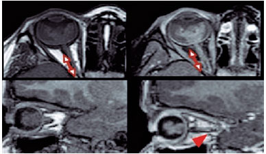
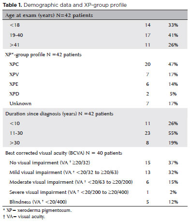
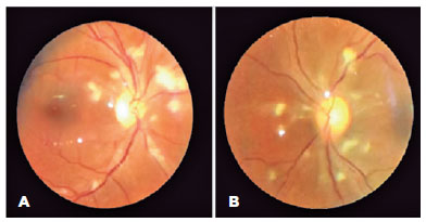
To assess Meibomian gland dysfunction using meibography in patients with xeroderma pigmentosum and correlate with ocular surface changes. This cross-sectional study evaluated patients with xeroderma pigmentosum. All patients underwent a comprehensive and standardized interview. The best-corrected visual acuity of each eye was determined. Detailed ophthalmic examination was conducted, including biomicroscopy examination of the ocular surface, Schirmer test type I, and meibography, and fundus examination was also performed when possible. Meibomian gland dysfunction was assessed by non-contact meibography using Oculus Keratograph® 5M (OCULUS Inc., Arlington, WA, USA). Saliva samples were collected using the Oragene DNA Self-collection kit (DNA Genotek Inc., Ottawa, Canada), and DNA was extracted as recommended by the manufacturer. Factors associated with abnormal meiboscores were assessed using generalized estimating equation models. A total of 42 participants were enrolled, and 27 patients underwent meibography. The meiboscore was abnormal in the upper eyelid in 8 (29.6%) patients and in the lower eyelid in 17 (62.9%). The likelihood of having abnormal meiboscores in the lower eyelid was 16.3 times greater than that in the upper eyelid.In the final multivariate model, age (p=0.001), mutation profile (p=0.006), and presence of ocular surface malignant tumor (OSMT) (p=0.014) remained significant for abnormal meiboscores. For a 1-year increase in age, the likelihood of abnormal meiboscores increased by 12%. Eyes with OSMT were 58.8 times more likely to have abnormal meiboscores than eyes without ocular surface malignant tumor.In the final model, age, xeroderma pigmentosum profile, previous cancer, and clinical alterations on the eyelid correlated with a meiboscore of ≥2.Meibomian gland dysfunction was common in patients with xeroderma pigmentosum, mainly in the lower eyelid. The severity of Meibomian gland dysfunction increases with age and is associated with severe eyelid changes.
Keywords: Meibomian glands/pathology; Meibomian glands/ diagnostic imaging; Photography; Xeroderma pigmentosum; Eyelid diseases/diagnostic imaging; Dry eye syndromes; DNA repair; Humans; Case report
 ABO is licensed under a Creative Commons Attribution-NonComercial 4.0 Internacional.
05-tab01.jpg)
11-tab01.jpg)
05-fig01.jpg)
10-fig01.jpg)
08-fig01.jpg)
11-fig01tb.jpg)
03-fig01.jpg)
01-fig01.jpg)
02-fig01.jpg)
14-fig01.jpg)