Showing of 1 until 14 from 188 result(s)
Search for: Paracoccidioidomycosis; Eyelid diseases; Differential diagnosis; Eyelids; Conjunctiva; Case report
Abstract
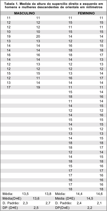
Objetivo: Determinar medidas anatômicas médias em descendentes de orientais (ambos os pais orientais da raça nipônica), a fim de determinar parâmetros a serem utilizados em cirurgias palpebrais nestes pacientes. Métodos: Foram aferidas as medidas da altura da prega palpebral, altura do supercílio e medida da pele pré-tarsal visível de 56 descendentes de orientais, num total de 112 pálpebras avaliadas. 16 (28,5%) indivíduos eram do sexo masculino e 40 (71,5%) do sexo feminino. A idade média foi 25,2 anos. Resultados: Não houve diferença estatisticamente significativa entre pálpebras direita e esquerda em ambos os sexos. A medida média da altura do supercílio foi de 13,6 mm (2,5 DP) no sexo masculino e 14,5 mm (2,2 DP) no sexo feminino (T= 1,77) (P= 0,083), diferença não-significativa estatisticamente. A altura média da pele pré-tarsal no sexo masculino foi 1,2 mm (1,2 DP) e no sexo feminino foi de 1,7 mm (1,6 DP); (T= 1,84) (P= 0,070), não-significativa estatisticamente. A altura média do sulco palpebral foi 3,9 mm (2,5 DP) no sexo masculino e 4,9 mm (2,9 DP) no sexo feminino (T= 1,95) (P= 0,060), não-significativa estatisticamente. Conclusões: Devido à equivalência estatística dos dados entre as pálpebras e entre ambos os sexos, as medidas médias obtidas podem servir como bom parâmetro anatômico em procedimentos cirúrgicos palpebrais em descendentes de orientais.
Keywords: Pálpebras orientais; Prega palpebral
Abstract
Objetivo: Avaliar as possíveis variáveis relacionadas à ceratose actínica e lesões malignas cutâneas nas pálpebras.
Métodos: Estudo prospectivo de pacientes com lesões palpebrais suspeitas de malignidade. Os participantes foram submetidos à biopsia por trépano (punch) de 2-mm em dois pontos opostos da lesão como método diagnóstico e os resultados foram comparados com o estudo histopatológico da peça excisada cirurgicamente. Aqueles que apresentaram ceratose actínica como resultado foram divididos em dois grupos (ceratose actínica associada com malignidade e ceratose actínica isolada) e foram comparados de acordo com as variáveis: idade, tempo de doença, maior diâmetro, área do tumor, classificação de Fitzpatrick, gênero, localização e acometimento da margem palpebral. A análise de cluster também foi realizada.
Resultados: Foram analisadas 174 lesões e 50 delas tinham ceratose actínica como componente do tumor. Ceratose actínica esteve associada ao Carcinoma Espinocelular em 22% dos casos e ao Carcinoma Basocelular em 38%, mostrando que ambos podem ter ceratose actínica adjacente. A análise estatística não encontrou diferença entre as variáveis. A análise de cluster identificou quatro grupos e mostrou que lesões malignas no canto medial tinham maiores diâmetro e área. Acometimento da margem na pálpebra inferior relacionou-se em 100% com malignidade, enquanto a ausência de acometimento da margem mostrou menor chance de malignidade.
Conclusões: Lesões maiores de ceratose actínica no canto medial e lesões com acometimento da margem palpebral inferior têm maiores chances de associação com malignidade.
Keywords: Ceratose actínica/patologia; Biópsia; Neoplasmas palpebrais; Pálpebra/lesões
Abstract
Objetivo: Investigar a superfície ocular e as glândulas meibomianas em pacientes com acne vulgar não tratada.
Métodos: Todos os indivíduos responderam ao questionário Ocular Surface Disease Index (OSDI) e tiveram avaliados o tempo de ruptura do filme lacrimal pelo método invasivo, a coloração da superfície ocular com fluoresceína e o teste de Schirmer II. Foram ainda avaliados o escore palpebral total e o de secreção das glândulas meibomianas. Foi realizada meibografia sem contato com um dispositivo topográfico corneano Sirius.
Resultados: Foram incluídos no estudo os olhos direitos de 35 voluntários com acne vulgar e 35 saudáveis. Os escores do Ocular Surface Disease Index e da coloração foram significativamente maiores no grupo com acne em comparação com o grupo controle (p=0,01 e p=0,003, respectivamente), mas o tempo de ruptura do filme lacrimal pelo método invasivo e as medidas do teste de Schirmer II foram significativamente menores (p=0,000 e p=0,003, respectivamente). O escore palpebral total e o escore de secreção das glândulas meibomianas também foram maiores no grupo com acne que no grupo controle (p=0,003 e p=0,000). Na avaliação morfológica das glândulas meibomianas, o espessamento, o afinamento, a tortuosidade e a presença de áreas fantasmas nas glândulas foram mais comuns no grupo acne vulgar que no grupo controle, com significância estatística (p=0,000, p=0,001, p=0,05 e p=0,006 respectivamente). A porcentagem da área de perdas das glândulas meibomianas foi significativamente mais alta no grupo com acne vulgar, tanto na meibografia superior quanto na inferior. A área de perda das glândulas meibomianas demonstrou uma correlação positiva com o escore palpebral total e com o escore de secreção das glândulas meibomianas.
Conclusão: A acne vulgar pode levar a uma predisposição para a disfunção das glândulas meibomianas e para danos na superfície ocular. Parece ser importante reconhecer precocemente as alterações das glândulas meibomianas e da superfície ocular, especialmente nos casos de acne vulgar para os quais se planeja o tratamento oral com isotretinoína.
Keywords: Acne vulgar/complicação; Acne vulgaris/tratamento farmacológico; Isotretinoína/uso terapêutico; Pálpebra; Túnica conjuntiva/patologia; Síndrome do olho seco; Glândula tarsal; Técnica de diagnóstico oftalmológico; Questionário
Abstract
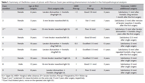
PURPOSE: This study was conducted to report the histopathological and clinical features of the Marcus Gunn phenomenon and other similar conditions of upper eyelid misfiring.
METHODS: This was a retrospective study of patients with congenital ptosis with Marcus Gunn phenomenon who have undergone surgical repair over a period of 12 years and another two patients with upper eyelid misfiring in association with extraocular movements to identify their histopathological findings as subtypes representing ocular congenital cranial dysinnervation disorder.
RESULTS: Among 136 patients with congenital ptosis, 11 (8%) patients with Marcus Gunn phenomenon or misfiring were identified, of whom 9 (6.6%) had typical known Marcus Gunn phenomenon and 2 (1.4%) had eyelid misfiring similar to Marcus Gunn phenomenon. In all patients, the histopathological changes of the excised levator muscle included overall loss and/or atrophy of muscle fibers and irregular-modified Gomori trichrome staining.
CONCLUSION: The Marcus Gunn phenomenon and similar misfiring conditions with synkinetic extraocular muscle movements share findings that are consistent with the neurogenic type of muscle atrophy. This result suggests a common underlying etiology with variable clinical findings, representing the ocular counterpart of congenital cranial dysinnervation disorder, which has been reported as ocular congenital cranial dysinnervation disorder.
Keywords: Eyelid diseases; Ocular motility disorders/surgery; Ophthalmologic surgical procedures
Abstract
PURPOSE: To evaluate the effect of upper eyelid ptosis repair with Muller muscle-conjunctival resection on meibomian gland function and ocular surface parameters.
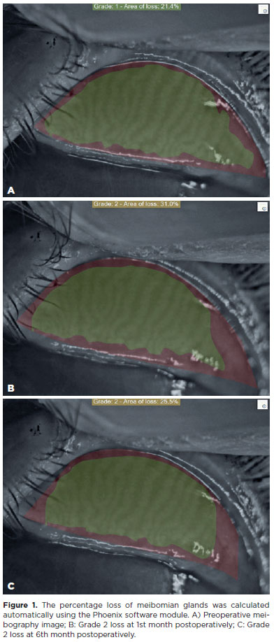
METHODS: Thirty-eight patients who underwent ptosis repair with Muller muscle-conjunctival resection were retrospectively reviewed. Meibomian gland loss, Ocular Surface Disease Index OXFORD score, meiboscore, and noninvasive keratograph break-up time were measured preoperatively and at 1st, 3rd, and 6th months postoperatively.
RESULTS: Noninvasive keratograph break-up time values decreased significantly at 1st and 3rd months postoperatively compared to the preoperative level, but were similar to the preoperative level at 6th months postoperatively (p<0.001 and p=0.628, respectively). Ocular surface disease index, OXFORD score, meibomian gland loss, and meiboscore values increased significantly in the 1st and 3rd postoperative months compared to the preoperative period, but these values decreased to preoperative levels in the 6th postoperative month (p<0.001 and p>0.05, respectively).
CONCLUSION: There is a transient deterioration in meibography findings and OSDI score in the early postoperative period after Muller muscle-conjunctival resection. Patients undergoing Muller muscle-conjunctival resection may require topical lubricants, especially in the first 3 postoperative months.
Keywords: Meibomian glands; Blepharoptosis; Preoperative period; Conjunctiva; Muscles; Eyelid diseases; Diagnostic techniques, ophthalmological
Abstract
PURPOSE: Ptosis is characterized by drooping of the upper eyelid, often requiring surgical intervention for functional and aesthetic purposes. Müller’s muscle conjunctival resection is a commonly utilized surgical technique to correct mild to moderate ptosis. This retrospective study aimed to evaluate the impact of Hering’s law on the outcomes of unilateral Müller’s muscle conjunctival resection surgery, particularly eyelid and brow symmetry.
METHODS: Thirty patients with unilateral ptosis underwent Müller’s muscle conjunctival resection. Pre- and postoperative assessments included ipsilateral and contralateral side margin-reflex distance and brow position, measured through digital image analysis.
RESULTS: We found significant improvements in postoperative margin-reflex distance measurements in the ipsilateral eyelid but not in the contralateral eyelid, indicating minimal influence of Hering’s law. Brow position showed a statistically significant increase on the contralateral side but not on the ipsilateral side.
CONCLUSION: Müller’s muscle conjunctival resection effectively restores symmetry in eyelid height and maintains brow symmetry. This is the first study to explore bilateral eyelid and brow symmetry after unilateral Müller’s muscle conjunctival resection surgery for mild to moderate ptosis. Further research should be conducted to understand the long-term effects of Müller’s muscle conjunctival resection on facial aesthetics, particularly in relation to brow position.
Keywords: Müller muscle conjunctiva resection; Hering’s law; Eyelids; Blepharoptosis; Reflex; Oculomotor muscles
Abstract
PURPOSE: Evaluation of lid contour and marginal peak point changes to compare outcomes of external levator advancement and Müller’s muscle conjunctival resection surgery in unilateral ptosis.
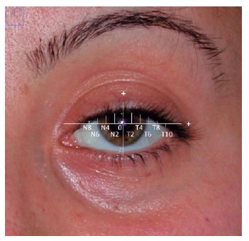
METHODS: We reviewed the charts of unilateral ptosis patients who underwent external levator advancement or Müller’s muscle conjunctival resection. Eyelid contour analysis was conducted on preoperative and 6-month postoperative digital images. This was performed with the multiple margin reflex distances technique, measuring the vertical distance from a line intersecting the center of the pupil to the eyelid margin at 10 positions at 2 mm intervals. The marginal peak point changes were analyzed digitally using the coordinates of the peak point according to the pupil center. Each position’s mean distance was compared preoperatively, postoperatively, and with the fellow eyelid.
RESULTS: Sixteen patients underwent external levator advancement and 16 patients had Müller’s muscle conjunctival resection. The mean margin reflex distance was improved by both techniques (1.46 vs. 2.43 mm and 1.12 vs. 2.25 mm, p=0.008 and p=0.0001 respectively) and approached that of the fellow eyelid (2.43 vs. 2.88 and 2.25 vs. 2.58 mm, p=0.23 and p=0.19, respectively). However, statistically significant lid margin elevation was limited to between the N6 and T6 points in the external levator advancement group. Whereas, significant elevation was achieved along the whole lid margin in the Müller’s muscle conjunctival resection group. The marginal peak point was shifted slightly laterally in the external levator advancement group (p=0.11).
CONCLUSIONS: Both techniques provide effective lid elevation, however, the external levator advancement’s effect lessens toward the canthi while Müller’s muscle conjunctival resection provides more uniform elevation across the lid margin. The margin reflex distance alone is not sufficient to reflect contour changes.
Keywords: Blepharoptosis; Eyelids; Conjunctiva; Oculomotor muscles; Image processing, computer-assisted; Treatment outcome
Abstract
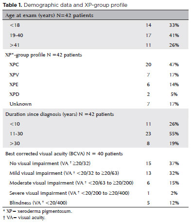
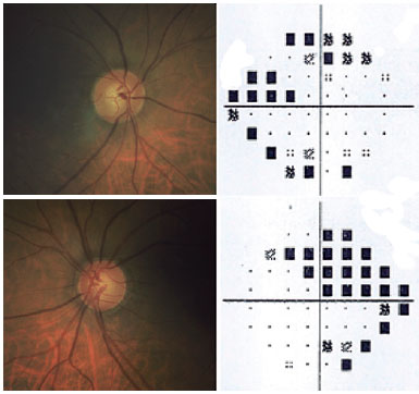
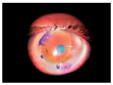
To assess Meibomian gland dysfunction using meibography in patients with xeroderma pigmentosum and correlate with ocular surface changes. This cross-sectional study evaluated patients with xeroderma pigmentosum. All patients underwent a comprehensive and standardized interview. The best-corrected visual acuity of each eye was determined. Detailed ophthalmic examination was conducted, including biomicroscopy examination of the ocular surface, Schirmer test type I, and meibography, and fundus examination was also performed when possible. Meibomian gland dysfunction was assessed by non-contact meibography using Oculus Keratograph® 5M (OCULUS Inc., Arlington, WA, USA). Saliva samples were collected using the Oragene DNA Self-collection kit (DNA Genotek Inc., Ottawa, Canada), and DNA was extracted as recommended by the manufacturer. Factors associated with abnormal meiboscores were assessed using generalized estimating equation models. A total of 42 participants were enrolled, and 27 patients underwent meibography. The meiboscore was abnormal in the upper eyelid in 8 (29.6%) patients and in the lower eyelid in 17 (62.9%). The likelihood of having abnormal meiboscores in the lower eyelid was 16.3 times greater than that in the upper eyelid.In the final multivariate model, age (p=0.001), mutation profile (p=0.006), and presence of ocular surface malignant tumor (OSMT) (p=0.014) remained significant for abnormal meiboscores. For a 1-year increase in age, the likelihood of abnormal meiboscores increased by 12%. Eyes with OSMT were 58.8 times more likely to have abnormal meiboscores than eyes without ocular surface malignant tumor.In the final model, age, xeroderma pigmentosum profile, previous cancer, and clinical alterations on the eyelid correlated with a meiboscore of ≥2.Meibomian gland dysfunction was common in patients with xeroderma pigmentosum, mainly in the lower eyelid. The severity of Meibomian gland dysfunction increases with age and is associated with severe eyelid changes.
Keywords: Meibomian glands/pathology; Meibomian glands/ diagnostic imaging; Photography; Xeroderma pigmentosum; Eyelid diseases/diagnostic imaging; Dry eye syndromes; DNA repair; Humans; Case report
Abstract
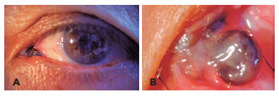
Um homem de 42 anos apresentou uma massa na conjuntiva palpebral inferior direita, próxima ao punctum, com evolução de 4 semanas. Uma biópsia excisional da lesão e o subsequente exame anatomopatológico revelaram que a massa era composta de células de Schwann com núcleos cônicos, cromatina fina e nucléolos não visíveis. Ao exame imuno-histoquímico, as células fusiformes mostraram-se difusa e fortemente positivas para a proteína S100. A imunocoloração também foi positiva para neurofilamentos e evidenciou os axônios. Considerando esses achados, o tumor foi diagnosticado como um neuroma circunscrito solitário. Procedeu-se uma investigação completa para neoplasia endócrina múltipla tipo 2b, entretanto, estigmas característicos e história familiar não foram detectados. Assim, o diagnóstico foi firmado como neuroma circunscrito solitário, condição rara cujo diagnóstico diferencial baseia-se no exame anatomopatológico e na avaliação imuno-histoquímica. Já que esse tumor pode estar relacionado à neoplasia endócrina múltipla tipo 2b, torna-se essencial, nesses casos, a investigação da neoplasia de forma sistemática.
Keywords: Neuroma; Neoplasia endócrina múltipla tipo 2b; Neoplasias da túnica conjuntiva; Diagnostico diferencial; Humanos; Relatos de casos
Abstract
O hamartoma combinado de retina e epitélio pigmentar da retina consiste em um tumor intraocular raro com comportamento benigno. O hamartoma combinado de retina e epitélio pigmentar da retina foi descrito na literatura apresentando dano retiniano variável, desde o envolvimento epirretiniano parcial até distorção completa das camadas retinianas e do epitélio pigmentar da retina. Relatamos o caso de uma menina de 8 anos com estrabismo de longa data que foi diagnosticada com hamartoma combinado de retina e epitélio pigmentar da retina, com base na avaliação de imagem multimodal. Exploramos os achados de imagem específicos de estudos usando tomografia de coerência óptica de domínio espectral, autofluorescência, angiografia por tomografia de coerência óptica e angiografia fluorescente.
Keywords: Hamartoma/diagnóstico; Epitélio pigmentado da retina; Neoplasia da retina; Tomografia de coerência óptica; Angiografia; Humanos; Relato de caso
Abstract
O diagnóstico de glaucoma de ângulo fechado secundário a cistos iridociliares é desafiador e não possui suporte da literatura compilada. Apresentamos um caso bilateral raro de glaucoma de ângulo fechado associado à íris pseudoplateau devido a cistos ciliares múltiplos e realizamos uma revisão sistemática da literatura de relatos de casos similares publicados entre novembro de 2006 e novembro de 2016. Apenas 19 relatos de casos apresentaram as modalidades de tratamento e na maioria deles foi necessário mais de uma abordagem terapêutica para controlar a pressão intra-ocular. Íris pseudoplateau atribuída a cistos iridociliários deve ser considerada no diagnóstico diferencial de pacientes com ângulos estreitos, particularmente aqueles com hipertensão ocular e glaucoma, em que o manejo é complexo. Além da gonioscopia, a biomicroscopia ultra-sônica é considerada o método conclusivo para o diagnóstico correto.
Keywords: Glaucoma de ângulo fechado/etiologia; Cistos/complicações; Corpo ciliar; Doenças da íris; Biomicroscopia ultra-sônica; Diagnóstico diferencial; Íris pseudoplateau
Abstract
A patient presented with corneoscleral thinning five months after the treatment of suspected ocular squamous surface neoplasia with mitomycin-C and interferon. For tectonic and aesthetic purposes, we decided to perform lamellar corneoscleral transplantation. The approach used established new tectonic support and corneal homeostasis. This technique might be an option in similar cases.
Keywords: Corneal diseases/diagnosis; Carcinoma, squamous cell; Conjunctival neoplasms/surgery; Interferon-alpha/therapeutic use; Mitomycin/therapeutic use; Ophthalmic solutions/administration & dosage; Humans; Case reports
Abstract
We present a rare case of primary caruncle basal cell carcinoma (BCC), a condition with limited occurrences. Our patient, an 80-year-old woman without prior ocular pathological history, presented a 2x2mm pedunculated blackish nodular lesion on the caruncle of her left eye, without local conjunctival or cutaneous involvement. Histological analysis following complete excision confirmed the presence of basal cell carcinoma within the caruncle. Over a span of 30 months, no recurrence has been observed. While scant cases are documented in the literature, we conducted a review of these instances. Despite its infrequent manifestation, this condition should be taken into account when evaluating caruncular tumors, given its tendency to invade the orbit. Complete excision with free surgical margins is the treatment of choice, and adjuvant radiotherapy or chemotherapy might be considered.
Keywords: Conjunctival diseases; Eye neoplasms; Sebaceous gland neoplasms; Conjunctival neoplasms; Carcinoma, basal cell; Diagnosis, differential; Humans; Case reports
Abstract
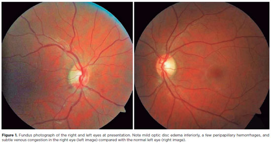
Optic neuritis is an important cause of unilateral and acute visual loss in young adults, but other differential diagnoses should be considered, especially when the disease has an atypical presentation. This report presents the case of a young woman with reduced visual acuity in her right eye, associated with optic disc edema and a relative afferent pupillary defect, that was initially misdiagnosed as optic neuritis and subsequently found to have paracentral acute middle maculopathy, possibly secondary to subtle impending central retinal vein occlusion. This case emphasizes the need to remember that retinal vascular diseases can occasionally mimic optic neuritis. Detailed anamnesis and ophthalmic examination can avoid unnecessary interventions.
Keywords: Optic disc; Papilledema; Optic neuritis; Retinal diseases; Diagnosis, differential; Visual acuity; Diagnostic errors
ABO is licensed under a Creative Commons Attribution-NonComercial 4.0 Internacional.

About
Issues
Editorial Board
Submission
Official publication of Conselho Brasileiro de Oftalmologia
Rua Casa do Ator, 1.117 - 2º andar - CEP: 04546-004
São Paulo - SP, Brazil
Phone: +55 11 3266-4000