Arq. Bras. Oftalmol. 1999;62 (6 )
:705-711
| DOI: 10.1590/S0004-27491999000600010
Abstract
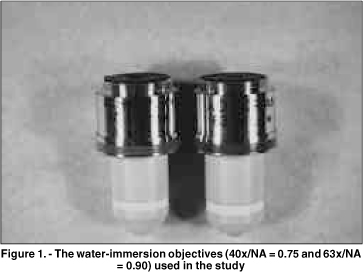
Objetivo: Este estudo prospectivo compara as imagens de microscopia confocal do epitélio corneano do coelho e do homem, obtidas através de 2 objetivas com aberturas numéricas (AN) diferentes. Métodos: Dez olhos de coelhos foram enucleados e fixados através de um suporte pneumático para garantir o melhor desempenho de cada objetiva. Cinco pacientes normais foram selecionados após consentimento. Os olhos de coelhos e dos pacientes foram previamente examinados na lâmpada de fenda. O exame de microscopia confocal (Tomey, Erlangen-Tennenlohe, Alemanha) foi realizado com as objetivas Achroplan 40x/AN = 0,75 e 63x/AN = 0,9 (Zeiss, Oberkochen, Alemanha). Imagens selecionadas do epitélio corneano foram avaliadas qualitativamente com relação ao tamanho, forma e refletividade das células. Resultados: As células no epitélio superficial dos coelhos e dos pacientes, previamente à descamação, tiveram uma refletividade maior que as células adjacentes. Este aspecto foi claramente observado somente com a objetiva 63x/AN = 0,9. As camadas basal e intermediária do epitélio em coelhos foram visualizadas somente através desta objetiva. Estas camadas nos pacientes tornaram-se mais nítidas com a objetiva de abertura numérica maior (63x/AN = 0,9). Conclusão: Uma objetiva de abertura numérica elevada produz melhor resolução dos cortes ópticos, facilitando a análise das camadas do epitélio no coelho e no homem.
Keywords: Corneal epithelium; Confocal microscopy; Epitélio corneano; Microscopia confocal
Arq. Bras. Oftalmol. 2022;85 (1 )
:1-6
| DOI: 10.5935/0004-2749.20220010
Abstract
OBJETIVO: Relatar as alterações no plexo nervoso corneano subbasal em pacientes com ceratite infecciosa de origem bacteriana utilizando a microscopia confocal in vivo.
MÉTODOS: Treze olhos de 13 pacientes com ceratite bacteriana unilateral e 12 indivíduos saudáveis como grupo controle foram incluídos prospectivamente no estudo. A microscopia confocal in vivo foi realizada em todos os pacientes em 2 momentos: na fase aguda da ceratite infecciosa e após 28 ± 0,6 meses da resolução da infecção.
RESULTADOS: A densidade dos nervos no plexo subbasal foi de 5,15 ± 1,03 mm/mm2 na fase aguda da
ceratite infecciosa (comparada com o grupo controle: 19,02 ± 1,78 mm/mm2, p<0,05). Apesar de significativa regeneração dos nervos corneanos ao longo de um intervalo de 28 meses após a resolução da infecção, a densidade dos nervos se manteve significativamente reduzida (9,73 ± 0,93 mm/mm2) quando comparada com o grupo controle (19,02 ± 1,78 mm/mm2, p<0,05). Além disso, as imagens obtidas com a microscopia confocal mostraram áreas de hiperreflectividade referente ao tecido corneano cicatricial com ramos de nervos, afinados e tortuosos, se regenerando nessas áreas.
CONCLUSÕES: Foi observado regeneração parcial dos nervos do plexo corneano subbasal durante os primeiro 28 meses após a resolução da fase aguda da ceratite infecciosa. Além disso, os nervos corneanos regenerados se mantiveram morfologicamente alterados quando comparados ao grupo controle. Esses resultados podem ser relevantes para o acompanhamento clínico e planejamento cirúrgico desses pacientes.
Keywords: Córnea/inervação; Nervo oftálmico; Infecções oculares virais; Ceratite herpética; Microscopia confocal
Arq. Bras. Oftalmol. 2020;83 (6 )
:473-477
| DOI: 10.5935/0004-2749.20200088
Abstract
Objetivos: Descrever os achados na angiografia por tomografia de coerência óptica associada à síndrome de Alport.
Métodos: Estudo descritivo de um serviço de referência em Oftalmologia (Hospital Evangélico de Vila Velha, Brasil). Os pacientes diagnosticados com síndrome de Alport, foram incluídos.
Resultados: O grupo de estudo foi composto por quatro pacientes (um feminino e três homens) com diagnóstico de síndrome de Alport. A acuidade visual no pior olho estava entre 20/40 a 20/60. Todos os pacientes do sexo masculino apresentaram lenticone anterior à biomiscroscopia. Os achados da retina observados incluíram pontos e manchas e alterações pigmentares na mácula. Na angiotomografia de coerência óptica, as camadas internas da retina de todos os pacientes apresentaram afinamento (especialmente na região temporal da mácula) e aumento da zona avascular foveal. Uma coroide espessa foi observada em ambos os olhos dos dois pacientes mais jovens.
Conclusões: Em pacientes com síndrome de Alport, as camadas internas da retina sofrem alterações devido à mutação do colágeno tipo IV. A angiotomografia de coerência óptica permite visualizar esses achados, tornando-o uma ferramenta útil na detecção de achados iniciais da retina associados à síndrome de Alport.
Keywords: Retina; Tomografia de coerência óptica; Angiofluoresceínografia/métodos; Nefrite hereditária
Arq. Bras. Oftalmol. 2025;88 (6 )
:1-5
| DOI: 10.5935/0004-2749.2024-0321
Abstract
PURPOSE: To report the ophthalmological signs, symptoms, and clinical management observed during an unprecedented outbreak of chemical ocular injuries related to cosmetic hair ointments in Brazil.
METHODS: This descriptive, cross-sectional study reviewed medical records of patients treated at the emergency center of Fundação Altino Ventura for chemical ocular trauma associated with cosmetic hair ointment use between February 2022 and February 2023. Records with incomplete medical information were excluded.
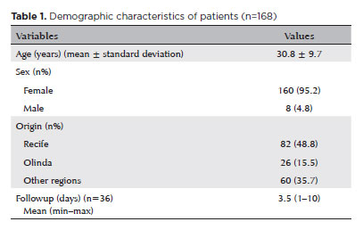
RESULTS: The study included 168 patients (95.2% [n=160] female), with a mean age of 30.8 ± 9.7 years. The most frequently reported symptoms at presentation were pain (167/168, 99.4%) and photophobia (92/168, 54.8%). Severe pain was reported by 137 patients (80%). Keratitis was present in 280 of 336 eyes (83.3%), conjunctival hyperemia in 256 eyes (76.4%), and corneal abrasions in 174 eyes (51.8%). A decrease in visual acuity (worse than 20/25) was documented in 18.5% (31/168) of cases. Lubricants, antibiotics, and re-epithelialization
ointments were prescribed to 64.8% (109/168) of the patients. Topical corticosteroids and oral vitamin C were administered to 34% (57/168) and 1.2% (2/168) of patients, respectively. Followup visits were required in 19% (33/168) of cases.
CONCLUSION: The outbreak of chemical ocular injuries linked to cosmetic ointments used for braiding and hair modeling in Brazil was marked by intense ocular pain, conjunctival hyperemia, keratitis, and corneal abrasions. Most patients were treated with lubricants, antibiotics, and re-epithelialization ointments, although approximately one-fifth required followup care, and one-third received additional treatment with either topical corticosteroids and/or oral vitamin C.
Keywords: Cosmetics; Hair preparations; Eye injuries; Burns, chemical; Eye burns; Keratitis; Cornea; Corneal diseases; Visual low.
Arq. Bras. Oftalmol. 2023;86 (4 )
:353-358
| DOI: 10.5935/0004-2749.20230055
Abstract
Objetivo: Examinar a eficácia da ceratectomia fototerapêutica para o tratamento de patologias variáveis que apresentarem opacidades anteriores da córnea, e avaliar a distribuição das indicações de ceratectomia fototerapêutica nos últimos 10 anos.
Métodos: Foram revisados retrospectivamente os prontuários de 276 pacientes, com 334 olhos tratados com ceratectomia fototerapêutica entre março de 2010 e o ano de 2020. As etiologias dos pacientes submetidos à ceratectomia fototerapêutica foram anotadas e suas alterações foram examinadas. Os resultados refrativos e de acuidade visual antes e após a operação foram registrados e analisados de acordo com a etiologia.
Resultados: A idade média dos pacientes foi de 40,7 ± 16,2 anos (faixa: 19-84). O tempo médio de acompanhamento foi de 25,5 ± 19,1 meses (faixa: 3-96). A ceratectomia fototerapêutica foi aplicada com mais frequência para distrofias estromais corneanas (44%, 151 olhos de 111 pacientes); entre as distrofias corneanas como um todo, a distrofia granular foi a indicação terapêutica mais comum desse procedimento. Ao contrário de outras indicações, nos últimos 10 anos houve um aumento na aplicação de ceratectomia fototerapêutica em casos de opacidade subepitelial persistente causada por conjuntivite adenoviral. Houve um aumento significativo na acuidade visual em todos os grupos, exceto para o grupo com defeito epitelial recorrente (p<0,05). A maior melhora na acuidade visual foi detectada em casos de distrofia estromal, no subgrupo das distrofias granulares.
Conclusão: Apesar das mudanças nas tendências de indicação, a ceratectomia fototerapêutica continua sendo uma abordagem terapêutica eficaz e confiável para tratar lesões da córnea anterior.
Keywords: Ceratectomia fotorrefrativa; Opacidade da córnea; Lesões da córnea; Distrofias da córnea; Fototerapia.
Arq. Bras. Oftalmol. 2026;89 (4 )
:1-7
| DOI: 10.5935/0004-2749.2024-0409
Abstract
PURPOSE: The purpose of this study is to look into the relationship between tear film osmolarity, tear crystallization, and corneal esthesiometry findings in Sjögren's syndrome patients.
METHODS: This cross-sectional observational study included 43 eyes from patients with a confirmed diagnosis of Sjögren's syndrome. Tear osmolarity was measured with an iPen osmometer, tear crystallization was graded using Roland's classification, and corneal sensitivity was evaluated with a Cochet–Bonnet aesthesiometer. Ocular symptoms were assessed using the Ocular Surface Disease Index questionnaire. Patients who had undergone keratoplasty or worn contact lenses within 4 hours of testing were excluded.
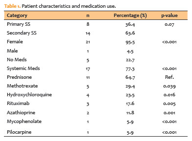
RESULTS: The cohort's mean tear osmolarity was 292.5±15.0 mOsm/L (median: 293 mOsm/L, IQR: 17.5). There was no significant difference between patients with primary Sjögren's syndrome (mean: 289.4 mOsm/L) and those with secondary Sjögren's syndrome (mean: 294.5 mOsm/L; p=0.413). Tear crystallization patterns were more severe in patients with primary Sjögren's syndrome (mean: 3.25, median: 3.5, IQR: 1.25) than in those with secondary Sjögren's syndrome (mean: 3.19, median: 3.0, IQR: 1.0), though the difference was not statistically significant (p=0.87). Corneal sensitivity was reduced by 3.5±1.7 mm (median: 4.0 mm, IQR: 2.13). Tear crystallization has a significant negative correlation with corneal sensitivity (r=−0.313, p=0.041), suggesting that poorer tear quality leads to decreased corneal sensitivity.
CONCLUSION: Tear crystallization patterns and corneal sensitivity were found to be significantly correlated in Sjögren's syndrome patients. The findings also indicate that systemic medication use may affect tear film quality.
Keywords: Sjogren's syndrome, Tear crystallization, Dry eye disease, Cornea, Tears, Osmolarity
Arq. Bras. Oftalmol. 2026;89 (3 )
:1-9
| DOI: 10.5935/0004-2749.2025-0259
Abstract
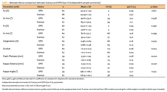
PURPOSE: To evaluate the reliability and comparability of a Scheimpflug-based tomographer relative to a Placido-based topographer and specular microscopy in healthy eyes.
METHODS: This cross-sectional study included 40 patients (80 eyes). Each eye underwent randomized imaging with a Scheimpflug-based tomographer, a Placido-based topographer, and Tomey EM-4000 specular microscopy. Three acquisitions per device were obtained. For interdevice comparisons, the best-quality scan per eye/device was selected, whereas all three scans were used for intradevice repeatability analyses. Unreliable scans were repeated (up to five attempts) and excluded if acceptable quality was not achieved, resulting in variable denominators. Between-device comparisons were performed using generalized estimating equations
with participant-level clustering and robust standard errors and were supplemented by Bland–Altman analysis.
RESULTS: The effective sample size varied by parameter (independent summaries: 59–67 eyes; paired comparisons: 48–51 eyes). In paired-eye analyses, the Scheimpflug-based tomographer measured slightly higher keratometry values than the Placido-based topographer (K1: 43.95 vs. 43.78 D, p=0.003; K2: 44.91 vs 44.73 D, p=0.002), more negative Q-values (p=0.001), smaller photopic pupil diameter (p<0.001), and shorter kappa distance (p<0.001). Mean absolute differences were 0.32 D for K1 and 0.30 D for K2, with high dispersion for angular metrics (kappa angle coefficient of variation: 195%).
CONCLUSIONS: The Scheimpflug-based tomographer provides reproducible corneal measurements in healthy eyes. However, systematic differences relative to the Placido-based topographer—particularly for keratometry, asphericity, and pupil and kappa metrics—suggest limited interchangeability. Consistent device use is recommended when these parameters inform clinical decision-making.
Keywords: Scheimpflug tomography; Placido topography; Specular microscopy; keratometry; Corneal imaging; Refractive surgical procedures; Lenses, intraocular
Arq. Bras. Oftalmol. 2024;87 (3 )
:1-8
| DOI: 10.5935/0004-2749.2023-0109
Abstract
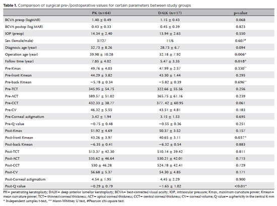
PURPOSES: This study aims to assess and compare the postoperative visual and topographic outcomes, complications, and graft survival rates following deep anterior lamellar keratoplasty and penetrating keratoplasty in patients with macular corneal dystrophy.
METHODS: In this study we enrolled 59 patients (23 male; and 36 female) with macular corneal dystrophy comprising 81 eyes. Out of these, 64 eyes underwent penetrating keratoplasty, while 17 eyes underwent deep anterior lamellar keratoplasty. The two groups were analyzed and compared based on best-corrected visual acuity, corneal tomography parameters, pachymetry, complication rates, and graft survival rates.
RESULTS: After 12 months, 70.6% of the patients who underwent deep anterior lamellar keratoplasty (DALK) and 75% of those who had penetrating keratoplasty (PK) achieved a best-corrected visual acuity of 20/40 or better (p=0.712). Following surgery, DALK group showed lower front Kmean (p=0.037), and Q values (p<0.01) compared to the PK group. Postoperative interface opacity was observed in seven eyes (41.2%) in the DALK group. Other topography values and other complications (graft rejection, graft failure, cataract, glaucoma, microbial keratitis, optic atrophy) did not show significant differences between the two groups. The need for regrafting was 9.4% and 11.8% in the PK and DALK groups, respectively (p=0.769). Graft survival rates were 87.5% and 88.2% for PK and DALK; respectively (p=0.88 by Log-rank test).
CONCLUSION: Both PK and DALK are equally effective in treating macular corneal dystrophy, showing similar visual, topographic, and survival outcomes. Although interface opacity occurs more frequently after DALK the visual results were comparable in both groups. Therefore, DALK emerges as a viable surgical choice for patients with macular corneal dystrophy without Descemet membrane involvement is absent.
Keywords: Macular corneal dystrophy; Corneal dystrophies; Hereditary; Keratoplasty; Penetrating; Corneal transplantation
Arq. Bras. Oftalmol. 2024;87 (3 )
:1-6
| DOI: 10.5935/0004-2749.2022-0220
Abstract
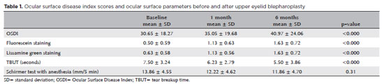
PURPOSE: This study aimed to investigate the effect of upper eyelid blepharoplasty with the removal of the skin and a strip of orbicularis oculi muscle on the ocular surface, tear film, and dry eye-related symptoms.
METHODS: Twenty-two eyes of 22 consecutive patients operated by a single surgeon (21 females; mean age, 61 years; age range, 41-75 years) were included. All subjects completed the Ocular Surface Disease Index questionnaire, underwent in vivo confocal microscopy, tear film breakup time measurements, the Schirmer test with anesthesia, and fluorescein and lissamine green staining measurements before, 1 month, and 6 months after upper blepharoplasty alone with preseptal orbicularis excision.
RESULTS: A significant increase in Ocular Surface Disease Index, and corneal fluorescein and lissamine green staining and a significant decrease in tear film breakup time were observed after 1 month (p=0.003, p=0.004, p=0.029, and p=0.024 respectively) and 6 months (p=0.001 for all findings). No significant difference in the Schirmer test score was observed during the follow-up. None of the in vivo confocal microscopy parameters showed significant changes during the study.
CONCLUSIONS: An increase in dry eye symptoms and a decrease in tear film stability along with ocular surface staining were observed in patients undergoing upper eyelid blepharoplasty.
Keywords: Blepharoplasty; Intravital microscopy; Lissamine green dyes; Fluorescein; Severity of illness index; Dry eye syndromes


 ABO is licensed under a Creative Commons Attribution-NonComercial 4.0 Internacional.


03-fig01.jpg)
02-fig01.jpg)
06-tab01.jpg)
15-fig01tb.jpg)
03-fig01.jpg)
05-fig01tb.jpg)