Arq. Bras. Oftalmol. 2022;85 (1 )
:77-81
| DOI: 10.5935/0004-2749.20220009
Abstract
Os autores relatam o caso de um homem diabético de 46 anos de idade em tratamento para mucormicose rino-orbital-cerebral com anfotericina B lipossomal e desbridamento cirúrgico. Piora clínica foi observada, com acometimento de motilidade ocular e acuidade visual de ausência de percepção luminosa, sendo evidenciada extensão radiológica da infecção, com invasão do seio cavernoso. Com base nos achados oftalmológicos, uma exenteração, procedimento potencialmente desfigurante, foi indicada, mas optamos por realizar amplo desbridamento cirúrgico e administração intraconal de anfotericina B por cateter. Melhora clínica e resolução da inflamação ocorreram após duas semanas de tratamento. Dessa forma, a mucormicose rino-orbital-cerebral foi efetivamente controlada com a administração intraconal de anfotericina B, evitando a exenteração. A intervenção deve ser considerada como um tratamento adjuvante em casos selecionados de mucormicose rino-orbital-cerebral antes que a exenteração seja realizada.
Keywords: Mucormicose; Celulite orbitária; Anfotericina B; Órbita; Infecções fúngicas
Arq. Bras. Oftalmol. 2022;85 (5 )
:513-516
| DOI: 10.5935/0004-2749.2020-0328
Abstract
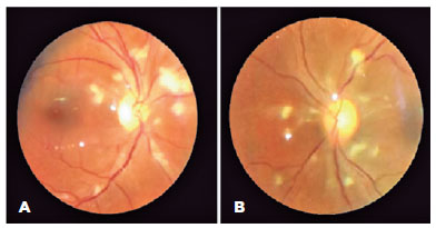
A manifestação oftálmica mais frequentemente relatada da infecção por SARS-CoV-2 é a conjuntivite. Trata-se de estudo de caso de retinopatia tipo Purtscher em uma paciente com coagulopatia grave associada ao COVID-19. Uma jovem com múltiplas comorbidades foi admitida por síndrome do desconforto respiratório agudo relacionado ao COVID-19. Seu quadro foi complicado pela fungemia. O exame oftálmico revelou pólo posterior bilateral, lesões intraretinianas e o fluconazol foi adicionado para tratar a retinite fúngica presumida. No decorrer de uma semana, manchas largas peripapilares de algodão e hemorragias sugestivas de retinopatia tipo Purtscher foram observadas. Os dímeros D, o fibrinogênio e a proteína c-reativa estavam acentuadamente elevados antes da nossa consulta, indicando um estado pró-trombótico e pró-inflamatório precedente. O duplex venoso subsequente revelou trombose venosa profunda nas veias subclávia direita e jugular interna. Os índices de fatores von Willebrand estavam marcadamente elevados, sugerindo coagulopatia grave associada ao COVID-19. A retinopatia tipo Purtscher, uma microangiopatia oclusiva rara foi descrita em várias condições pró-inflamatórias e pró-trombóticas. Para nosso conhecimento, este é o primeiro relatório de retinopatia tipo Purtscher com coagulopatia associada ao COVID-19.
Keywords: Doença retiniana; Infecção por coronavírus; Síndrome respiratória aguda grave; Relato de caso.
Arq. Bras. Oftalmol. 2022;85 (5 )
:517-519
| DOI: 10.5935/0004-2749.20220063
Abstract
Reportamos o caso de homem previamente hígido, 48 anos, com paralisia isolada do nervo abducente 18 dias após infecção pelo novo coronavírus (COVID-19) confirmada por reação cadeia polimerase de transcriptase reversa. A principal queixa do paciente na admissão era diplopia. O exame ocular revelou paralisia do sexto nervo craniano do olho esquerdo. Esotropia incomitante no exame inicial media 30 dioptrias prismáticas. Abdução estava limitada com adução completa no olho esquerdo. O paciente foi submetido a investigação clínica e neurológica com exame de neuroimagem, incluindo análise de amostra do líquido cefalorraquidiano para descartar causas infecciosas. Optou-se por abordagem conservadora com terapia ortóptica e prisma de Fresnel. Oito meses após a infecção pelo COVID-19, o paciente evoluiu com regressão do estrabismo e informou recuperação completa do quadro. Este relato sugere que paralisia isolada do nervo abducente causada por SARS-CoV-2 pode melhorar com abordagem conservadora.
Keywords: Infecções por coronavirus; Doenças do nervo abducente; Estrabismo; Vírus da SARS; Transtornos da motilidade ocular; Humanos; Relatos de casos
Arq. Bras. Oftalmol. 2022;85 (3 )
:301-305
| DOI: 10.5935/0004-2749.20220043
Abstract
A sífilis é uma doença reemergente e potencialmente grave. Por sua onipresença e pleomorfismo, é denominada “grande imitadora”. Relatamos caso de paciente jovem com sífilis secundária, que se apresentou com coriorretinopatia placóide sifilítica posterior aguda bilateral, simultaneamente a periostite craniana sifilítica. A despeito de realce paquimeníngeo observado na ressonância magnética, acreditamos que este tenha sido uma extensão do processo ósseo e não, uma meningite em si, uma vez que o exame do líquido cefalorraquidiano estava completamente normal. Tratamento com penicilina cristalina intravenosa resultou em completa resolução dos sinais, sintomas e achados de imagem. A sífilis secundária é o estágio de maior bacteremia e maior transmissibilidade da doença, apresentando-se principalmente com quadros mucocutâneos, mas também, menos frequentemente, com envolvimento de outros órgãos. Elevada suspeição e uma abordagem pragmática são necessárias para o diagnóstico, uma vez que essa doença pode afetar vários órgãos, como no caso relatado, em que foram acometidos olhos, ossos e pele.
Keywords: Sífilis/complicações; Neurossífilis; Infecções oculares bacterianas; Uveíte posterior; Coriorretinite; Periostite; Relato de caso
Arq. Bras. Oftalmol. 2022;85 (5 )
:520-523
| DOI: 10.5935/0004-2749.20220065
Abstract
A regeneração aberrante nas paralisias do terceiro nervo, ligando a contração do reto medial ao músculo levantador da pálpebra, é uma grande oportunidade para fazer um planejamento cirúrgico para tratar tanto a ptose quanto o desvio horizontal em um procedimento único. Relatamos uma ptose grave associada à exotropia corrigida com sucesso com uma única cirurgia de estrabismo horizontal devido à regeneração aberrante e discutimos as bases do planejamento cirúrgico.
Keywords: Doença do nervo oculomotor/cirurgia; Estrabismo; Blefaroptose; Movimento ocular/fisiologia; Procedimento cirúrgico oftalmológico; Regeneração nervosa; Humanos; Relato de caso
Arq. Bras. Oftalmol. 2022;85 (2 )
:182-185
| DOI: 10.5935/0004-2749.20220028
Abstract
As manifestações neurológicas de COVID-19 não são bem compreendidas. Relatamos o caso de um homem febril de 44 anos que apresentou visão dupla e dor de cabeça dois dias após os sintomas iniciais de fadiga, fraqueza muscular generalizada e perda de apetite. O paciente foi posteriormente diagnosticado com COVID-19 e paresia transitória do nervo abducente, não apresentando sintomas respiratórios ou achados neurológicos específicos adicionais. Como o número de casos tem aumentado em todo o mundo, sugerimos que os médicos tenham um índice de suspeita maior para COVID-19 em pacientes com neuropatias cranianas, mesmo em casos leves sem sintomas respiratórios típicos.
Keywords: Diplopia; Síndrome Respiratória Aguda Grave; Oftalmoplegia; Infecção por coronavírus; Doença do nervo abducente; SARS-CoV-2; Pandemia; Humanos; Relato de caso
Arq. Bras. Oftalmol. 2024;87 (4 )
:1-3
| DOI: 10.5935/0004-2749.2021-0495
Abstract
A falência primária do enxerto é uma complicação conhecida que pode ocorrer após o transplante penetrante de córnea. O tratamento usual dessa complicação é com um novo transplante penetrante. Apresentamos um caso em que foi usado o transplante endotelial de membrana de Descemet (DMEK - do inglês Descemet membrane endothelial keratoplasty) para o tratamento da falência primária após o transplante penetrante. Uma paciente submetida a transplante penetrante evoluiu com falência primária do enxerto a despeito do uso intenso de corticoide tópico e uma prova terapêutica de antivirais. Três meses após a cirurgia inicial, foi optado pela realização do transplante endotelial de membrana de Descemet sob o transplante penetrante. Houve um clareamento precoce e progressivo do enxerto com melhora importante da visão. Após um mês, a visão sem correção era de 20/40 melhorando para 20/20 com refração. O transplante endotelial de membrana de Descemet pode ser uma alternativa a um novo transplante penetrante como tratamento da falência primária.
Keywords: Doenças da córnea; Transplante da córnea/efeitos adversos; Rejeição do enxerto; Ceratoplastia penetrante; Lâmina limitante posterior; Ceratoplastia endotelial com remoção da lâmina limitante posterior; Infecções oculares virais; Humanos; Relatos de casos
Arq. Bras. Oftalmol. 2024;87 (4 )
:1-4
| DOI: 10.5935/0004-2749.2022-0107
Abstract
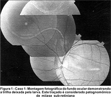
Neonato de 7 semanas, do sexo masculino, nascido de parto cesárea, apresentou sorologia positiva para dengue com hemorragias retinianas e pré-retinianas, opacidades vítreas e manchas algodonosas. O paciente e sua mãe haviam apresentado sorologias positivas para Non Structural Protein 1 através de ELISA. Achados na retina e no vítreo melhoraram em um período de dezesseis semanas. O exame de tomografia de coerência óptica de domínio espectral demonstrou arquitetura macular preservada. Neste relato de caso, sugerimos que alterações na retina e no vítreo podem ser os achados oculares aparentes em neonatos com infecção vertical por dengue, e que estes podem se resolver sem maiores sequelas estruturais.
Keywords: Dengue; Complicações infecciosas na gravidez; Transmissão vertical de doenças infecciosas; Manifestações oculares; Hemorragia retiniana; Hemorragia vítrea; Humanos; Doenças do recém-nascido; Relatos de casos
 ABO is licensed under a Creative Commons Attribution-NonComercial 4.0 Internacional.
01-fig01.jpg)
08-fig01.jpg)
14-fig01tb.jpg)
11-fig01tb.jpg)
14-fig01.jpg)
01-fig01.jpg)
03-fig01.jpg)
04-fig01.jpg)
05-fig01.jpg)
04-fig01.jpg)
02-fig01.jpg)