Arq. Bras. Oftalmol. 2020;83 (6 )
:473-477
| DOI: 10.5935/0004-2749.20200088
Abstract
Objetivos: Descrever os achados na angiografia por tomografia de coerência óptica associada à síndrome de Alport.
Métodos: Estudo descritivo de um serviço de referência em Oftalmologia (Hospital Evangélico de Vila Velha, Brasil). Os pacientes diagnosticados com síndrome de Alport, foram incluídos.
Resultados: O grupo de estudo foi composto por quatro pacientes (um feminino e três homens) com diagnóstico de síndrome de Alport. A acuidade visual no pior olho estava entre 20/40 a 20/60. Todos os pacientes do sexo masculino apresentaram lenticone anterior à biomiscroscopia. Os achados da retina observados incluíram pontos e manchas e alterações pigmentares na mácula. Na angiotomografia de coerência óptica, as camadas internas da retina de todos os pacientes apresentaram afinamento (especialmente na região temporal da mácula) e aumento da zona avascular foveal. Uma coroide espessa foi observada em ambos os olhos dos dois pacientes mais jovens.
Conclusões: Em pacientes com síndrome de Alport, as camadas internas da retina sofrem alterações devido à mutação do colágeno tipo IV. A angiotomografia de coerência óptica permite visualizar esses achados, tornando-o uma ferramenta útil na detecção de achados iniciais da retina associados à síndrome de Alport.
Keywords: Retina; Tomografia de coerência óptica; Angiofluoresceínografia/métodos; Nefrite hereditária
Arq. Bras. Oftalmol. 2025;88 (3 )
:1-8
| DOI: 10.5935/0004-2749.2024-0104
Abstract
PURPOSE: This study aimed to characterize retinitis pigmentosa associated with the eyes shut homolog gene, which causes hereditary retinal degeneration.
METHODS: The anatomical and functional findings of retinitis pigmentosa in patients with variants of the eyes shut homolog gene were characterized and compared using multimodal imaging and genetic analysis of the variants. Clinical data such as visual acuity, lens status, and refraction were obtained from medical records. Patients underwent an ophthalmic examination, including static visual field, microperimetry, optical coherence tomography, fundus autofluorescence, and fundus photography.
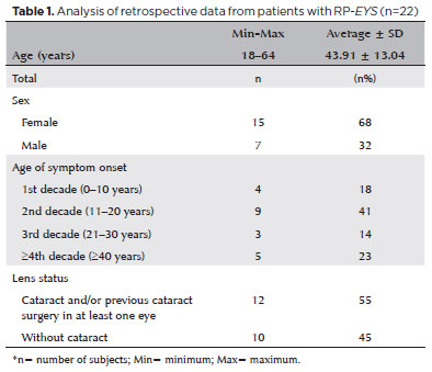
RESULTS: Twenty-two patients were included in the study. Several anatomical and functional characteristics of retinitis pigmentosa-eyes shut homolog were identified, including the presence of cataracts, cystoid macular edema, epiretinal membrane, and a tubular visual field. Genetic results revealed 26 distinct variants in the cohort, with 7 novel variants not previously documented or reported in the scientific literature or databases.
CONCLUSION: The findings demonstrate that eyes shut homolog-retinitis pigmentosa manifests in specific patterns, starting in adolescence with mild progression and advancing with age. The integration of multimodal imaging and genetic analysis has provided a detailed understanding of the anatomical and functional features of retinitis pigmentosa-eyes shut homolog. Seven novel variants of the eyes shut homolog gene have been identified. These findings enhance the understanding of eyes shut homolog-related retinitis pigmentosa characteristics of by detailing the spectrum of mutations in this gene within the Brazilian population.
Keywords: Retinal diseases/diagnostic imaging; Retinitis pigmentosa/genetics; Retinal degeneration; Eye proteins/genetics; Eye diseases, hereditary/genetics; Genes, recessive; Phenotype; Multimodal imaging; Tomography, optical coherence/methods; Fluorescein angiogr
Arq. Bras. Oftalmol. 2024;87 (3 )
:1-8
| DOI: 10.5935/0004-2749.2023-0109
Abstract
PURPOSES: This study aims to assess and compare the postoperative visual and topographic outcomes, complications, and graft survival rates following deep anterior lamellar keratoplasty and penetrating keratoplasty in patients with macular corneal dystrophy.
METHODS: In this study we enrolled 59 patients (23 male; and 36 female) with macular corneal dystrophy comprising 81 eyes. Out of these, 64 eyes underwent penetrating keratoplasty, while 17 eyes underwent deep anterior lamellar keratoplasty. The two groups were analyzed and compared based on best-corrected visual acuity, corneal tomography parameters, pachymetry, complication rates, and graft survival rates.
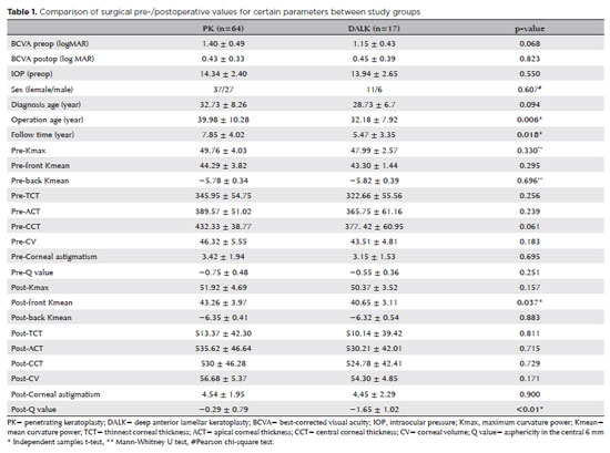
RESULTS: After 12 months, 70.6% of the patients who underwent deep anterior lamellar keratoplasty (DALK) and 75% of those who had penetrating keratoplasty (PK) achieved a best-corrected visual acuity of 20/40 or better (p=0.712). Following surgery, DALK group showed lower front Kmean (p=0.037), and Q values (p<0.01) compared to the PK group. Postoperative interface opacity was observed in seven eyes (41.2%) in the DALK group. Other topography values and other complications (graft rejection, graft failure, cataract, glaucoma, microbial keratitis, optic atrophy) did not show significant differences between the two groups. The need for regrafting was 9.4% and 11.8% in the PK and DALK groups, respectively (p=0.769). Graft survival rates were 87.5% and 88.2% for PK and DALK; respectively (p=0.88 by Log-rank test).
CONCLUSION: Both PK and DALK are equally effective in treating macular corneal dystrophy, showing similar visual, topographic, and survival outcomes. Although interface opacity occurs more frequently after DALK the visual results were comparable in both groups. Therefore, DALK emerges as a viable surgical choice for patients with macular corneal dystrophy without Descemet membrane involvement is absent.
Keywords: Macular corneal dystrophy; Corneal dystrophies; Hereditary; Keratoplasty; Penetrating; Corneal transplantation


 ABO is licensed under a Creative Commons Attribution-NonComercial 4.0 Internacional.


02-fig01.jpg)
08-fig01.jpg)
10-fig01.jpg)
01-fig01.jpg)
02-fig01.jpg)
03-fig01.jpg)