Arq. Bras. Oftalmol. 2020;83 (4 )
:277-282
| DOI: 10.5935/0004-2749.20200049
Abstract
Objetivo: Este estudo foi realizado para avaliar os resultados do cross-linking corneano acelerado em córneas ceratocônicas com os valores mais baixos de paquimetria <400 μm.
Métodos: O estudo incluiu 28 olhos de 24 pacientes. As acuidades visuais não corrigidas e melhor corrigidas (logMAR), leituras ceratométricas mais planas e íngremes, espessura corneana central no ponto mais fino, aberrações corneanas de mais alta ordem e a sensibilidade ao contraste foram avaliadas antes e em 1, 3, 6, 12 e 24 meses após a realização do do cross-linking.
Resultados: A média da acuidade visual melhor corrigida e a sensibilidade ao contraste aumentaram (p=0,02, p=0,03, respectivamente), enquanto a média da acuidade visual não corrigida não diferiu significativamente (p>0,05) aos 24 meses após o cross-linking, comparada com medidas antes do procedimento. Embora a leitura da média da ceratometria mais plana não tenha apresentado alteração significativa (p=0,58), a leitura ceratométrica mais íngreme diminuiu quando comparada ao seu valor antes do cross-linking (p=0,001). Não foi observada alteração na média da espessura corneana central no ponto mais fino aos 24 meses após o cross-linking em comparação com seu valor antes do procedimento (p=0,12).
Conclusão: O cross-linking corneano acelerado nos olhos ceratocônicos com córneas finas pode interromper a progressão do ceratocone nas córneas mais finas que 400 μm 24 meses após o tratamento.
Keywords: Ceratocone; Paquimetria corneana; Reagentes para ligações cruzadas; Riboblavina/uso terapêutico
Arq. Bras. Oftalmol. 2021;84 (4 )
:324-329
| DOI: 10.5935/0004-2749.20210046
Abstract
OBJETIVO: O ceratocone na população pediátrica apresenta algumas particularidades em relação à população adulta. O maior desafio é devido à doença ser geralmente mais severa e rapidamente progressiva em crianças.
MÉTODOS: Este artigo utiliza uma análise retrospectiva para relatar o uso do crosslinking em jovens menores de 18 anos e sua evolução pelo menos 24 meses após o procedimento. Foram estudados 12 olhos de 10 pacientes, e dados como acuidade visual com e sem correção, ceratometria máxima, espessura corneana, espessura foveal e microscopia endotelial avaliados no pré e pós-operatórios. O crosslinking corneano foi realizado em todos os pacientes pelo mesmo cirurgião.
RESULTADOS: Observou-se uma tendência de redução do valor do Kmax e melhora da acuidade visual corrigida em todos os momentos de pós operatório. Com relação à paquimetria, observou-se afinamento corneano do ponto mais fino, nos primeiros quatro meses de pós-operatório.
CONCLUSÃO: Resultados encorajadores foram obtidos com relação à estabilização da doença, progressão e segurança do procedimento, corroborando com as conclusões de outros autores. A importância do diagnóstico precoce e do acompanhamento a curto prazo do paciente deve ser destacada.
Keywords: Ceratocone/diagnóstico; Ceratocone/tratamento farmacológico; Córnea; Doenças da córnea; Topografia da córnea; Colágeno/metabolismo; Raios ultravioleta; Reagentes para ligações cruzadas/uso terapêutico; Riboflavina/uso terapêutico; Acuidade visual; Adolesc
Arq. Bras. Oftalmol. 2025;88 (1 )
:1-4
| DOI: 10.5935/0004-2749.2023-0056
Abstract
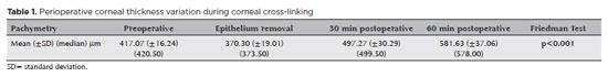
PURPOSE: This study aimed to analyze variations in intraoperative corneal thickness during corneal cross-linking in patients with keratoconus and to investigate its possible correlation with presurgical maximal keratometry (Kmax) and pachymetry.
METHODS: This was a prospective case series. We used a method similar to the Dresden protocol, with the application of hydroxypropyl methylcellulose 0.1% hypo-osmolar riboflavin in corneas between 330 and 400 µm after epithelium removal. Corneal thickness was measured using portable calipers before and immediately after epithelium removal, and 30 and 60 min after the procedure.
RESULTS: The 30 patients in this study were followed up for one year. A statistically significant difference was observed in pachymetry values during the intraoperative period (p<0.0001) and an increase of 3.05 µm (95%CI: 0.56–5.54) for each diopter was seen after epithelium removal (p0.019). We found an average Kmax difference of −2.12 D between men and women (p0.013). One year after treatment, there was a statistically significant reduction in pachymetry (p<0.0001) and Kmax (p0.0170) values.
CONCLUSIONS: A significant increase in pachymetry measurements was seen during the procedure, and most patients showed a regression in Kmax and pachymetry values one year after surgery.
Keywords: Corneal pachymetry; corneal topography; cross-linking reagents/therapeutic use; hypromellose derivatives; keratoconus/surgery; riboflavin/therapeutic use
Arq. Bras. Oftalmol. 2025;88 (6 )
:1-7
| DOI: 10.5935/0004-2749.2025-0120
Abstract
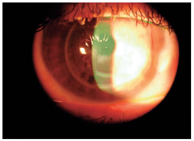
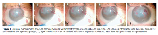
PURPOSE: To describe the technique and outcomes of intrastromal autologous blood injection in patients with severe corneal hydrops.
METHODS: Nineteen patients with corneal hydrops underwent intrastromal autologous blood injection. Postoperative assessments included best-corrected visual acuity and time to resolution of corneal edema
RESULTS: Corneal edema resolved within 1 week in 5 patients, within 1 month in 11, and within 3 months in 3. The mean duration of edema persistence was 37.94 ± 33.05 days (range, 6–124). Corneal thickness decreased from 2.06 ± 0.71-mm preoperatively to 1.34 ± 0.65-mm at day 7, 0.85 ± 0.56-mm at day 30, and 0.57 ± 0.13-mm at day 90 (p<0.001). Descemet’s membrane (DM) detachment decreased from 1.01 ± 0.75-mm to 0.44 ± 0.57-mm, 0.24 ± 0.36-mm, and 0.08 ± 0.11-mm on postoperative days 7, 30, and 90, respectively (p<0.001). DM break size decreased from 1.12 ± 1.19-mm to 0.62 ± 0.84-mm at 3 months (p<0.005). Three patients developed hematocornea; no other major complications were observed. At 3 months, mean best-corrected visual acuity improved from 2.37 ± 0.66 to 0.41 ± 0.17 logMAR with hard contact lenses (p<0.001).
CONCLUSIONS: Intrastromal autologous blood injection is an effective treatment for severe corneal hydrops, promoting faster edema resolution and visual improvement with minimal complications.
Keywords: Corneal edema; Corneal diseases; Edema; Visual acuity; keratoconus.
Arq. Bras. Oftalmol. 2025;88 (3 )
:1-7
| DOI: 10.5935/0004-2749.2023-0309
Abstract
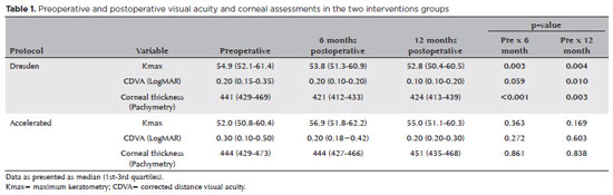
PURPOSE: Keratoconus presents certain peculiarities in pediatric patients when compared with adults. The greatest challenge in children is that the disease is more severe and faster in progression. In this retrospective study, we aimed to compare the accelerated and Dresden protocols for corneal crosslinking in patients aged <18 years who were followed-up for at least 12 months.
METHODS: A total of 36 eyes from 27 patients were included in the study. The best corrected and uncorrected visual acuity, maximal keratometry, corneal thickness, foveal thickness, and endothelial microscopy findings were evaluated at baseline and during the postoperative period at one, three, and six months. Thereafter, the patients were evaluated at one, three, six and twelve months postoperative. Corneal crosslinking was performed in all patients via the Dresden protocol (n=21 eyes) or the accelerated protocol (n=15 eyes). Data between the two groups were compared and XY statistical analysis was used.
RESULTS: Both protocols were effective in halting keratoconus progression. No patient had progression at the 12-month follow-up. A significant reduction in Kmax and improvement in the corrected distance visual acuity were observed only in the Dresden protocol group. Although the Dresden protocol was superior to the accelerated protocol in reducing Kmax (p=0.002), there was no significant difference in corrected distance visual acuity between the two groups.
CONCLUSION: The accelerated protocol is as efficient as the Dresden protocol in stabilizing keratoconus progression. Although the Dresden protocol was superior to the accelerated protocol in reducing the Kmax, it did not produce better clinical results. Thus, the accelerated protocol is an efficient option. Furthermore, considering the advantages of reduced surgical time, the accelerated protocol is effective in halting keratoconus progression in the pediatric age group.
Keywords: Keratoconus; Corneal diseases; Ultraviolet rays; Cross-linking reagents; Visual acuity
Arq. Bras. Oftalmol. 2025;88 (5 )
:1-7
| DOI: 10.5935/0004-2749.2024-0217
Abstract
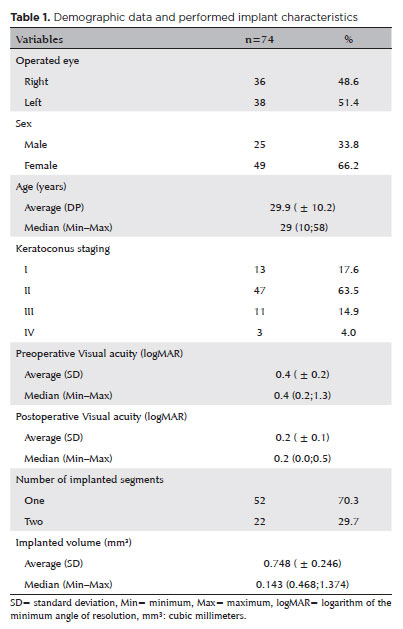
PURPOSE: This study aimed to evaluate the influence of intrastromal corneal ring segment implants on the intraocular pressure measurements using Goldmann applanation tonometry, rebound tonometry, and noncontact tonometry in keratoconic corneas and analyze the intertonometer agreement.
METHODS: We enrolled 74 eyes in this observational and prospective study. Each participant had a complete eye examination, corneal analysis with Scheimpflug Tomography (Pentacam®), and intraocular pressure evaluation with Goldmann applanation tonometry, rebound tonometry, and noncontact tonometry, before and after intrastromal corneal ring segment implantation (on postoperative days 1, 7, 45, and 90). Intertonometer agreement was assessed using Bland-Altman analysis.
RESULTS: The mean age was 29.9 ± 10.2 years, and 47 (63.5%) eyes had keratoconus grade II. Intraocular pressures were higher for noncontact tonometry preoperatively and on 90 postoperative day (mean ± SD: 12.4 ± and 12.1 ± 2.2 mmHg, respectively), followed by Goldmann applanation tonometry (11.1 ± 3.0 and 11.2 ± 2.7 mmHg, respectively), and were lower for rebound tonometry (9.7 ± and 9.4 ± 3.2 mmHg, respectively). The variation from the Goldmann tonometry on 7 postoperative day to the baseline (p=0.022) and that of noncontact tonometry on 90 postoperative day to the baseline (p=0.021) were statistically significant. The rebound tonometry underestimated intraocular pressure when compared with the Goldmann applanation tonometry by a mean of 1.47 ± 5.19 mmHg. Noncontact tonometry, when compared with Goldmann applanation tonometry, overesti-mated intraocular pressure by a mean of 1.23 ± 4.15 mmHg.
CONCLUSION: Despite statistically significant differences between some postoperative periods, the intraocular pressure measurement differences may not be clinically relevant.
Keywords: Keratoconus; Intraocular pressure; Cornea; Corneal stroma; Postoperative period; Tonometry ocular; Prostheses and implants
Arq. Bras. Oftalmol. 2024;87 (3 )
:1-8
| DOI: 10.5935/0004-2749.2022-0004
Abstract
OBJETIVO: Examinar os efeitos do tratamento de reticulação unilateral do colágeno corneano na acuidade visual e os achados topográficos em olhos não tratados de pacientes com ceratocone progressivo bilateral.
MÉTODOS: Foram rastreados retrospectivamente pacientes com ceratocone progressivo submetidos a tratamento de reticulação. Foram incluídos no estudo 188 olhos não tratados de 188 pacientes tratado unilateralmente com reticulação padrão ou acelerada e que recusaram o procedimento de reticulação no outro olho. A acuidade visual e os achados topográficos dos olhos não tratados foram obtidos no pré- e pós-operatório no 1o, 3o, 6o, 12o, 24o, 30o e 36o mês.
RESULTADOS: As alterações ao longo do tempo foram semelhantes para as variáveis examinadas nos olhos não tratados de pacientes tratados com métodos de reticulação padrão e acelerado (p>0,05). No 12º mês, 136 olhos não tratados (95,8%) estavam estáveis, de acordo com os critérios de progressão. Apenas quatro olhos (8%) mostraram progressão no 24o mês. Nenhuma progressão foi observada nos 16 pacientes que tiveram um acompanhamento de 36 meses.
CONCLUSÕES: O estudo mostrou que os olhos não tratados de pacientes com ceratocone progressivo bilateral não apresentaram taxas de progressão significativas após o tratamento unilateral com reticulação.
Keywords: Topografia da córnea; Reagentes de ligações cruzadas; Ceratocone; Fármacos fotossensibilizantes; Colágeno/uso terapêutico; Fotoquimioterapia/métodos; Acuidade visual
Arq. Bras. Oftalmol. 2024;87 (4 )
:1-6
| DOI: 10.5935/0004-2749.2022-0128
Abstract
Objetivo: Relatar um experimento projetado para determinar alterações anatômicas em córneas porcinas após a colocação de um novo implante depolímero na córnea.
Métodos: Foi utilizado olho de porco ex vivo. Um novo agente modelador biocompatível, de colágeno tipo 1, com 6mm de diâmetro foi moldado com excimer laser em sua face posterior, para criar três formatos planocôncavos. Os implantes foram inseridos dentro de um bolsão, dissecado manualmente, a 200 micrômetros (µm). Foram definidos três grupos de tratamento: grupo A (n=3), teve a profundidade máxima de ablação de70 µm; o grupo B (n=3), profundidade máxima de ablação de 64 µm; e o grupo C (n=3), profundidade máxima de ablação de 104 µm, com buraco central. O grupo controle, D (n=3), foi incluído, com a criação do bolsão estromal, porém sem inserir o material. A avaliação desses olhos foi realizada por tomografia de coerência óptica (OCT) e por tomografia corneana.
Resultados: A tomografia corneana mostrou uma tendência para diminuição da ceratometria média em todos os 4 grupos. A tomografia de coerência óptica mostrou córneas com implantes localizados no estroma anterior e aplanamento visível, enquanto as córneas não mudaram qualitativamente o formato no grupo controle.
Conclusões: O novo implante de biomaterial planocôncavo descrito aqui foi capaz de remodelar a córnea em modelo de animal ex vivo, resultando no aplanamento corneano. Novos estudos são necessários usando modelos animais in vivo para confirmar tais achados.
Keywords: Córnea; Cirurgia da córnea a laser; Substância própria; Proteses e implantes; Lasers de excimer; Materiais biocompatíveis; Animais; Suínos
Arq. Bras. Oftalmol. 2024;87 (3 )
:1-7
| DOI: 10.5935/0004-2749.2023-0049
Abstract
PURPOSE: To investigate the association of pre-photorefractive keratectomy Schirmer-1 test value with post-photorefractive keratectomy central corneal epithelial thickness, ocular surface disease index score, and uncorrected distance visual acuity.
METHODS: Patients were categorized according to preoperative Schirmer-1 value: the normal Schirmer Group (n=54; Schirmer-1 test value, >10 mm) and the low Schirmer Group (n=52; Schirmer-1 test value, between 6 and 10 mm). We analyzed ablation depth, visual acuity, result of Schirmer-1 test (with anesthesia), tear film break-up time, ocular surface disease index score, central corneal epithelial thickness, and spherical equivalent refraction.
RESULTS: We found significant differences between the groups in Schirmer-1 test value, tear film break-up time, and ocular surface disease index score, both preoperatively and postoperatively (p<0.001). The preoperative central corneal epithelial thicknesses of the two groups were similar (p>0.05). After photorefractive keratectomy, the Schirmer-1 test value and spherical equivalent refraction decreased in both groups (p<0.05), and ocular surface disease index scores and central corneal epithelial thickness values increased in the low Schirmer Group (p<0.001) but not in the normal Schirmer Group (p>0.05). The postoperative central corneal epithelial thicknesses of the low Schirmer Group were significantly higher than those of the normal Schirmer Group (p<0.001). Postoperative uncorrected distance visual acuity did not differ significantly between the two groups (p>0.05).
CONCLUSIONS: In patients with low Schirmer-1 test values before photorefractive keratectomy, the corneal epithelium thickened and ocular surface complaints increased during the postoperative period. However, changes in the corneal epithelium did not affect the postoperative uncorrected distance visual acuity. To reduce postoperative problems on the ocular surface in these patients, we recommend that dry eye be treated before photorefractive keratectomy.
Keywords: Epithelium, corneal; Cornea; Photorefractive keratectomy; Schirmer test; Visual acuity
Arq. Bras. Oftalmol. 2025;88 (5 )
:1-7
| DOI: 10.5935/0004-2749.2024-0368
Abstract
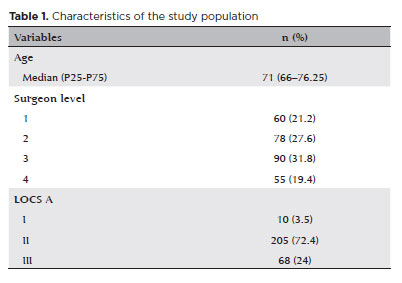
PURPOSE: To compare endothelial corneal cell changes following cataract surgery performed by phacoemulsification with intraocular lens implantation, conducted by surgeons with varying levels of experience.
METHODS: Two hundred and eighty-three eyes diagnosed with cataract were included. Lens opacity was classified into three categories (I, II, and III). Surgeons were categorized into four experience levels (1, 2, 3, and 4), based on years of practice and lifetime surgeries performed. Corneal endothelial characteristics were assessed using non-contact specular microscopy, with measurements taken before surgery and 30-60 days post-surgery.
RESULTS: Pre- and postoperative endothelial analysis showed no significant differences between surgeon levels regarding visual acuity achieved, corneal thickness, and endothelial hexagonality. However, the central endothelial cell density index showed a significantly greater reduction among level 1 surgeons (p=0.026). Grade II cataracts exhibited significant variations in the central endothelial cell density (p=0.011) and average cell size, with level 1 surgeons showing the largest increases (p=0.024).
CONCLUSIONS: The analysis revealed significant differences in visual acuity and endothelial indices between surgeon experience levels, with less experienced surgeons showing greater variations and poorer performance. Clinical protocols should consider these data to establish safer training protocols.
Keywords: Cataract extraction; Phacoemulsification; Endothelium; corneal; Lens implantation, intraocular; Visual acuity; Internship and residency; Surgeons
Arq. Bras. Oftalmol. 2024;87 (2 )
:1-5
| DOI: 10.5935/0004-2749.2022-0273
Abstract
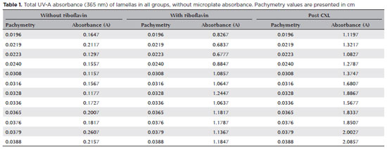
PURPOSE: To determine the absorbance coefficient of the thin porcine cornea to ultraviolet-A radiation (365 nm) submitted for crosslinking.
METHODS: This in vitro, benchtop experiment using cadaver tissue study analyzed 12 porcine corneal lamellas, which were obtained using a microkeratome after mechanical de-epithelization and separated into three thickness groups: 180, 300, and 360 µm. The corneal thickness values were measured by anterior-segment optical coherence tomography. All lamellas had ultraviolet-A (365 nm) absorbance measured with a 96-well plate spectrophotometer using an ultraviolet transparent microplate before riboflavin instillation and pre- and post-crosslinking according to the Dresden protocol.
RESULTS: The ultraviolet absorbance profiles of the 180, 300, and 360 µm groups were obtained as a-coefficients of 12.85, 76.55, and 120.27, respectively. A theoretical formula was calculated though a statistical analysis that demonstrated the correlation between stromal lamellar thickness and ultraviolet absorbance.
CONCLUSIONS: Corneal thickness and ultraviolet-A spectral absorbance of corneal lamellas showed linear correlation. These findings can potentially contribute to the optimization of ultraviolet-A application during crosslinking, making the treatment of corneas with thickness <400 µm safe and personalized energy delivery for each corneal thickness.
Keywords: Ultraviolet light; Spectrophotometry; Crosslinking; Keratoconus; Corneal thickness
 ABO is licensed under a Creative Commons Attribution-NonComercial 4.0 Internacional.
02-tab01tb.jpg)
03-tab01.jpg)
08-tab01.jpg)
09-fig01.jpg)
17-equ01.jpg)
09-fig01.jpg)