Arq. Bras. Oftalmol. 2024;87 (3 )
:1-4
| DOI: 10.5935/0004-2749.2021-0235
Abstract
Paciente do sexo masculino, 33 anos, apresentou ceratite infecciosa subaguda unilateral 4 semanas após a cirurgia. A inflamação da córnea foi resistente aos regimes de antibióticos tópicos padrão. A aba da córnea foi derretida e seccionada durante o levantamento e amostragem para diagnóstico. A melhora clínica só foi alcançada após levantamento do retalho, raspagem e diagnóstico microbiológico de micobactérias atípicas e tratamento com amicacina fortificada tópica, claritromicina e claritromicina sistêmica.
Keywords: Córnea/microbiologia; Úlcera da córnea; Infecções oculares bacterianas; Mycobacterium abscessus; Procedimentos cirúrgicos refrativos; Ceratomileuse assistida por excimer laser in situ; Amicacina/uso terapêutico; Claritromicina/uso terapêutico; Humanos; Re
Arq. Bras. Oftalmol. 2024;87 (4 )
:1-5
| DOI: 10.5935/0004-2749.2023-0066
Abstract
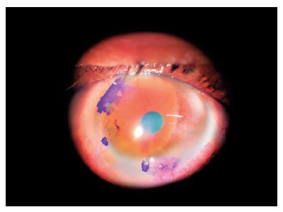
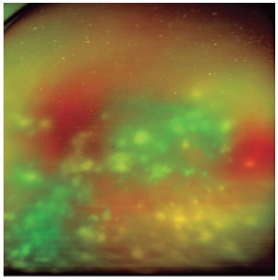
Endophthalmitis is a severe form of purulent inflammation caused by the infection of the intraocular tissues or fluids. This infection infrequently occurs through endogenous routes, which are often correlated with major risk factors. Escherichia coli, a gram-negative rod, can cause endophthalmitis through hematogenous spread. We here report a 59-year-old man who presented to our service with acute visual impairment in his left eye, preceded by floaters. He was taking sirolimus and azathioprine for a transplanted kidney, had undergone catheterization for bladder atresia, and had a history of recurrent E. coli urinary tract infections. On evaluation, the left eye exhibited visual acuity of hand motion, anterior chamber reaction (3+/4+), and intense vitritis (4+/4+) with white flake clusters, which prevented appropriate retinal evaluation. Pars plana vitrectomy was performed, and the culture yielded E. coli. The present case highlights the importance of identifying the signs and symptoms of infection early so that diagnosis and treatment of endophthalmitis can be promptly initiated.
Keywords: Endophthalmitis; Escherichia coli; Escherichia coli infections; Eye infections, Bacterial; Sepsis; Vitrectomy; Anti-bacterial agents/therapeutic use; Humans; Case reports
Arq. Bras. Oftalmol. 2024;87 (3 )
:1-4
| DOI: 10.5935/0004-2749.2021-0377
Abstract
Este relato de caso apresenta um paciente feminino de 33 anos encaminhado para um serviço especializado de retina devido à leve perda de visão em olho direito. A acuidade visual foi de 20/25 no olho direito e 20/50 no olho esquerdo, o equivalente esférico foi de -12,75 dioptrias e -14,75 dioptrias, respectivamente. Avaliações multimodais revelaram isquese peripapilar nas camadas internas e externas da retina, descolamento vítreo posterior grau II e fundo tesselado. Avaliação funcional com perimetria Humphrey e microperimetria MP-3 revelaram sensibilidade macular normais e diminuição da sensibilidade na região peripapilar, especialmente no olho direito. Potencial visual evocado de padrão reverso apresentou no olho direito latência e amplitude N75 e P100 dentro dos valores normais para verificação de 1º. Entretanto, a amplitude foi baixa para a de 15´. Pacientes alto míopes com esfiloma posterior envolvendo o nervo óptico são suscetíveis à tração da hialoide posterior. Portanto a tração vitreopapilar resultante pode causar comprometimento da visão.
Keywords: Miopia degenerativa; Retinosquise; Descolamento retiniano; Tomografia de coerência óptica; Humanos; Relatos de casos
Arq. Bras. Oftalmol. 2024;87 (4 )
:1-5
| DOI: 10.5935/0004-2749.2023-0221
Abstract
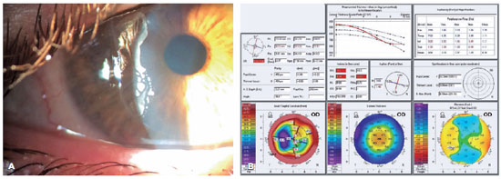
We present a case of a patient complaining of monocular diplopia due to a decentered ablation after LASIK. The patient underwent a wavefront-guided retreatment, which resulted in an epithelial ingrowth complication. Additionally, the patient developed cataract, with cataract surgery requiring reliable biometric measurements. Therefore, we opted for corneal treatment and corneal surface regularization. Although we attempted to lift the flap and wash the interface initially, the procedure proved unsuccessful, thereby necessitating immediate flap amputation. Once the corneal surface was regularized in the seventh postoperative month, transepithelial photorefractive keratectomy was successfully performed to homogenize the ocular surface, thereby significantly improving the patient's corrected visual acuity and resolving monocular diplopia. The surface and corneal curvature stabilized by the fifth month after the procedure. Phacoemulsification was then performed along with the implantation of a toric monofocal lens, which was selected using an appropriate formula, resulting in an excellent uncorrected visual acuity.
Keywords: Refractive surgical procedures; Surgical flap/surgery; Keratomileusis laser In situ/methods; Biometry; Corneal topography; Lasers, Excimer/adverse effects; Dipoplia/etiologia; Visual acuity; Humans; Case reports
 ABO is licensed under a Creative Commons Attribution-NonComercial 4.0 Internacional.
09-fig01.jpg)
11-fig01.jpg)
10-fig01.jpg)
11-fig01tb.jpg)
12-fig01.jpg)
10-fig01.jpg)
02-fig01.jpg)
03-fig01.jpg)
03-fig01.jpg)
04-fig01.jpg)
02-fig01.jpg)