Showing of 1 until 15 from 206 result(s)
Search for: Diagnostic techniques, ophthalmological; Preoperative care; Cataract extraction; Lens implantation, intraocular; Refractive errors
Abstract
Objetivo: Avaliar a qualidade óptica medindo o índice de dispersão objetiva de luz e os parâmetros de qualidade óptica objetiva (Razão de Strehl e Função de Transferência de Modulação) em indivíduos com emetropia e ametropia.
Métodos: Estudo prospectivo, transversal, incluindo 408 olhos. O grupo ametrópico era de olhos com melhor acuidade visual corrigida de 0,0 logMAR ou melhor e apresentando, pelo menos, um erro refrativo de 0,25 D ou mais. Os pacientes foram submetidos a exame com lâmpada de fenda, acuidade visual, refração e qualidade óptica com o HD Analyzer.
Resultados: O índice de dispersão objetiva de luz médio foi de 0,62 ± 0,63, 0,77 ± 0,70, 0,74 ± 0,30, 0,93 ± 0,55, 0,85 ± 0,61 e a média da Razão de Strehl e de Função de Transferência de Modulação foram 38,17 ± 10,4, 37,37 ± 10,06, 29,84 ± 9,71, 33,2 ± 12,11 e 33,13 ± 10,09 em olhos emetrópicos, míopes, hipermétropes, equivalente esférico ≥0 e equivalente esférico <0 respectivamente. Foram encontradas diferenças significativas em todas as variáveis entre olhos emetrópicos e com hipermetropia corrigida, equivalente esférico ≥ 0 e equivalente esférico <0 (p<0,05).
Conclusão: Em condições com lentes corrigidas (com armações de prova), os olhos emetrópicos e com miopia simples apresentaram qualidade óptica significativamente melhor em comparação com os olhos hipermétropes e astigmáticos. O significado clínico destes resultados deve ser estudado posteriormente.
Keywords: Erro de refração; Emetropia; Dispositivo óptico; Técnica de diagnóstico oftalmológico/instrumentação; Refração ocular; Acuidade visual

Abstract
Objetivo: Investigar picos de pressão intraocular em pacientes fácicos e pseudofácicos com glaucoma primário de ângulo aberto no teste de sobrecarga hídrica.
Método: Quarenta pacientes com glaucoma primário de ângulo aberto foram avaliados; vinte eram fácicos e vinte eram pseudofácicos. Um olho (selecionado aleatoriamente) foi incluído no estudo, todos os pacientes foram submetidos às curvas da pressão intraocular imediatamente após o teste de sobrecarga hídrica.
Resultados: Observou-se uma diferença estatisticamente significante na média dos picos nas curvas da pressão intraocular para os pacientes fácicos e pseudofácicos (p=0,045). Houve diferença estatisticamente significante nos picos da pressão intraocular no teste de sobrecarga hídrica entre os grupos, sendo observados valores mais altos nos pacientes fácicos (p=0,004).
Conclusão: Os picos da pressão intraocular no teste de sobrecarga hídrica foram maiores no grupo fácico que no grupo pseudofácico.
Keywords: Glaucoma de ângulo aberto; Técnicas de diagnóstico oftalmológico; Água potável; Pressão intraocular
Abstract
OBJETIVO: O objetivo deste estudo foi analisar a segurança do implante de lente intraocular primária em um grande número de olhos em crianças <24 meses.
MÉTODOS: Foram revisados os prontuários de pacientes com idade entre 5-24 meses, submetidos a implante primário de lente intraocular no saco capsular. Uma lente intraocular acrílica de três peças dobrável foi implantada pelo mesmo cirurgião usando uma única técnica cirúrgica. Pacientes que tiveram <1 ano de acompanhamento após a cirurgia foram excluídos. Os principais resultados incluíram medidas de acuidade visual, mudança miópica, complicações pós operatórias e cirurgias adicionais.
RESULTADOS: Foram analisados 68 pacientes (93 olhos). A média de idade dos pacientes no momento da cirurgia foi de 15,06 ± 6,19 (5 a 24) meses, e o equivalente esférico 1 mês após a cirurgia foi de 3,62 ± 2,32 D. Após 5,67 ± 3,10 anos, o equivalente esférico foi de -0,09 ± 3,22 D, e a acuidade visual corrigida à distância foi de 0,33 ± 0,33 e 0,64 ± 0,43 logMAR em casos bilaterais e casos unilaterais, respectivamente (p=0,000). A maior mudança míopica foi observado em bebês submetidos à cirurgia aos 5 e 6 meses de idade. As complicações mais frequentes incluíram opacificação do eixo visual e corectopia. Glaucoma e descolamento de retina não foram relatados.
CONCLUSÃO: O implante primário de lente intraocular no saco capsular em crianças de 5-24 meses é seguro e está associado à baixas taxas de eventos adversos e cirurgias adicional.
Keywords: Catarata pediátrica; Lente intraocular; Implante primário LIO; Mudança miópica; Catarata congênita
Abstract
Objetivos: Rever características epidemiológicas de crianças submetidas a cirurgia de catarata, em centro de referência no estado de São Paulo, Brasil, e fatos associados a atrasos no tratamento.
Métodos: Um total de 240 olhos submetidos a cirurgia de catarata, em 178 crianças, foram revisados neste estudo transversal observacional. Os seguintes aspectos foram analisados: características clínicas e epidemiológicas, sinais apontados pelos pais, teste do reflexo vermelho, olho operado e idade no diagnóstico e na cirurgia.
Resultados: A média de idades na primeira visita e cirurgia de catarata foi de 48.9 meses (DP=50,0 meses) e 64.5 meses (DP=55.4 meses), respectivamente. O sinal mais importante apontado pelos pais foi a leucocoria. O teste do reflexo vermelho foi realizado em dois terços das crianças com resultados anormais em 28%. Histórico familiar de catarata foi evidente em 30 (20,9%) crianças (n=144). Os achados mais prevalentes em termos de histórico de problemas oculares foram: cirurgias oculares prévias em 37 (16,6%) olhos (n= 223), alterações do segmento anterior em 20 (9,0%) olhos (n=221), estrabismo em 21 (9,5%) olhos (n=220) e nistagmo em 38 (24,4%) crianças (n=156).
Conclusões: Uma das causas para o atraso na admissão pode ter sido a falha em realizar o teste do reflexo vermelho, apesar de não ter sido possível verificar se todas as crianças foram submetidas ao exame. A hereditariedade foi o fator mais importante quanto à causa da catarata nessas crianças. A presença de estrabismo e nistagmo mais uma vez aponta para o diagnóstico tardio. Ausência de programas de referência e centros oftalmológicos especializados em crianças, além do número restrito de profissionais de apoio treinados na área e especialistas em oftalmologia pediátrica, foram as barreiras mais importantes para o tratamento adequado da catarata em crianças.
Keywords: Catarata/ congênito; Extração de catarata; Técnicas de diagnóstico oftalmológico ; Baixa visão; Atenção terciária à saúde; Criança
Abstract
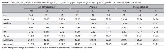
PURPOSE: The volume of the vitreous chamber varies with the size of the eye. The space created in the vitreous cavity by a vitrectomy is called the vitrectomized space. The volume of the vitrectomized space is strongly correlated with the axial length of the eye. This study aims to present guidelines for estimating the using participants stratified by axial length, sex, and history of cataract surgery.
METHODS: This retrospective, observational, cross-sectional study included 144 randomly selected participants who underwent vitrectomies between 2013 and 2023. Before surgery, the axial lengths of participants' eyes were measured using optical biometrics. The axial lengths of the eyes in our sample were between 20-32 mm. In all cases, a complete vitrectomy was performed, followed by complete fluid-air exchange and injection of a balanced saline solution. The volume infused was recorded.
RESULTS: The median (interquartile range; range) volume of the vitrectomized space was 6.1 (3.8; 3.1-11.3) mL in men and 6.1 (3.3; 3.2-11.2) mL in women (p=0.811). The median volume of the vitrectomized space was 5.9 (3.6; 3.1-11.2) mL in patients with phakic lenses and 6.25 (3.6; 3.3-11.3) mL in those with pseudophakic lenses (p=0.533). A positive correlation was found between the axial length and the volume of the vitrectomized space in this sample (r=0.968; p<0.001). In a cubic polynomial regression, the coefficient of determination was 0.948. Similar results were observed in both sexes and in both phakic and pseudophakic patients. The estimated cubic polynomial regression equation for this sample was VVS = 0.000589052857847605 × AL3 - 0.025114926401582700 × AL2 + 0.685961117595624000 × AL - 5.088226672620790000.
CONCLUSION: We developed this axial length estimation of the volume of vitrectomized space as a guideline for the determination of vitrectomized space volume using axial length.
Keywords: Cataract extraction; Retinal perforations/surgery; Epiretinal membrane/surgery; Vitreous body; Axial length, eye; Vitrectomy; Biometry/methods; Diagnostic techniques, ophthalmological; Guidelines as topic.
Abstract
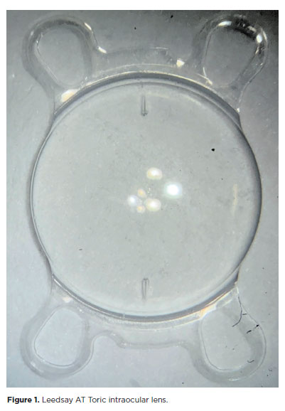
PURPOSE: The purpose of this study was to assess visual outcomes and patient satisfaction following cataract surgery involving the implantation of quad-loop intraocular lenses, including trifocal, bifocal, and toric variants.
METHODS: Information was obtained from both physical and electronic medical records of patients who underwent phacoemulsification cataract surgery with implantation of different intraocular lenses between January 1, 2022, and December 31, 2023. The study included individuals aged over 18 who received bilateral implantation of bifocal, trifocal, or monofocal toric intraocular lenses. Visual acuity was assessed at various postoperative time points using the logMAR scale. Quantitative variables were analyzed using mean and standard deviation.
RESULTS: A total of 92 eyes received premium intraocular lenses: 4 bifocal, 32 trifocal, 52 toric monofocal, and 4 trifocal toric lenses. The average preoperative corrected visual acuity was logMAR 0.478 ± 0.259. On the first postoperative day, the average uncorrected visual acuity was logMAR 0.301 ± 0.207. By day 30, 67.4% of eyes achieved uncorrected distance visual acuity of logMAR 0.2 or better. Patient satisfaction was high, with few reports of glare or halos.
CONCLUSION: Quad-loop intraocular lenses-including trifocal, bifocal, and toric models-demonstrated effective improvement in visual acuity and high levels of patient satisfaction. These lenses represent a suitable option for enhancing visual outcomes after cataract surgery. Additional studies with larger cohorts are recommended to confirm these results.
Keywords: Cataract extraction; Aberrometry/methods; Lenses, intraocular; Lens implantation, intraocular; Prosthesis design
Abstract
PURPOSE: Endophthalmitis is one of the most important adverse events after cataract surgery, as it can lead to total vision loss. This study aimed to describe the occurrence of endophthalmitis after phacoemulsification with intraocular lens implantation in patients treated in a community setting in Porto Velho, Rondônia, Brazil.
METHODS: This retrospective cohort study was conducted using a database of 649 medical records of patients who underwent surgery and were followed for three months. Poisson regression analysis was used to estimate relative risks and 95% confidence intervals (95% CIs).
RESULTS: The incidence of confirmed endophthalmitis was 11.94% (95% CI, 9.50-14.76), while the incidence of confirmed and probable cases was 20.50% (95% CI, 17.52-23.73). For confirmed cases, bilateral surgery and the use of lens model 3 were identified as risk factors for endophthalmitis, whereas age over 70 yr and preoperative antibiotic use were protective factors. For confirmed and probable cases, brown and yellow skin color, bilateral surgery, and the use of lens model 3 were also identified as risk factors. Gram-negative bacteria were the predominant etiological agents, and corneal edema was the main clinical manifestation. The mean duration of treatment was eight days, and 27.12% of patients used antibiotics.
CONCLUSION: The incidence observed was substantially higher than that reported in the literature, with a predominance of Gram-negative agents and an association with bilateral surgeries and the Eyeol intraocular lens model. These findings reinforce the need for continuous epidemiological surveillance and the implementation of specific biosafety and infection control protocols during cataract surgery campaigns.
Keywords: Endophthalmitis; Disease outbreaks; Phacoemulsification; Lens implantation, intraocular; Lenses, intraocular; Cataract; Risk factors; Anti-bacterial agents
Abstract
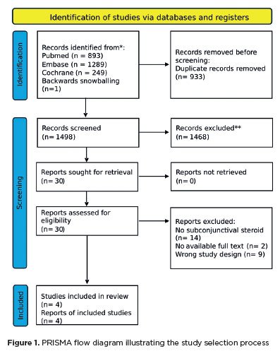
The advantages and disadvantages of using perioperative subconjunctival steroid injections in dropless cataract surgery continue to be debated. A systematic review of PubMed, EMBASE, and the Cochrane Central database identified five studies—two randomized controlled trials and three non-randomized studies—encompassing 70,751 eyes. Among these, 12,319 eyes (17.4%) received subconjunctival steroid injections, while 58,432 eyes (82.6%) were managed with topical steroids. The Cochrane Collaboration’s RoB 2 tool was applied for bias assessments in randomized controlled trials, and heterogeneity was assessed using the I² statistics. No statistically significant differences were found between the two groups regarding macular edema (p=0.249), visual acuity (p=0.73), or laser flare count (p=0.45). Both subconjunctival injections and topical steroids demonstrated comparable efficacy and safety in controlling postoperative inflammation after cataract surgery. Additional research is warranted to validate these conclusions.
Keywords: Cataract extraction; Phacoemulsification; Lens implantation, intraocular; Postoperative care; Intravitreal injections; Anti-inflammatory agents, non-steroidal/administration & dosage; Glucocorticoids; Triamcinolone acetonide; Research design; Randomiz
Abstract
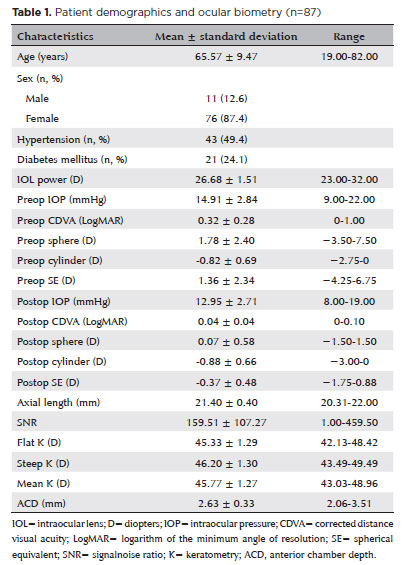
PURPOSE: To compare the refractive prediction error of Hill-radial basis function 3.0 with those of 3 conventional formulas and 11 combination methods in eyes with short axial lengths.
METHODS: The refractive prediction error was calculated using 4 formulas (Hoffer Q, SRK-T, Haigis, and Hill-RBF) and 11 combination methods (average of two or more methods). The absolute error was determined, and the proportion of eyes within 0.25-diopter (D) increments of absolute error was analyzed. Furthermore, the intraclass correlation coefficients of each method were computed to evaluate the agreement between target refractive error and postoperative spherical equivalent.
RESULTS: This study included 87 eyes. Based on the refractive prediction error findings, Hoffer Q formula exhibited the highest myopic errors, followed by SRK-T, Hill-RBF, and Haigis. Among all the methods, the Haigis and Hill-RBF combination yielded a mean refractive prediction error closest to zero. The SRK-T and Hill-RBF combination showed the lowest mean absolute error, whereas the Hoffer Q, SRK-T, and Haigis combination had the lowest median absolute error. Hill-radial basis function exhibited the highest intraclass correlation coefficient, whereas SRK-T showed the lowest. Haigis and Hill-RBF, as well as the combination of both, demonstrated the lowest proportion of refractive surprises (absolute error >1.00 D). Among the individual formulas, Hill-RBF had the highest success rate (absolute error ≤0.50 D). Moreover, among all the methods, the SRK-T and Hill-RBF combination exhibited the highest success rate.
CONCLUSIONS: Hill-radial basis function showed accuracy comparable to or surpassing that of conventional formulas in eyes with short axial lengths. The use and integration of various formulas in cataract surgery for eyes with short axial lengths may help reduce the incidence of refractive surprises.
Keywords: Cataract; Lenses, intraocular; Axial length, eye; Refractive errors; Artificial intelligence
Abstract
PURPOSE: To evaluate the effect of upper eyelid ptosis repair with Muller muscle-conjunctival resection on meibomian gland function and ocular surface parameters.
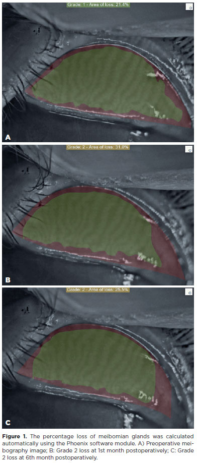
METHODS: Thirty-eight patients who underwent ptosis repair with Muller muscle-conjunctival resection were retrospectively reviewed. Meibomian gland loss, Ocular Surface Disease Index OXFORD score, meiboscore, and noninvasive keratograph break-up time were measured preoperatively and at 1st, 3rd, and 6th months postoperatively.
RESULTS: Noninvasive keratograph break-up time values decreased significantly at 1st and 3rd months postoperatively compared to the preoperative level, but were similar to the preoperative level at 6th months postoperatively (p<0.001 and p=0.628, respectively). Ocular surface disease index, OXFORD score, meibomian gland loss, and meiboscore values increased significantly in the 1st and 3rd postoperative months compared to the preoperative period, but these values decreased to preoperative levels in the 6th postoperative month (p<0.001 and p>0.05, respectively).
CONCLUSION: There is a transient deterioration in meibography findings and OSDI score in the early postoperative period after Muller muscle-conjunctival resection. Patients undergoing Muller muscle-conjunctival resection may require topical lubricants, especially in the first 3 postoperative months.
Keywords: Meibomian glands; Blepharoptosis; Preoperative period; Conjunctiva; Muscles; Eyelid diseases; Diagnostic techniques, ophthalmological
Abstract
PURPOSE: To compare endothelial corneal cell changes following cataract surgery performed by phacoemulsification with intraocular lens implantation, conducted by surgeons with varying levels of experience.
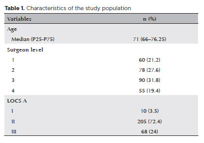
METHODS: Two hundred and eighty-three eyes diagnosed with cataract were included. Lens opacity was classified into three categories (I, II, and III). Surgeons were categorized into four experience levels (1, 2, 3, and 4), based on years of practice and lifetime surgeries performed. Corneal endothelial characteristics were assessed using non-contact specular microscopy, with measurements taken before surgery and 30-60 days post-surgery.
RESULTS: Pre- and postoperative endothelial analysis showed no significant differences between surgeon levels regarding visual acuity achieved, corneal thickness, and endothelial hexagonality. However, the central endothelial cell density index showed a significantly greater reduction among level 1 surgeons (p=0.026). Grade II cataracts exhibited significant variations in the central endothelial cell density (p=0.011) and average cell size, with level 1 surgeons showing the largest increases (p=0.024).
CONCLUSIONS: The analysis revealed significant differences in visual acuity and endothelial indices between surgeon experience levels, with less experienced surgeons showing greater variations and poorer performance. Clinical protocols should consider these data to establish safer training protocols.
Keywords: Cataract extraction; Phacoemulsification; Endothelium; corneal; Lens implantation, intraocular; Visual acuity; Internship and residency; Surgeons
Abstract
A cicloplegia é crucial para um exame oftalmológico pediátrico acurado. Este documento visa a fornecer uma recomendação para cicloplegia e midríase pediátrica para oftalmologistas brasileiros. Foi desenvolvido com base em revisão literária, na experiência clínica de especialistas brasileiros, por meio de questionários, e no consenso do comitê de especialistas da Sociedade Brasileira de Oftalmologia Pediátrica (SBOP). De acordo com as melhores evidências, este comitê recomenda o uso de uma gota de ciclopentolato 1%, mais uma gota de tropicamida 1% em crianças maiores de 6 meses e duas gotas de tropicamida 1% com intervalo de 0-5 minutos para menores de 6 meses. A midríase pode ser potencializada por uma gota de fenilefrina 2,5%. Para o rastreamento da retinopatia da prematuridade, a recomendação é tropicamida 0,5 ou 1%, duas ou três vezes, com 5 minutos de intervalo, e 2,5% de fenilefrina, preferencialmente uma vez. O uso prévio de proxymetacaína 0,5% é sempre recomendado.
Keywords: Midríase; Refração ocular; Recém-nascido; Criança; Técnicas de diagnóstico oftalmológico.
Abstract
We present a case report detailing the successful phacoemulsification surgery with artificial iris implantation for two individuals with oculocutaneous albinism. These women suffered from cataracts, resulting in reduced visual acuity and heightened photophobia due to iris pigmentary epithelium deficiency. The patients underwent phacoemulsification along with prosthetic artificial iris implantation into the posterior chamber. This intervention resulted in improved visual acuity, reduced photophobia and glare, and an overall enhanced quality of life. Our report highlights two cases of successful phacoemulsification and artificial iris implantation in patients with oculocutaneous albinism and cataracts, leading to improved visual acuity, reduced photophobia, and enhanced quality of life. Notably, there are no prior records in South American literature of cataract surgery combined with artificial iris implantation for oculocutaneous albinism patients up to the time of this publication.
Keywords: Cataract extraction; Albinism, oculocutaneous; Lens implantation, intraocular
Abstract
Um homem de 59 anos apresentou embaçamento visual unilateral no olho esquerdo. Sua acuidade visual nesse olho era no nível de movimentos da mão. O paciente havia se submetido a uma cirurgia de facoemulsificação em que foi feita a implantação intraestromal de uma lente intraocular de câmara posterior. Foi feita a extração dessa lente intraestromal intraocular e uma nova lente intraocular foi implantada. A melhor acuidade visual corrigida final foi de 20/40 pela tabela de Snellen. Com este relato de caso, os autores desejam apontar que uma incisão de degrau único em córnea clara, quando combinada com a injeção de uma lente ocular através da incisão, pode levar a um direcionamento incorreto da lente intraocular para dentro do estroma corneano. Portanto, recomenda-se uma construção cuidadosa da incisão ao se remover uma lente intraocular direcionada incorretamente.
Keywords: Implante de lente intraocular; Lentes intraocular; Facoemulsificação; Cicatrização; Catarata; Acuidade visual
Abstract
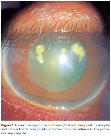
Aging and face sagging have many causes, and various techniques are used for treatment, including noninvasive procedures, such as focused ultrasound, which uses the principle of collagen regeneration by coagulative necrosis of the dermis layers using radiofrequency, but this procedure has complications. We reported a case of a 54-year-old female patient who complained of poor visual acuity in her right eye three days after a focused ultrasound facial aesthetic procedure, with the best visual acuity of 20/60. Biomicroscopy of the right eye revealed an acute cataract with three points of fibrosis extending from the posterior to the anterior capsule. The patient underwent phacoemulsification surgery with visual rehabilitation and improved vision of 20/20. We hypothesized that the occurrence of acute cataract was related to the inappropriate use of focused ultrasound.
Keywords: Cosmetic techniques; Skin aging; Rejuvenation; Ultrasonic therapy/adverse effects; High-intensity focused ultrasound ablation/methods; Cataract/etiology; Phacoemulsification; Lens implantation, intraocular; Visual acuity; Humans; Middle age; Female; Case
ABO is licensed under a Creative Commons Attribution-NonComercial 4.0 Internacional.

About
Issues
Editorial Board
Submission
Official publication of Conselho Brasileiro de Oftalmologia
Rua Casa do Ator, 1.117 - 2º andar - CEP: 04546-004
São Paulo - SP, Brazil
Phone: +55 11 3266-4000