Arq. Bras. Oftalmol. 2020;83 (5 )
:410-416
| DOI: 10.5935/0004-2749.20200080
Abstract
Objetivo: Avaliar as espessuras internas da retina e da coroide em pacientes com retinite pigmentosa precoce.
Métodos: Foram analisadas imagens de tomografia de coerência óptica de domínio espectral de 35 pacientes com retinite pigmentosa e 40 indivíduos saudáveis. Medimos a espessura do complexo de células maculares e ganglionares. Realizamos medições da espessura da coroide na região subfoveal e a 500 µm, 1000 µm e 1500 µm do centro da fóvea.
Resultados: Pacientes com retinite pigmentosa apresentaram espessuras maculares e da coroide significativamente mais finas em todas as medições e suas medidas individuais da espessura do complexo de células ganglionares foram inferiores às de indivíduos saudáveis. A espessura média do complexo de células ganglionares foi significativamente menor nos pacientes com retinite pigmentosa do que nos controles. A espessura macular média foi significativamente correlacionada com as espessuras médias do complexo das células de coroide e das células ganglionares médias. Não encontramoscorrelação entre a espessura media da coroide e a espessura media do complexo de células ganglionares.
Conclusões: A coroide foi levemente afetada em nossos pacientes com retinite pigmentosa precoce. A tendência à significância na retina interna foi possivelmente causada por uma boa acuidade visual.
Keywords: Coroide/anatomia & histologia; Retina/anatomia & histologia; Células ganglionares da retina; Retinite pigmentosa; Tomografia de coerência óptica
Arq. Bras. Oftalmol. 2025;88 (3 )
:1-8
| DOI: 10.5935/0004-2749.2024-0104
Abstract
PURPOSE: This study aimed to characterize retinitis pigmentosa associated with the eyes shut homolog gene, which causes hereditary retinal degeneration.
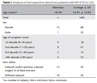
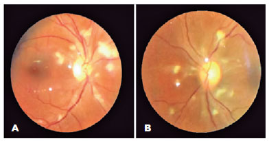
METHODS: The anatomical and functional findings of retinitis pigmentosa in patients with variants of the eyes shut homolog gene were characterized and compared using multimodal imaging and genetic analysis of the variants. Clinical data such as visual acuity, lens status, and refraction were obtained from medical records. Patients underwent an ophthalmic examination, including static visual field, microperimetry, optical coherence tomography, fundus autofluorescence, and fundus photography.
RESULTS: Twenty-two patients were included in the study. Several anatomical and functional characteristics of retinitis pigmentosa-eyes shut homolog were identified, including the presence of cataracts, cystoid macular edema, epiretinal membrane, and a tubular visual field. Genetic results revealed 26 distinct variants in the cohort, with 7 novel variants not previously documented or reported in the scientific literature or databases.
CONCLUSION: The findings demonstrate that eyes shut homolog-retinitis pigmentosa manifests in specific patterns, starting in adolescence with mild progression and advancing with age. The integration of multimodal imaging and genetic analysis has provided a detailed understanding of the anatomical and functional features of retinitis pigmentosa-eyes shut homolog. Seven novel variants of the eyes shut homolog gene have been identified. These findings enhance the understanding of eyes shut homolog-related retinitis pigmentosa characteristics of by detailing the spectrum of mutations in this gene within the Brazilian population.
Keywords: Retinal diseases/diagnostic imaging; Retinitis pigmentosa/genetics; Retinal degeneration; Eye proteins/genetics; Eye diseases, hereditary/genetics; Genes, recessive; Phenotype; Multimodal imaging; Tomography, optical coherence/methods; Fluorescein angiogr
 ABO is licensed under a Creative Commons Attribution-NonComercial 4.0 Internacional.
09-fig01tb.jpg)
09-fig01.jpg)
13-fig01.jpg)
04-fig01.jpg)
11-fig01tb.jpg)

12-fig01.jpg)
03-fig01.jpg)
03-fig01.jpg)
02-fig01.jpg)
02-fig01.jpg)
15-fig01.jpg)