Arq. Bras. Oftalmol. 2022;85 (1 )
:7-12
| DOI: 10.5935/0004-2749.20220002
Abstract
Objetivo: A degeneração macular relacionada à idade é a causa mais comum de cegueira em países desenvolvidos e muitos fatores etiológicos têm-lhe sido atribuídos. O objetivo do presente estudo foi investigar a relação entre os níveis séricos de vitamina D e a degeneração macular relacionada à idade.
Métodos: Os dados de 114 pacientes com degeneração macular relacionada à idade foram analisados retrospectivamente. Foram alocados no Grupo Controle 102 pacientes sem registro de outras doenças além do erro refrativo. A acuidade visual melhor corrigida, os achados do exame de fundo de olho e os da tomografia de coerência óptica de domínio espectral foram analisados. Os pacientes foram alocados em grupos de acordo com a classificação do Age-Related Eye Disease Study (Estudo da Doença Ocular Relacionada à Idade). Os níveis séricos de vitamina D 25(OH) foram medidos. A espessura foveal central e a espessura da coroide subfoveal foram medidas com tomografia de coerência óptica.
Resultados: Os níveis de vitamina D 25(OH) em pacientes com degeneração macular relacionada à idade e em indivíduos saudáveis pareados por idade e sexo foram 14,6 ± 9,8 ng/mL e 29,14 ± 15,1 ng/mL, respectivamente. Os níveis de vitamina D foram significativamente menores no Grupo da Degeneração Macular relacionada à idade em comparação com o Grupo Controle (p>0,001). O valor da espessura da coroide subfoveal foi menor em pacientes com degeneração macular relacionada à idade (p>0,001). Foi encontrada uma fraca correlação positiva entre o nível de vitamina D 25(OH) e a espessura da coroide (r=0,357, p=0,01). O nível de vitamina D 25(OH), quando avaliado de acordo com os estágios da degeneração macular relacionada à idade, revelou ser menor na doença em estágio avançado (p=0,01). Constatou-se um risco aumentado de desenvolvimento de membrana neovascular da coroide e de fibrose sub-retiniana com a diminuição dos níveis de vitamina D.
Conclusões: A diminuição significativa dos níveis de vitamina D 25(OH) na degeneração macular relacionada à idade em estágio avançado sugere a presença de uma correlação significativa entre a deficiência de vitamina D e o desenvolvimento dessa patologia. Mais estudos são necessários para investigar se a suplementação de vitamina D tem ou não influência no desenvolvimento e progressão da degeneração macular relacionada à idade.
Keywords: Membrana neovascular da coroide; Degeneração macular; Tomografia de coerência óptica; Fibrose; Retina; Vitamina D; Deficiência de vitamina D
Arq. Bras. Oftalmol. 2025;88 (3 )
:1-5
| DOI: 10.5935/0004-2749.2023-0174
Abstract
PURPOSE: To compare objective and subjective intraocular pressure measurements immediately after cataract surgery and intraocular pressure measurements between less experienced surgeons (Group 1) and experienced surgeons (Group 2).
METHODS: Surgeons were asked to estimate the IOP after corneal sealing after surgery based on their tactile perception of eye tension (subjective intraocular pressure) Objective intraocular pressure was measured using a Perkins tonometer while patients were still in the surgical field. Objective intraocular pressure was compared to subjective intraocular pressure. Results from less experienced surgeons were compared to more experienced surgeons.
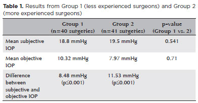
RESULTS: The study comprised 81 surgeries (81 eyes) performed by 27 surgeons. The mean objective intraocular pressure (9.14 mmHg; SD=5.86) was statistically significantly lower (p<0.001) than the mean subjective intraocular pressure (19.21 mmHg; SD=4.82). Hypotony (intraocular pressure <6mmHg) was observed in 25 eyes (30.86%). The mean subjective intraocular pressure was 18.8 mmHg (SD=5.19) for less experienced surgeons and 19.5 mmHg (SD=4.46) for more experienced, without statistically significant difference (p=0.541). No statistically significant difference (p=0.71) was observed when comparing objective intraocular pressure in Group 1 (10.32 mmHg; SD=6.65) and Group 2 (7.97 mmHg; SD=4.7).
CONCLUSION: Objective intraocular pressure was significantly lower than subjective intraocular pressure, regardless of surgeons' experience. This study showed that the subjective method is unreliable compared to the gold standard (Perkins tonometer) and does not improve with surgeons' experience. Establishing standard training methods is paramount to developing surgeons' skills.
Keywords: Cataract; Intraocular pressure; Hypotony, Tonometry; Eye diseases; Training
Arq. Bras. Oftalmol. 2025;88 (2 )
:1-9
| DOI: 10.5935/0004-2749.2023-0326
Abstract
PURPOSE: To evaluate the predictive value of initial intraocular pressure difference of the detached and fellow eyes of patients with complex rhegmatogenous retinal detachment on postoperative persistent ocular hypotony.
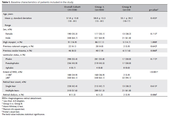
METHODS: This retrospective observational study included 538 eyes of 538 unilateral complex rhegmatogenous retinal detachment patients with a proliferative vitreoretinopathy grade of C-1 or higher, treated with silicone oil endotamponade following pars plana vitrectomy. The patients were divided into Group A (patients having silicone oil removal without ocular hypotony; n=504) and Group B (patients with persistent ocular hypotony following silicone oil removal [n=8, 23.5%] and with retained silicone oil [n=26, 76.5%] due to the risk of persistent ocular hypotony; total n=34). Ocular hypotony was defined as an intraocular pressure of <6 mmHg on two or more occasions. Patients' demographics, including age, sex, and follow-up time, and ocular characteristics, including ocular surgical and trauma history, initial and final best-corrected visual acuity, intraocular pressure and initial intraocular pressure difference of the detached and fellow eyes, and anatomical success rates and postoperative complications, were retrospectively collected from the electronic patient files.
RESULTS: The initial intraocular pressure was significantly lower in the detached eyes of Group B than in Group A (8.3 ± 3.5 vs. 12.9 ± 3.3, p<0.001). Also, the initial intraocular pressure difference was significantly higher in Group B than in Group A (8.9 ± 3.2 vs. 2.2 ± 2.7mmHg, p<0.001). The receiver operating characteristic curve analysis showed that the cutoff value of the initial intraocular pressure difference was 7.5mmHg for the risk of persistent ocular hypotony. The most influential factors on postoperative persistent ocular hypotony in the binary logistic regression analysis were the initial intraocular pressure difference and the need for a retinectomy.
CONCLUSION: In eyes with complex rhegmatogenous retinal detachment treated with pars plana vitrectomy and silicone oil tamponade, the initial intraocular pressure difference could be of value in predicting postoperative persistent ocular hypotony and could guide surgeons on the decision of silicone oil removal.
Keywords: Hypotony; Intraocular pressure; Pars plana vitrectomy; Retinal detachment; Silicone oils; Ocular hypotension; Visual acuity
 ABO is licensed under a Creative Commons Attribution-NonComercial 4.0 Internacional.
04-tab01tb.jpg)