Arq. Bras. Oftalmol. 2020;83 (6 )
:490-496
| DOI: 10.5935/0004-2749.20200090
Abstract
Objetivo: Comparar a espessura central foveal, a da camada de fibras nervosas da retina e a da coróide subfoveal através da tomografia de coerência óptica swept-source em crianças de 5 anos de idade com história de retinopatia da prematuridade (RP) tratada com bevacizumabe intravítreo, ou com fotocoagulação a laser, com crianças em regressão espontânea da retinopatia da prematuridade, e com crianças saudáveis da mesma idade.
Métodos: Um total de 79 crianças foi dividido em quatro grupos. Grupo 1: crianças que receberam tratamento com bevacizumabe intravítreo. Grupo 2: crianças que foram tratadas com fotocoagulação a laser. Grupo 3: crianças que tiveram regressão espontânea da retinopatia da prematuridade . Grupo 4: crianças da mesma idade saudáveis e nascidas a termo. As funções visuais e o status refrativo foram avaliados aos 5 anos de idade. A análise de tomografia de coerência óptica foi feita por um dispositivo do tipo swept-source (DRI-OCT Triton; Topcon, EUA).
Resultados: Haviam 12 crianças (15,2%) no grupo 1, 23 crianças (29,1%) no grupo 2, 30 crianças (38%) no grupo 3 e 14 crianças (17,7%) no grupo 4. A distribuição por sexo foi semelhante em todos os grupos (p=0,420). A acuidade visual com a melhor correção mostrou-se significativamente maior no grupo 4 em comparação com os grupos 1, 2 e 3 (respectivamente, p=0,035, p=0,001 e p=0,001). Os resultados dos erros de refração foram semelhantes em todos os grupos (p=0,119). A espessura foveal central mostrou-se significativamente maior no grupo 2 do que no grupo 1 (p=0,023). Não foram observadas diferenças significativas entre os grupos quanto à espessura da camada de fibras nervosas da retina e à espessura da coroide subfoveal (p>0,05).
Conclusões: Os desfechos visuais funcionais foram melhores nas crianças saudáveis nascidas a termo, em comparação com aqueles observados nas crianças com história de retinopatia da prematuridade tratada ou com regressão espontânea. O tratamento com laser teve um efeito significativo na espessura foveal central em crianças de 5 anos de idade, nascidas prematuras, como revelado pela tomografia de coerência óptica swept-source.
Keywords: Retinopatia da prematuridade/tratamento farmacológico; Tomografia de coerência óptica; Bevacizumab/uso terapêutico; Fotocoagulação; Recém-nascido
Arq. Bras. Oftalmol. 2022;85 (4 )
:364-369
| DOI: 10.5935/0004-2749.20220049
Abstract
Objetivos: Avaliar duas unidades de terapia intensiva neonatais do Paraná e identificar os fatores de risco que levam ao desenvolvimento da retinopatia da prematuridade nestas unidades neonatais.
Metodos: Foi realizado um estudo de coorte, prospectivo, com avaliação dos bebês prematuros examinados no período de 12 meses com idade gestacional ≤32 semanas e/ou com peso de nascimento ≤1500 gramas, internados na unidade de cuidados intensivos neonatais do Hospital do Trabalhador e do Hospital infantil Waldemar Monastier, que recebe neonatos transportados das maternidades de todo o estado do Paraná.
Resultados: A incidência de retinopatia da prematuridade foi maior no Hospital Infantil Waldemar Monastier, entre os prematuros que necessitaram de transporte do local de nascimento para a unidade de cuidados intensivos (52,2% vs 29,6%), Os fatores de risco associados ao desenvolvimento da doença foram; Maior número de dias de internamento, baixa idade gestacional ao nascimento, maior tempo de uso de oxigênio, uso de drogas vasoativas, ausência de uso de corticoide pré-natal, presença de hemorragia intracraniana e qualquer tipo de alteração da glicemia.
Conclusão: Os cuidados neonatais precoces e o transporte do recém-nascido pré- termo podem influenciar a ocorrência e o prognostico da retinopatia da prematuridade.
Keywords: Retinopatia da prematuridade; Recém-nascido; Recém-nascido prematuro; Doenças do prematuro; Cegueira
Arq. Bras. Oftalmol. 2022;85 (2 )
:136-143
| DOI: 10.5935/0004-2749.20220022
Abstract
Objetivo: Estimar a epidemiologia do pterígio; sua correlação com sintomas de olho seco e com potenciais preditores sistêmicos e oculares.
Métodos: Estudo transversal, de base populacional, no qual foram realizadas visitas domiciliares aleatórias a 600 participantes, com 40 anos ou mais de idade, em Ribeirão Preto-SP (n=420) e Cassia dos Coqueiros-SP (n=180), Brasil. Uma entrevista estruturada com um questionário detalhado foi usada para coletar informações sobre demografia e possíveis fatores de risco. Em um segundo momento, participantes aleatórios com pterígio (n=63) ou não (n=110) foram avaliados quanto a alterações na superfície ocular.
Resultados: A frequência de pterígio em Ribeirão Preto foi de 21%; 15.7% entre as mulheres e 32.1% entre os homens (p=0,0002). Em Cássia dos Coqueiros, essa taxa foi de 19.4%; onde 17.3% eram mulheres e 25.5% eram homens (p=0,28). A média de idade naqueles afetados pelo pterígio foi superior à dos participantes sem pterígio, 65,6 ± 10,5 e 61,2 ± 12,0 anos, respectivamente (p=0,02). Houve uma correlação positiva entre o pterígio e história prévia de radioterapia e quimioterapia (p<0,0001 para ambos). Houve maior coloração de fluoresceína na córnea e maior coloração de lissamina verde na conjuntiva em olhos com pterígio (p=0,0003 e 0,0001, respectivamente).
Conclusão: Encontramos uma alta frequência de pterígio em duas populações adultas brasileiras, principalmente em homens e idosos. Danos na superfície ocular e história prévia de radioterapia e/ou quimioterapia foram associados ao pterígio.
Keywords: Pterígio/epidemiologia; Síndrome do olho seco; Prevalência; Fatores de risco
Arq. Bras. Oftalmol. 2022;85 (5 )
:485-489
| DOI: 10.5935/0004-2749.20220058
Abstract
Objetivo: Avaliar o efeito da dilatação da pupila sobre a pressão intraocular em recém-nascidos pré-termo e a termo.
Métodos: Este estudo prospectivo envolveu 55 olhos de 28 bebês pré-termo e 38 olhos de 20 bebês a termo. Os bebês foram divididos em dois grupos, pré-termo e a termo, de acordo com a idade gestacional ao nascimento: grupo pré-termo <37 semanas; grupo a termo ≥37 semanas. A dilatação da pupila foi feita com tropicamida 0,5% e fenilefrina 2,5%. As medições da pressão intraocular foram realizadas com Icare PRO (Icare Finland Oy, Helsinki, Finlândia) antes e depois da dilatação da pupila. O teste t pareado foi usado para comparar as medidas antes e depois da dilatação da pupila.
Resultados: A alteração média da pressão intraocular foi de -1,04 ± 3,03 mmHg (+6,20/-11,40 mmHg) no grupo pré-termo e -0,39 ± 2,81 mmHg (+4,60/-9,70 mmHg) no grupo a termo. Uma diferença estatisticamente significativa na pressão intraocular foi observada apenas no grupo pré-termo após a dilatação da pupila (p=0,01).
Conclusão: Após a dilatação da pupila, pode ocorrer alteração inesperada da pressão intraocular em recém-nascidos, principalmente em bebês pré-termo.
Keywords: Lactente; Recém-nascido; Recém-nascido prematuro; Pressão Intraocular; Pupila; Fenilefrina; Tropicamida; Dilatação
Arq. Bras. Oftalmol. 2025;88 (4 )
:1-8
| DOI: 10.5935/0004-2749.2024-0214
Abstract
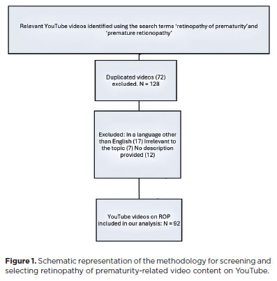
PURPOSE: This study aimed to evaluate the quality and reliability of YouTube videos as an educational resource about retinopathy of prematurity.
METHODS: Videos were sourced from YouTube using the search terms "retinopathy of prematurity" and "premature retinopathy" with the default settings. Each video was assessed on the following metrics: views, likes, dislikes, comments, upload source, country of origin, view ratio, like ratio, and video power index. The quality and reliability of the videos were evaluated by two independent researchers using the DISCERN questionnaire, the JAMA benchmarks, the Global Quality Score scale, the Health on the Net Code of Conduct, and the Ensuring Quality Information for Patients scale.
RESULTS: The study assessed 92 videos, the majority of which (42 videos, 45.7%) originated from the United States. Most of the videos focused on screening, pathophysiology, and diagnosis of retinopathy of prematurity (61.9%). The primary contributors were medical organizations (19 videos, 20.6%), nonacademic health channels (19 videos, 20.6%), and physicians (15 videos, 16.3%). Significant differences were found between the DISCERN (p=0.003), JAMA (p=0.001), Global Quality Score (p=0.003), Health on the Net Code of Conduct (p=0.006), and Ensuring Quality Information for Patients (p=0.001) scores among different video sources. However, the key video metrics did not differ. Using the DISCERN and Global Quality Score scales, the overall YouTube video content on retinopathy of prematurity was rated as moderate in quality. Using the Health On the Net Code of Conduct and Ensuring Quality Information for Patients scales, it was rated as high quality. Strong correlations were observed between the scores on all of the scales (p<0.001).
CONCLUSION: Videos from medical organizations and healthcare centers were of a higher quality than those from nonmedical sources. Despite the varied foci of each evaluation scale, the strong correlation between them indicates that they provide reliable and comprehensive assessments of the quality of informational content.
Keywords: Retinopathy of prematurity; YouTube; Information dissemination/methods; Online education; Internet access; Social media/instrumentation; Information seeking behavior; Internet/statistics & numerical data; Consumer health information; Social networking
Arq. Bras. Oftalmol. 2026;89 (2 )
:1-8
| DOI: 10.5935/0004-2749.2025-0175
Abstract
PURPOSE: Endophthalmitis is one of the most important adverse events after cataract surgery, as it can lead to total vision loss. This study aimed to describe the occurrence of endophthalmitis after phacoemulsification with intraocular lens implantation in patients treated in a community setting in Porto Velho, Rondônia, Brazil.
METHODS: This retrospective cohort study was conducted using a database of 649 medical records of patients who underwent surgery and were followed for three months. Poisson regression analysis was used to estimate relative risks and 95% confidence intervals (95% CIs).
RESULTS: The incidence of confirmed endophthalmitis was 11.94% (95% CI, 9.50-14.76), while the incidence of confirmed and probable cases was 20.50% (95% CI, 17.52-23.73). For confirmed cases, bilateral surgery and the use of lens model 3 were identified as risk factors for endophthalmitis, whereas age over 70 yr and preoperative antibiotic use were protective factors. For confirmed and probable cases, brown and yellow skin color, bilateral surgery, and the use of lens model 3 were also identified as risk factors. Gram-negative bacteria were the predominant etiological agents, and corneal edema was the main clinical manifestation. The mean duration of treatment was eight days, and 27.12% of patients used antibiotics.
CONCLUSION: The incidence observed was substantially higher than that reported in the literature, with a predominance of Gram-negative agents and an association with bilateral surgeries and the Eyeol intraocular lens model. These findings reinforce the need for continuous epidemiological surveillance and the implementation of specific biosafety and infection control protocols during cataract surgery campaigns.
Keywords: Endophthalmitis; Disease outbreaks; Phacoemulsification; Lens implantation, intraocular; Lenses, intraocular; Cataract; Risk factors; Anti-bacterial agents
Arq. Bras. Oftalmol. 2025;88 (6 )
:1-7
| DOI: 10.5935/0004-2749.2025-0083
Abstract
PURPOSE: To examine how ophthalmological features, screen exposure duration, and break habits among office employees affect ocular surface parameters.
METHODS: This single-center cross-sectional study involved two assessments on the same day: one before and one after a visual display terminal task. During the initial assessment, information on screen use was gathered, and refractive error, anterior segment examination, tear breakup time, and Schirmer test measurements were conducted. Participants tracked their screen usage and break durations throughout the day. At the end of the workday, tear breakup time and Schirmer I tests were repeated. Baseline and follow-up results were compared, and regression analysis was performed to identify factors linked to tear breakup time reduction.
RESULTS: The study enrolled 60 female office employees. Their mean screen time was 269.26 ± 70.21 min, with an average break duration of 151.93 ± 46.24 min. Tear breakup time at the second assessment (6.38 ± 2.70) was significantly lower than at baseline (8.62 ± 2.73) (p<0.001), whereas Schirmer test scores showed no significant change (p>0.05). Tear breakup time reduction was noted in 54 participants (90.0%), with a significant association between tear breakup time decrease percentage and screen exposure (p=0.001, r=0.463). Regression analysis showed that uncorrected or undercorrected refractive error was an independent risk factor for a ≥30% tear breakup time reduction, while taking more frequent short breaks (<15 min) acted as a protective factor.
CONCLUSIONS: Taking more frequent short breaks (<15 min) and correcting refractive errors help prevent intra-day tear breakup time decline during visual display terminal use. Structuring breaks to support tear film stability is advisable for occupations that require regular visual display terminal tasks.
Keywords: Tear film; Screen time; Tear breakup time; Office workers; Protective factors; Lacerations; Refractive errors; Risk factors.
Arq. Bras. Oftalmol. 2025;88 (2 )
:1-7
| DOI: 10.5935/0004-2749.2023-0265
Abstract
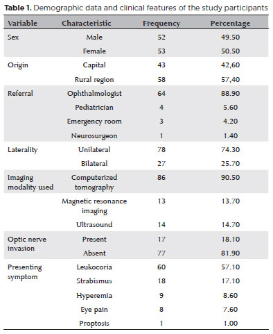
PURPOSE: Although Brazil has a high prevalence of retinoblastoma, there is a lack of epidemiological data on the disease. Thus, in this study, we aimed to evaluate the epidemiological profile of patients diagnosed with retinoblastoma in the ophthalmology department of a pediatric tertiary referral hospital in Ceara, Brazil.
METHODS: A descriptive and cross-sectional study was conducted by retrospectively analyzing the clinical and socioeconomic data from the medical records of pediatric patients followed-up at the hospital between 2007 and 2021. Retinoblastoma was diagnosed on the basis of a fundoscopic or histopathologic examination.
RESULTS: The data of 105 patients were included in the study, and the mean patient age at the time of diagnosis was 1.7 years. Most of the patients were women (50.5%) and hailed from rural areas (57.4%), which was associated with a higher tumor stage. Of the 150 patients, 57.1% initially presented with leukocoria. Ocular hyperemia was associated with more advanced stages of retinoblastoma (p=0.004). Bilateral involvement was observed in 25.7% of the patients and at a significantly younger age (p=0.009). The presence of retinal detachment, vascularized lesions, and vitreous seeds significantly increased the likelihood of requiring enucleation.
DISCUSSION: This study presents an epidemiological description of retinoblastoma in Brazil, which highlights the significance of early detection. Delayed diagnosis is associated with a poorer visual prognosis and higher mortality rate, particularly in patients with unilateral disease. Risk factors for a more severe disease were retinal detachment, vascularized lesions, and vitreous seeds. The correlation between histopathological features and clinical outcomes was limited.
CONCLUSION: Further studies are required to assess the influence of ocular hyperemia, fundoscopic assessment, and histopathologic findings on the prognosis of retinoblastoma. Moreover, it is critical to devise interventions to reduce the time-to-diagnosis in rural areas.
Keywords: Retinoblastoma; Retinal neoplasms; Epidemiology; Prevalence; Risk factors; Delayed diagnosis; Child
Arq. Bras. Oftalmol. 2024;87 (4 )
:1-5
| DOI: 10.5935/0004-2749.2022-0335
Abstract
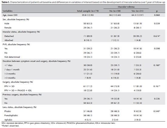
PURPOSE: To clarify the postoperative incidence of macular edema in patients undergoing surgery to repair rhegmatogenous retinal detachment and identify the associated risk factors.
METHODS: In this prospective, observational study, 79 patients who underwent surgery to correct rhegmatogenous retinal detachment using pars plana vitrectomy with silicone oil injection were analyzed. Patients were followed up postoperatively at 7, 30, 90, 180, and 365 days. At each visit, optical coherence tomography was performed to assess the presence or absence of macular edema. were analyzed as possible risk factors for macular edema: age, sex, macular status (attached or detached), presence of vitreoretinal proliferation, history of previous intraocular surgery, reported time of symptoms suggestive of rhegmatogenous retinal detachment up to the date of surgery, and the surgical modality performed.
RESULTS: The 1-year macular edema prevalence rate was 26.6%. In the adjusted analysis, older patients had a higher risk of macular edema, and each 1-year increase in age increased the risk of macular edema by 6% (95% confidence interval = 1.00-1.12). The macular status, vitreoretinal proliferation, the surgical technique used, prior intraocular surgery, and the intraocular lens status were not identified as risk factors. However, the incidence of macular edema increased up to 180 days after surgery, peaking at 10.6%, and then decreased until 365 days after surgery.
CONCLUSION: Macular edema was a common complication after surgery to treat rhegmatogenous retinal detachment, with its incidence peaking between 30 and 180 days after surgery. Age was an important risk factor for macular edema in this cohort.
Keywords: Macular edema; Retinal detachment; Vitrectomy; Tomography, optical coherence; Incidence; Risk factors
Arq. Bras. Oftalmol. 2025;88 (5 )
:1-8
| DOI: 10.5935/0004-2749.2024-0328
Abstract
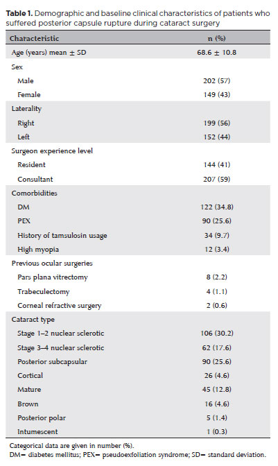
PURPOSE: Posterior capsule rupture is defined as an intraoperative posterior capsule tear resulting in vitreous loss. This study aimed to analyze the clinical characteristics, preoperative risk factors, intraoperative management strategies, and postoperative complications associated with posterior capsule rupture during phacoemulsification surgery.
METHODS: This was a retrospective observational cohort study of the medical records for 25,224 phacoemulsification surgeries performed at our tertiary eye care center between 2017 and 2022. We collected and collated the demographic characteristics and clinical findings of the patients in our cohort. Intraoperative management strategies and postoperative outcomes over a 1-year followup period were also recorded.
RESULTS: Posterior capsule rupture occurred in 351 eyes (351 patients), giving an overall posterior capsule rupture rate of 1.3%. The mean patient age was 68.6 ± 10.8 years. Pseudoexfoliation syndrome, mature cataracts, brown cataracts, and surgery performed by a resident were identified as risk factors for posterior capsule rupture (p<0.05 for each; the risk ratios were 2.70, 2.15, 2.44, 1.34, respectively). The most common intraoperative complications were dislocated lens fragments in the vitreous (8%) and iris damage (7.1%). The mean best-corrected visual acuity improved from 1.31 ± 0.84 (logMAR) postoperatively to 0.51 ± 0.56 at the end of the 1-year follow-up period (p<0.001). Corneal edema (55.6%) and elevated intraocular pressure (33.3%) were the most common early postoperative complications. Persistently elevated intraocular pressure (11.1%) and cystoid macular edema (5.1%) were the most common late postoperative complications.
CONCLUSION: Posterior capsule rupture is a common complication of phacoemulsification surgery that requires prolonged postoperative follow-up and a multidisciplinary approach. Despite the increased incidence of complications when rupture occurs, appropriate intraoperative and postoperative management can lead to satisfactory visual outcomes.
Keywords: Cataract extraction; Phacoemulsification; Posterior capsule rupture; Corneal edema; Risk factors; Postoperative complications; Intraoperative complications
Arq. Bras. Oftalmol. 2025;88 (3 )
:1-6
| DOI: 10.5935/0004-2749.2024-0170
Abstract
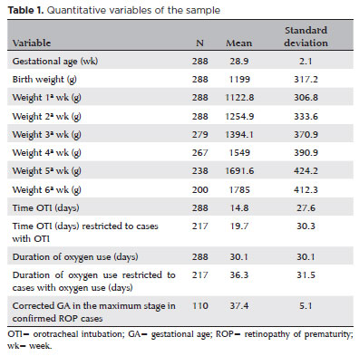
PURPOSE: To assess the sensitivity and specificity of the retinopathy of prematurity score (ROPScore) and weight, insulin-like growth factor-1, retinopathy of prematurity algorithm in predicting the risk of developing severe retinopathy of prematurity (prethreshold type 1) in a sample of preterm infants in Brazil.
METHODS: Retrospective analysis of medical records of preterm infants (n=288) with birth weight of ≤1500 g and/or gestational age of 23-32 weeks in a neonatal unit in Southern Brazil from May 2013 to December 2020 (92 months).
RESULTS: The incidence of confirmed severe retinopathy of prematurity was 6.6%. ROPScore showed a 100% sensitivity, 44.6% specificity (95% confidence interval [CI] 38.7-50.6), 11.3% positive predictive value (95% CI 6.5-16.1), and 100% negative predictive value in predicting severe retinopathy of prematurity. The weight, insulin-like growth factor-1, retinopathy of prematurity algorithm demonstrated a 78.9% sensitivity (95% CI 60.6-97.3), 51.3% specificity (95% CI 45.3-57.3), 10.3% positive predictive value (95% CI 5.3-15.2), and 97.2% negative predictive value (95% CI 94.5-99.9).
CONCLUSION: ROPScore identified all patients at risk for severe retinopathy of prematurity. These findings support incorporating ROPScore into Brazilian guidelines to optimize retinopathy of prematurity screening and reduce unnecessary ophthalmologic examinations. Weight, insulin-like growth factor-1, retinopathy of prematurity's suboptimal performance in this Brazilian sample highlights the need for country-specific algorithm adjustments.
Keywords: Retinopathy of prematurity; ROPScore, WINROP; Prediction algorithm; Infant, premature
Arq. Bras. Oftalmol. 2024;87 (5 )
:1-7
| DOI: 10.5935/0004-2749.2023-0296
Abstract
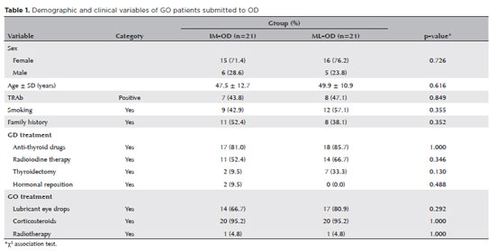
PURPOSE: To compare inferomedial wall orbital decompression to balanced medial plus lateral wall orbital decompression in patients with Graves’ orbitopathy in the inactive phase with regard to exophthalmos reduction and the effects on quality of life.
METHODS: Forty-two patients with inactive Graves’ orbitopathy were randomly divided into two groups and submitted to one of two orbital decompression techniques: inferomedial wall orbital decompression or medial plus lateral wall orbital decompression. Preoperative and postoperative assessments included Hertel’s exophthalmometry and a validated Graves’ orbitopathy quality of life questionnaire. The results of the two groups were compared.
RESULTS: Compared to preoperative measurement, exophthalmos reduction was statistically significant in both groups (p<0.001) but more so in patients undergoing medial plus lateral wall orbital decompression (p=0.010). Neither orbital decompression techniques increased the visual functioning subscale score on the Graves’ orbitopathy quality of life questionnaire (inferomedial wall orbital decompression p=0.362 and medial plus lateral wall orbital decompression p=0.727), but a statistically significant difference was observed in the score of the appearance subscale in patients submitted to medial plus lateral wall orbital decompression (p=0.006).
CONCLUSIONS: Inferomedial wall orbital decompression is a good alternative for patients who do not require large exophthalmos reduction. However, medial plus lateral wall orbital decompression offers greater exophthalmos reduction and greater improvement in appearance (higher Graves’ orbitopathy quality of life questionnaire scores), making it a suitable option for esthetic-functional rehabilitation.
Keywords: Graves’ ophthalmopathy; Quality of life; Exophthalmos; Strabismus; Diplopia; Decompression, surgical
 ABO is licensed under a Creative Commons Attribution-NonComercial 4.0 Internacional.
05-fig01tb.jpg)
06-tab01.jpg)
08-tab01tb.jpg)
01-tab01.jpg)
12-fig01.jpg)
06-fig01.jpg)