Arq. Bras. Oftalmol. 2024;87 (1 )
:1-3
| DOI: 10.5935/0004-2749.2021-0246
Abstract
Este artigo relata o caso de um paciente do sexo masculino, 11 anos de idade, com história de proptose e baixa de acuidade visual progressiva. Ao exame oftalmológico apresentava melhor acuidade visual de 20/25 em olho direito e percepção de luz em olho esquerdo. Existia exotropia e limitação à adução no olho esquerdo. À campimetria automatizada, observou-se quadrantopsia temporal inferior em olho direito e escotoma total em olho esquerdo. À ressonância magnética, evidenciou-se lesão expansiva em trajeto do nervo óptico esquerdo estendendo-se até região do tronco encefálico, com acometimento quiasmático. O objetivo deste artigo é relatar o glioma de vias ópticas, bem como discutir os achados e sua interligação com a literatura atual.
Keywords: Glioma; Neoplasias do nervo óptico; Quiasma óptico; Astrocitoma; Imageamento por ressonância magnética; Acuidade Visual; Relatos de casos; Humanos.
Arq. Bras. Oftalmol. 2023;86 (3 )
:1-4
| DOI: 10.5935/0004-2749.20230016
Abstract
A disseminação metastática ocular de tumores sistêmicos é incomum, ocorrendo principalmente na coroide e em pacientes idosos. O câncer de pulmão é considerado o principal tumor metastático ocular em homens, contudo, outras doenças oculares, como as uveítes e lesões retinianas, podem mimetizar os implantes secundários tumorais nos tecidos oculares. O aspecto fundoscópico das neoplasias da coroide pode apresentar similaridade com outros processos infecciosos, especialmente o tuberculoma de coroide. Dessa forma, a investigação clínica detalhada é de grande importância no diagnóstico de pacientes com massas coroideanas, especialmente quando configuram a primeira manifestação de uma doença sistêmica e grave. Relatamos um caso raro de metástase coroideana como primeira manifestação clínica do carcinoma de células renais em um homem jovem, mimetizando um tuberculoma de coroide.
Keywords: Neoplasias renais/complicações; Metástase neoplásica; Carcinoma de células renais; Neoplasias da coroide/etiologia; Humanos; Relatos de casos
Arq. Bras. Oftalmol. 2024;87 (3 )
:1-4
| DOI: 10.5935/0004-2749.2021-0235
Abstract
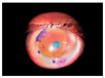
Paciente do sexo masculino, 33 anos, apresentou ceratite infecciosa subaguda unilateral 4 semanas após a cirurgia. A inflamação da córnea foi resistente aos regimes de antibióticos tópicos padrão. A aba da córnea foi derretida e seccionada durante o levantamento e amostragem para diagnóstico. A melhora clínica só foi alcançada após levantamento do retalho, raspagem e diagnóstico microbiológico de micobactérias atípicas e tratamento com amicacina fortificada tópica, claritromicina e claritromicina sistêmica.
Keywords: Córnea/microbiologia; Úlcera da córnea; Infecções oculares bacterianas; Mycobacterium abscessus; Procedimentos cirúrgicos refrativos; Ceratomileuse assistida por excimer laser in situ; Amicacina/uso terapêutico; Claritromicina/uso terapêutico; Humanos; Re
Arq. Bras. Oftalmol. 2024;87 (4 )
:1-5
| DOI: 10.5935/0004-2749.2023-0221
Abstract
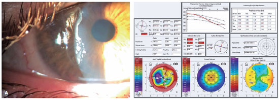
We present a case of a patient complaining of monocular diplopia due to a decentered ablation after LASIK. The patient underwent a wavefront-guided retreatment, which resulted in an epithelial ingrowth complication. Additionally, the patient developed cataract, with cataract surgery requiring reliable biometric measurements. Therefore, we opted for corneal treatment and corneal surface regularization. Although we attempted to lift the flap and wash the interface initially, the procedure proved unsuccessful, thereby necessitating immediate flap amputation. Once the corneal surface was regularized in the seventh postoperative month, transepithelial photorefractive keratectomy was successfully performed to homogenize the ocular surface, thereby significantly improving the patient's corrected visual acuity and resolving monocular diplopia. The surface and corneal curvature stabilized by the fifth month after the procedure. Phacoemulsification was then performed along with the implantation of a toric monofocal lens, which was selected using an appropriate formula, resulting in an excellent uncorrected visual acuity.
Keywords: Refractive surgical procedures; Surgical flap/surgery; Keratomileusis laser In situ/methods; Biometry; Corneal topography; Lasers, Excimer/adverse effects; Dipoplia/etiologia; Visual acuity; Humans; Case reports
Arq. Bras. Oftalmol. 2024;87 (3 )
:1-4
| DOI: 10.5935/0004-2749.2022-0357
Abstract
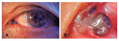
We present a rare case of primary caruncle basal cell carcinoma (BCC), a condition with limited occurrences. Our patient, an 80-year-old woman without prior ocular pathological history, presented a 2x2mm pedunculated blackish nodular lesion on the caruncle of her left eye, without local conjunctival or cutaneous involvement. Histological analysis following complete excision confirmed the presence of basal cell carcinoma within the caruncle. Over a span of 30 months, no recurrence has been observed. While scant cases are documented in the literature, we conducted a review of these instances. Despite its infrequent manifestation, this condition should be taken into account when evaluating caruncular tumors, given its tendency to invade the orbit. Complete excision with free surgical margins is the treatment of choice, and adjuvant radiotherapy or chemotherapy might be considered.
Keywords: Conjunctival diseases; Eye neoplasms; Sebaceous gland neoplasms; Conjunctival neoplasms; Carcinoma, basal cell; Diagnosis, differential; Humans; Case reports
Arq. Bras. Oftalmol. 2024;87 (2 )
:1-3
| DOI: 10.5935/0004-2749.2022-0091
Abstract
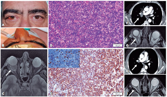
Hepatitis C virus infection may be implicated in 12.7% of ocular adnexal marginal zone lymphomas. We present the first case of an orbital-systemic mucosa-associated lymphoid tissue lymphoma that responded to hepatitis C virus medical treatment. A 62-year-old male with a right-sided orbital mass was diagnosed with stage IIA orbital marginal zone lymphoma in addition to hepatitis C virus infection based on clinical, imaging, laboratory, and histological examinations. The systemic and orbital responses were achieved 1 year after undergoing hepatitis C virus treatment with glecaprevir/pibrentasvir. The association between the hepatitis C virus infection and orbital-systemic mucosa-associated lymphoid tissue lymphoma is relevant. Accordingly, patients with orbital mucosa-associated lymphoid tissue lymphoma should be assessed for hepatitis C virus seroreactivity for therapeutic and prognostic purposes.
Keywords: Orbital disease; Orbital neoplasms; Lymphoma, B-cell marginal zone; Hepacivirus; Hepatitis C; Humans; Case reports
Arq. Bras. Oftalmol. 2025;88 (2 )
:1-4
| DOI: 10.5935/0004-2749.2024-0007
Abstract
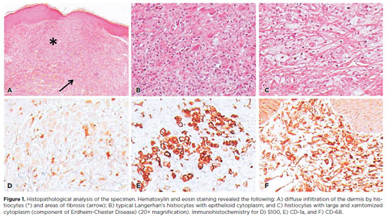
Langerhans cell histiocytosis comprises a heterogeneous range of clinical manifestations secondary to clonal proliferation of histiocytes, characterized by the accumulation of these cells in various organs and tissues. The ophthalmological component commonly involved is the orbit. Herein, we report a rare case of Langerhans cell histiocytosis with eyelid involvement, which resulted in severe ocular surface complications, which subsequently significantly impacted the patient's quality of life. This case report highlights the fact that despite being rare, Langerhans cell histiocytosis should be included in the differential diagnosis of eyelid lesions. Furthermore, a multidisciplinary approach with a systemic overview is crucial for managing the ocular complications.
Keywords: Dry eye syndromes; Erdheim-Chester disease/drug therapy; Human; Female; Case reports
 ABO is licensed under a Creative Commons Attribution-NonComercial 4.0 Internacional.
09-fig01.jpg)
13-fig01.jpg)
11-fig01.jpg)
11-fig01tb.jpg)
02-fig01.jpg)
03-fig01.jpg)
02-fig01.jpg)
04-fig01.jpg)
14-fig01.jpg)