Arq. Bras. Oftalmol. 2023;86 (2 )
:131-136
| DOI: 10.5935/0004-2749.20230036
Abstract
Objetivo: Avaliar o impacto dos tumores córneo-conjuntivais na superfície ocular e na qualidade de vida dos pacientes antes e após o tratamento cirúrgico.
Métodos: Este estudo prospectivo conduziu uma avaliação pré-operatória e com 30 e 90 dias de pós-operatório de pacientes com diagnóstico de tumores de córnea e conjuntiva. Os dados demográficos foram coletados no pré-operatório. Os questionários Health Survey Short-Form (SF-12) e Ocular Surface Disease Index (OSDI) foram aplicados para avaliar a qualidade de vida dos pacientes e a percepção de suas funções relacionadas à visão. Os testes tear break-up time (TBUT) e Schirmer foram realizados para avaliação da superfície ocular. A extensão do tumor foi medida usando o programa ImageJ.
Resultados: Vinte e três pacientes foram incluídos. A média de idade foi de 52,8 ± 17,3 anos (27-79 anos). O tipo mais comum de tumor foi o carcinoma de células escamosas (61,5%). A acuidade visual dos pacientes melhorou significativamente em 1 mês e 3 meses (p=0,018 e p=0,036, respectivamente). Não houve diferenças significativas entre os testes tear break-up time e Schirmer no pré-operatório e com 3 meses de pós-operatório (p=0,150 e p=0,490, respectivamente). Os escores do SF-12 demonstraram que o componente mental apresentou diferença estatisticamente significante entre o pré-operatório e no 30 e 90 dias de pós-operatório (p=0,008 e p=0,026, respectivamente). A extensão do tumor foi de 868,7 ± 344,9 pixels (intervalo, 224,6-1481,6 pixels) e foram significativamente correlacionados com o componente mental de SF-12 no pré-operatório (p=0,011), 30 (p=0,017) e 90 dias de pós-operatório (p=0,012), e o componente emocional no 30º dia de pós-operatório (p=0,016).
Conclusão: Pacientes com tumores córneo-conjuntivais melhoraram os sintomas oculares, a acuidade visual e o componente emocional da qualidade de vida após a excisão cirúrgica do tumor.
Keywords: Neoplasias oculares; Neoplasias da túnica conjuntiva; Doenças da córnea; Acuidade visual; Qualidade de vida.
Arq. Bras. Oftalmol. 2022;85 (2 )
:136-143
| DOI: 10.5935/0004-2749.20220022
Abstract
Objetivo: Estimar a epidemiologia do pterígio; sua correlação com sintomas de olho seco e com potenciais preditores sistêmicos e oculares.
Métodos: Estudo transversal, de base populacional, no qual foram realizadas visitas domiciliares aleatórias a 600 participantes, com 40 anos ou mais de idade, em Ribeirão Preto-SP (n=420) e Cassia dos Coqueiros-SP (n=180), Brasil. Uma entrevista estruturada com um questionário detalhado foi usada para coletar informações sobre demografia e possíveis fatores de risco. Em um segundo momento, participantes aleatórios com pterígio (n=63) ou não (n=110) foram avaliados quanto a alterações na superfície ocular.
Resultados: A frequência de pterígio em Ribeirão Preto foi de 21%; 15.7% entre as mulheres e 32.1% entre os homens (p=0,0002). Em Cássia dos Coqueiros, essa taxa foi de 19.4%; onde 17.3% eram mulheres e 25.5% eram homens (p=0,28). A média de idade naqueles afetados pelo pterígio foi superior à dos participantes sem pterígio, 65,6 ± 10,5 e 61,2 ± 12,0 anos, respectivamente (p=0,02). Houve uma correlação positiva entre o pterígio e história prévia de radioterapia e quimioterapia (p<0,0001 para ambos). Houve maior coloração de fluoresceína na córnea e maior coloração de lissamina verde na conjuntiva em olhos com pterígio (p=0,0003 e 0,0001, respectivamente).
Conclusão: Encontramos uma alta frequência de pterígio em duas populações adultas brasileiras, principalmente em homens e idosos. Danos na superfície ocular e história prévia de radioterapia e/ou quimioterapia foram associados ao pterígio.
Keywords: Pterígio/epidemiologia; Síndrome do olho seco; Prevalência; Fatores de risco
Arq. Bras. Oftalmol. 2025;88 (6 )
:1-5
| DOI: 10.5935/0004-2749.2024-0321
Abstract
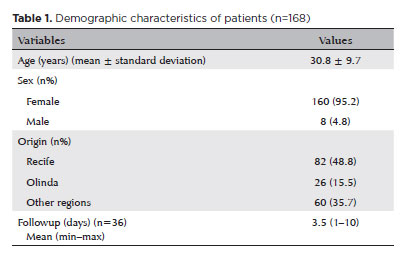
PURPOSE: To report the ophthalmological signs, symptoms, and clinical management observed during an unprecedented outbreak of chemical ocular injuries related to cosmetic hair ointments in Brazil.
METHODS: This descriptive, cross-sectional study reviewed medical records of patients treated at the emergency center of Fundação Altino Ventura for chemical ocular trauma associated with cosmetic hair ointment use between February 2022 and February 2023. Records with incomplete medical information were excluded.
RESULTS: The study included 168 patients (95.2% [n=160] female), with a mean age of 30.8 ± 9.7 years. The most frequently reported symptoms at presentation were pain (167/168, 99.4%) and photophobia (92/168, 54.8%). Severe pain was reported by 137 patients (80%). Keratitis was present in 280 of 336 eyes (83.3%), conjunctival hyperemia in 256 eyes (76.4%), and corneal abrasions in 174 eyes (51.8%). A decrease in visual acuity (worse than 20/25) was documented in 18.5% (31/168) of cases. Lubricants, antibiotics, and re-epithelialization
ointments were prescribed to 64.8% (109/168) of the patients. Topical corticosteroids and oral vitamin C were administered to 34% (57/168) and 1.2% (2/168) of patients, respectively. Followup visits were required in 19% (33/168) of cases.
CONCLUSION: The outbreak of chemical ocular injuries linked to cosmetic ointments used for braiding and hair modeling in Brazil was marked by intense ocular pain, conjunctival hyperemia, keratitis, and corneal abrasions. Most patients were treated with lubricants, antibiotics, and re-epithelialization ointments, although approximately one-fifth required followup care, and one-third received additional treatment with either topical corticosteroids and/or oral vitamin C.
Keywords: Cosmetics; Hair preparations; Eye injuries; Burns, chemical; Eye burns; Keratitis; Cornea; Corneal diseases; Visual low.
Arq. Bras. Oftalmol. 2021;84 (6 )
:576-581
| DOI: 10.5935/0004-2749.20210095
Abstract
Objetivo: Comparar a acuidade visual prevista pelo Escore de Trauma Ocular com a acuidade visual final dos pacientes vítimas de trauma ocular aberto atendidos em hospital no sul do Brasil.
Métodos: Foram analisados 120 olhos de 119 vítimas de trauma ocular aberto. Foi realizado um estudo observacional e retrospectivo em hospital universitário. Foram extraídos dados de prontuários relacionados a idade, sexo, olho acometido e mecanismo de trauma, bem como dados para o cálculo do Escore de Trauma Ocular (acuidade visual inicial, presença de ruptura de globo, perfuração, endoftalmite, descolamento de retina, defeito pupilar aferente) e acuidade visual final.
Resultado: Houve concordância entre a acuidade visual prevista pelo Escore de Trauma Ocular e a acuidade visual final prevista no presente estudo. A análise isolada das variáveis demonstrou significância para acuidade visual inicial (p<0,001), para descolamento de retina (p=0,001) e para defeito pupilar aferente (p<0,004). Não houve diferença significativa entre a acuidade visual final do estudo original do Escore de Trauma Ocular. e na população abordada no presente estudo.
Conclusão: O Escore de Trauma Ocular pode ser aplicado à população estudada no presente estudo como ferramenta de determinação do prognóstico visual em vítimas de trauma ocular aberto. As variáveis mais significativas são acuidade visual inicial, descolamento de retina e defeito pupilar aferente. Estudos prospectivos com amostras maiores são necessários para comprovar tal hipótese.
Keywords: Índices de gravidade do trauma; Acuidade visual; Traumatismos oculares; Prognóstico
Arq. Bras. Oftalmol. 2022;85 (4 )
:377-381
| DOI: 10.5935/0004-2749.20220016
Abstract
Objetivo: Determinar a incidência de emergências oculares em um centro oftalmológico de referência no Brasil.
Métodos: O banco de dados de prontuários da Fundação Altino Ventura, Recife, Brasil, foi analisado retrospectivamente e incluiu pacientes atendidos, entre janeiro de 2017 e janeiro de 2018, na sala de emergência oftalmológica. Foram excluídos os prontuários com dados incompletos e com quadros ambulatoriais. Apenas o primeiro atendimento na emergência foi considerado para análise.
Resultados: Em um ano, 134.788 pacientes (idade média de 38,7 ± 22 anos [0-99 anos]) foram admitidos na emergência da Fundação Altino Ventura. Os diagnósticos mais frequentes foram conjuntivite (52.732 casos [37,3%]), blefarite (7.213 casos [5,1%]) e corpo estranho na córnea/conjuntiva (6.925 casos [4,9%]). Corpo estranho na córnea/conjuntiva e trauma ocular foram cerca de 8 vezes e 2 vezes mais incidente em indivíduos do sexo masculino, respectivamente (p<0,001 em ambos). Triquíase e blefarite afetaram ~2 vezes mais pacientes do sexo feminino, respectivamente (p<0,001 em ambos). Corpo estranho na córnea/conjuntiva e trauma ocular afetaram mais pacientes em idade produtiva (>15 anos), enquanto úlcera, blefarite e triquíase da córnea afetaram mais pacientes idosos. Todos os grupamentos de diagnóstico (doenças infecciosas, trauma ocular, corpos estranhos, retinopatias, doenças das pálpebras, doenças da córnea, crise glaucomatosa e doenças neurooftalmológicas) foram mais incidentes na primavera (valor de p<0,001).
Conclusão: As emergências oftalmológicas mais comuns no presente estudo foram as doenças infecciosas e o corpo estranho. Porém, a incidência das emergências oculares são fluências pela faixa etária e gênero do paciente, além da época do ano.
Keywords: Emergência; Oftalmopatia; Transtorno da visão; Conjuntivite; Corpo estranho; Traumatismo oculare; Estações do ano; Brasil
Arq. Bras. Oftalmol. 2025;88 (6 )
:1-6
| DOI: 10.5935/0004-2749.2025-0153
Abstract
PURPOSE: This clinical study aimed to assess the effectiveness of microemulsion artificial tears containing povidone and propylene glycol in the management of dry eye disease. Secondary objectives included evaluating improvements in tear-film stability, measured by tear break-up time and corneal staining scores, along with the tolerability and safety of the formulation.
METHODS: This was a prospective, single-arm interventional study involving 30 participants (52 eyes) diagnosed with dry eye disease. Participants self-administered the investigational eye drops twice daily for 28 consecutive days. Primary and secondary outcomes included changes in the Ocular Surface Disease Index, tear break-up time, and corneal staining scores. Adverse events were documented throughout the study period.
RESULTS: Significant improvements in Ocular Surface Disease Index scores were observed, reflecting a reduction in dry eye disease symptoms. Tear break-up time increased notably between baseline and follow-up assessments, with the proportion of eyes exhibiting tear break-up time ≥10 srising from 25.0% to 63.5%. Additionally, the percentage of eyes with a corneal staining score of zero improved from 23.1% to 69.2%. Conjunctival staining also decreased, with the proportion of eyes with scores of 2 and 3 dropping from 11.5% to 3.8% and 5.8% to 0%, respectively.
CONCLUSIONS: The findings suggest that povidone and propylene glycol-based artificial tears significantly enhance tear-film stability and alleviate symptoms in patients with mild to moderate dry eye disease, with minimal adverse effects. This formulation represents a safe and effective short-term treatment option for dry eye disease management.
Keywords: Artificial tears; Dry eye disease; Tear-film stability; Propylene glycol; Povidone; Visual acuity; Surveys and questionnaires
Arq. Bras. Oftalmol. 2025;88 (6 )
:1-7
| DOI: 10.5935/0004-2749.2025-0120
Abstract
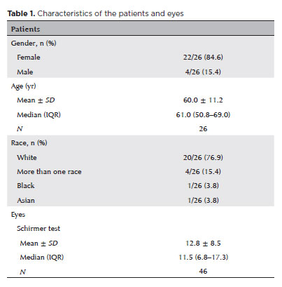
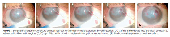
PURPOSE: To describe the technique and outcomes of intrastromal autologous blood injection in patients with severe corneal hydrops.
METHODS: Nineteen patients with corneal hydrops underwent intrastromal autologous blood injection. Postoperative assessments included best-corrected visual acuity and time to resolution of corneal edema
RESULTS: Corneal edema resolved within 1 week in 5 patients, within 1 month in 11, and within 3 months in 3. The mean duration of edema persistence was 37.94 ± 33.05 days (range, 6–124). Corneal thickness decreased from 2.06 ± 0.71-mm preoperatively to 1.34 ± 0.65-mm at day 7, 0.85 ± 0.56-mm at day 30, and 0.57 ± 0.13-mm at day 90 (p<0.001). Descemet’s membrane (DM) detachment decreased from 1.01 ± 0.75-mm to 0.44 ± 0.57-mm, 0.24 ± 0.36-mm, and 0.08 ± 0.11-mm on postoperative days 7, 30, and 90, respectively (p<0.001). DM break size decreased from 1.12 ± 1.19-mm to 0.62 ± 0.84-mm at 3 months (p<0.005). Three patients developed hematocornea; no other major complications were observed. At 3 months, mean best-corrected visual acuity improved from 2.37 ± 0.66 to 0.41 ± 0.17 logMAR with hard contact lenses (p<0.001).
CONCLUSIONS: Intrastromal autologous blood injection is an effective treatment for severe corneal hydrops, promoting faster edema resolution and visual improvement with minimal complications.
Keywords: Corneal edema; Corneal diseases; Edema; Visual acuity; keratoconus.
Arq. Bras. Oftalmol. 2025;88 (3 )
:1-7
| DOI: 10.5935/0004-2749.2023-0309
Abstract
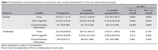
PURPOSE: Keratoconus presents certain peculiarities in pediatric patients when compared with adults. The greatest challenge in children is that the disease is more severe and faster in progression. In this retrospective study, we aimed to compare the accelerated and Dresden protocols for corneal crosslinking in patients aged <18 years who were followed-up for at least 12 months.
METHODS: A total of 36 eyes from 27 patients were included in the study. The best corrected and uncorrected visual acuity, maximal keratometry, corneal thickness, foveal thickness, and endothelial microscopy findings were evaluated at baseline and during the postoperative period at one, three, and six months. Thereafter, the patients were evaluated at one, three, six and twelve months postoperative. Corneal crosslinking was performed in all patients via the Dresden protocol (n=21 eyes) or the accelerated protocol (n=15 eyes). Data between the two groups were compared and XY statistical analysis was used.
RESULTS: Both protocols were effective in halting keratoconus progression. No patient had progression at the 12-month follow-up. A significant reduction in Kmax and improvement in the corrected distance visual acuity were observed only in the Dresden protocol group. Although the Dresden protocol was superior to the accelerated protocol in reducing Kmax (p=0.002), there was no significant difference in corrected distance visual acuity between the two groups.
CONCLUSION: The accelerated protocol is as efficient as the Dresden protocol in stabilizing keratoconus progression. Although the Dresden protocol was superior to the accelerated protocol in reducing the Kmax, it did not produce better clinical results. Thus, the accelerated protocol is an efficient option. Furthermore, considering the advantages of reduced surgical time, the accelerated protocol is effective in halting keratoconus progression in the pediatric age group.
Keywords: Keratoconus; Corneal diseases; Ultraviolet rays; Cross-linking reagents; Visual acuity
Arq. Bras. Oftalmol. 2025;88 (3 )
:1-11
| DOI: 10.5935/0004-2749.2024-0049
Abstract
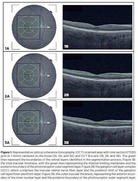
PURPOSE: This study aimed to evaluate the total macular thickness as well as the thickness of the inner and outer retinal layers in patients with Parkinson's disease. It also aimed to verify the correlation of these parameters with motor symptoms and cognitive function.
METHODS: A total of 46 eyes of 23 patients with Parkinson's disease and 40 eyes of 20 healthy controls were included in the study. The patients' cognitive, functional, and nonmotor symptoms were evaluated using the Katz Index of Independence and Pfeffer's Activities of Daily Living, Mini-Mental State Examination, Frontal Assessment Battery, Schwab and England Staging Scales, and Movement Disorders Society Nonmotor Symptoms Scale. The macular thickness measurements obtained via total, inner, and outer optical coherence tomography were recorded. Furthermore, the correlation of the parameters of optical coherence tomography with cognitive, functional, and nonmotor symptoms was assessed.
RESULTS: The scores of the Katz Index of Independence and Pfeffer's Activities of Daily Living as well as the Movement Disorders Society Nonmotor Symptoms Scale were significantly lower in patients with Parkinson's disease than in healthy controls. Moreover, the former had greater total macular thickness. The temporal and inferior outer sectors were significantly greater for the ganglion cell complex thickness in patients. A significant correlation was observed between the total macular thickness and the Movement Disorder Society-Unified Parkinson's Disease Rating Scale, Parte III (MDS-UPDRS-III) values. Contrarily, there was a negative correlation between the outer macular thickness and the MDS-UPDRS-III values. Meanwhile, the total macular thickness and ganglion cell complex thickness were significantly correlated with the scores of the Mini-Mental State Examination, Schwab and England Staging Scale, Frontal Assessment Battery, and Katz Index of Independence and Pfeffer's Activities of Daily Living. In addition, the Schwab and England scale was correlated with the outer macular thickness.
CONCLUSION: The total and inner macular thicknesses at the temporal and inferior outer sectors were greater in patients with Parkinson's disease than in the control group. These findings indicate that macular thickness may be greater in those with Parkinson's disease, particularly when associated with mild motor symptoms. In addition, the parameters of the total, inner, and outer optical coherence tomography were significantly associated with motor and nonmotor symptoms as well as cognitive function impairment.
Keywords: Parkinson's disease; Tomography, optical coherence; Neurodegenerative diseases; Cognitive dysfunction; Cognition; Motor perception; Visual acuity; Retina
Arq. Bras. Oftalmol. 2024;87 (3 )
:1-7
| DOI: 10.5935/0004-2749.2022-0374
Abstract
PURPOSE: To describe a 2019 acute toxoplasmosis outbreak in the city of São Paulo, Brazil, and to evaluate the laboratory serological profile for toxoplasmosis for three consecutive years. The ophthalmological manifestations of the patients involved in the outbreak were also studied.
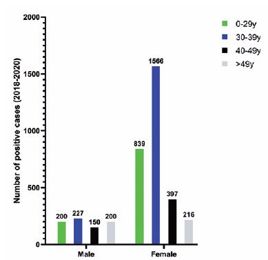
METHODS: A cross-sectional descriptive study of a toxoplasmosis outbreak in São Paulo, Brazil, between February and May 2019. Epidemiological data were described, as were the observed ocular manifestations. As part of this study the number of patients with positive IgM toxoplasmosis serology was obtained from a large laboratory network (DASA) for three consecutive years, including the year of the outbreak (2018, 2019, 2020).
RESULTS: Eighty-three individuals were identified in the outbreak and two clusters were studied. The clinical picture of at least 77% of the patients, the epidemiological analysis, and the short incubation period (5-8 days) suggested contamination by oocysts. Serological laboratory data analysis revealed an increase of positive toxoplasmosis IgM in 2019 of 73% compared to the previous year. Ophthalmological examination revealed that at least 4.8% of the patients developed toxoplasmic retinochoroiditis, none of whom had been treated during the acute systemic disease.
CONCLUSION: Our findings indicate vegetable contamination as the possible source of this outbreak, a high prevalence of toxoplasmosis in São Paulo during the outbreak period, and a drop in the number of tests during the COVID-19 pandemic. Retinochoroiditis was observed in at least 4.8% of the cases. We confirm the need to implement effective means for the prevention, diagnosis, and treatment of the disease. This may involve raising awareness among the population of the importance of vegetable hygiene, and improved quality control of food and water.
Keywords: Toxoplasmosis/etiology; Food parasitology; Water/parasitology; Uveitis, posterior/parasitology; Chorioretinitis/parasitology; Visual acuity; Disease outbreaks; Eye manifestations; Humans.
Arq. Bras. Oftalmol. 2024;87 (2 )
:1-5
| DOI: 10.5935/0004-2749.2021-0395
Abstract
Objetivos: Avaliar a segurança e eficácia a longo prazo da vitreólise com Nd:YAG laser para moscas volantes sintomáticas, uma vez que permanece como um procedimento controverso devido a falta de evidência científica robusta sobre a manutenção dos resultados e ocorrência de efeitos adversos.
Métodos: Este estudo é uma extensão observacional de um ensaio clínico prospectivo, randomizado, duplo cego, previamente publicado. Oito de treze pacientes que foram submetidos a vitreólise com YAG laser foram acompanhados para uma reavaliação tardia, dezoito meses após o procedimento, para avaliar a eficácia e segurança do procedimento.
Resultados: Todos os pacientes mantiveram a melhora na sintomatologia notada ao final do procedimento original, com 25% dos casos apresentando melhora completa, e uma proporção semelhante (37,5%) demonstrando melhora significativa ou parcial. A melhora objetiva na opacidade foi similar ao achado no seguimento original de 6 meses. O questionário de qualidade de vida NEI-VFQ 25 não demonstrou diferença estatisticamente significativa nas respostas entre o sexto e o décimo oitavo mês de acompanhamento. Nenhum efeito adverso foi notado no exame clínico ou reportado pelos pacientes.
Conclusão: A eficácia da vitreólise observada ao sexto mês do acompanhamento foi mantida até o décimo oitavo mês, com todos os pacientes notando algum grau de melhora quando comparado ao estado pré procedimento. Nenhum efeito adverso tardio foi notado. Um ensaio clínico randomizado maior é necessário para confirmar a segurança do procedimento.
Keywords: Terapia a laser; Lasers de estado sólido; Vitrectomia; Corpo vítreo; Cirurgia vitreorretiniana; Acuidade visual; Doenças oculares; Qualidade de vida; Inquéritos e questionários
 ABO is licensed under a Creative Commons Attribution-NonComercial 4.0 Internacional.
13-tab01.jpg)
08-tab01tb.jpg)
09-tab01.jpg)
01-tab01.jpg)
01-fig01tb.jpg)