Arq. Bras. Oftalmol. 2025;88 (4 )
:1-5
| DOI: 10.5935/0004-2749.2024-0167
Abstract
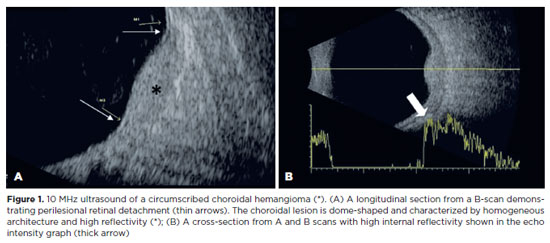
PURPOSE: To evaluate if color Doppler can detect internal blood flow in circumscribed choroidal hemangioma.
METHODS: This cross-sectional study examined seven eyes of seven participants with circumscribed choroidal hemangiomas, with or without prior treatment. B-scan ultrasound and color Doppler were used to assess the dimensions, topographical distribution, and internal blood flow of the affected eyes.
RESULTS: The sample included seven patients (five female) with a median age of 61 (62.29 ± 13.83) years. There were seven eyes with circumscribed choroidal hemangiomas in the patient sample. Color Doppler detected internal vascular flow in all cases (100%). The lesions had an average diameter/thickness ratio of >2 mm and an average thickness of <5 mm and were predominantly located superiorly and supero-temporally.
CONCLUSION: Internal blood flow was detected in circumscribed choroidal hemangiomas using color Doppler. Detection was unaffected by the patient's treatment status.
Keywords: Ultrasonography, doppler, collor; Choroidal neoplasms; Hemangioma
Arq. Bras. Oftalmol. 2022;85 (4 )
:370-376
| DOI: 10.5935/0004-2749.20220032
Abstract
Objetivos: Investigar a incidência, fatores de risco e desfechos visuais do desenvolvimento da membrana epirretiniana após reparo do descolamento regmatogênico da retina.
Métodos: Trata-se de um estudo retrospectivo de 309 olhos submetidos à cirurgia inicial para descolamento regmatogênico da retina primário sem complicações. Os exames foram realizados no pré-operatório aos 1, 3, 6 e 12 meses pós-operatórios. Os pacientes foram divididos em dois grupos, dependendo da presença ou ausência de membrana epirretiniana.
Resultados: A incidência de membrana epirretiniana pós-operatória foi de 28,5%; 42,7% desses pacientes apresentaram desenvolvimento grave da membrana epirretiniana e, portanto, foram submetidos à remoção desta membrana. A regressão logística mostrou que as lágrimas retinianas gigantes (RC: 2,66; 95% IC: 1,045 - 6,792, p=0,040) e lágrimas em ferradura (RC: 0,534; 95% IC: 0,295-0,967, p=0,039), foram preditores significativos de membrana epirretiniana pós-operatória. A coloração com acetonida de triancinolona foi significativamente associada à prevenção da membrana epirretiniana (p=0,022). Trinta e quatro pacientes apresentaram acuidade visual melhorada, ou igual, ou acuidade visual final melhor corrigida; 4 olhos foram avaliados na consulta final de acompanhamento e apresentaram redução da acuidade visual melhor corrigida.
Conclusão: Nossa análise demonstra que as lágrimas de ferradura e as lágrimas retinianas gigantes representam fatores de risco para a membrana epirretiniana pós-operatória. A coloração com acetonida de triancinolona teve um efeito preventivo significativo na membrana epirretiniana no pós-operatório. Além disso, uma segunda rodada de vitrectomia pars plana, incluindo remoção da membrana, levou a uma melhora significativa da acuidade visual final melhor corrigida na última consulta de acompanhamento, embora a recuperação tenha sido limitada.
Keywords: Membrana epirretiniana; Descolamento da retina; Recurvamento da esclera; Acuidade visual; Vitrectomia
Arq. Bras. Oftalmol. 2025;88 (2 )
:1-9
| DOI: 10.5935/0004-2749.2023-0326
Abstract
PURPOSE: To evaluate the predictive value of initial intraocular pressure difference of the detached and fellow eyes of patients with complex rhegmatogenous retinal detachment on postoperative persistent ocular hypotony.
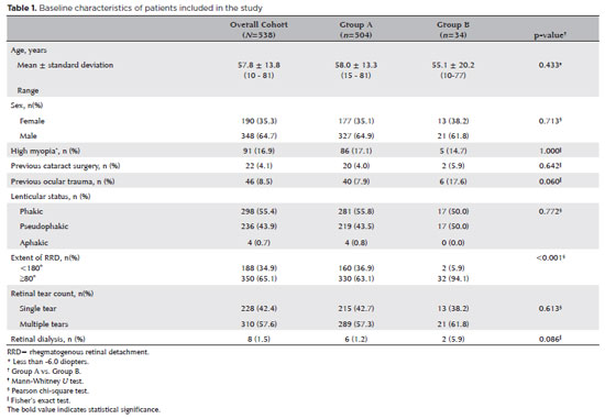
METHODS: This retrospective observational study included 538 eyes of 538 unilateral complex rhegmatogenous retinal detachment patients with a proliferative vitreoretinopathy grade of C-1 or higher, treated with silicone oil endotamponade following pars plana vitrectomy. The patients were divided into Group A (patients having silicone oil removal without ocular hypotony; n=504) and Group B (patients with persistent ocular hypotony following silicone oil removal [n=8, 23.5%] and with retained silicone oil [n=26, 76.5%] due to the risk of persistent ocular hypotony; total n=34). Ocular hypotony was defined as an intraocular pressure of <6 mmHg on two or more occasions. Patients' demographics, including age, sex, and follow-up time, and ocular characteristics, including ocular surgical and trauma history, initial and final best-corrected visual acuity, intraocular pressure and initial intraocular pressure difference of the detached and fellow eyes, and anatomical success rates and postoperative complications, were retrospectively collected from the electronic patient files.
RESULTS: The initial intraocular pressure was significantly lower in the detached eyes of Group B than in Group A (8.3 ± 3.5 vs. 12.9 ± 3.3, p<0.001). Also, the initial intraocular pressure difference was significantly higher in Group B than in Group A (8.9 ± 3.2 vs. 2.2 ± 2.7mmHg, p<0.001). The receiver operating characteristic curve analysis showed that the cutoff value of the initial intraocular pressure difference was 7.5mmHg for the risk of persistent ocular hypotony. The most influential factors on postoperative persistent ocular hypotony in the binary logistic regression analysis were the initial intraocular pressure difference and the need for a retinectomy.
CONCLUSION: In eyes with complex rhegmatogenous retinal detachment treated with pars plana vitrectomy and silicone oil tamponade, the initial intraocular pressure difference could be of value in predicting postoperative persistent ocular hypotony and could guide surgeons on the decision of silicone oil removal.
Keywords: Hypotony; Intraocular pressure; Pars plana vitrectomy; Retinal detachment; Silicone oils; Ocular hypotension; Visual acuity
Arq. Bras. Oftalmol. 2024;87 (4 )
:1-5
| DOI: 10.5935/0004-2749.2022-0335
Abstract
PURPOSE: To clarify the postoperative incidence of macular edema in patients undergoing surgery to repair rhegmatogenous retinal detachment and identify the associated risk factors.
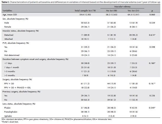
METHODS: In this prospective, observational study, 79 patients who underwent surgery to correct rhegmatogenous retinal detachment using pars plana vitrectomy with silicone oil injection were analyzed. Patients were followed up postoperatively at 7, 30, 90, 180, and 365 days. At each visit, optical coherence tomography was performed to assess the presence or absence of macular edema. were analyzed as possible risk factors for macular edema: age, sex, macular status (attached or detached), presence of vitreoretinal proliferation, history of previous intraocular surgery, reported time of symptoms suggestive of rhegmatogenous retinal detachment up to the date of surgery, and the surgical modality performed.
RESULTS: The 1-year macular edema prevalence rate was 26.6%. In the adjusted analysis, older patients had a higher risk of macular edema, and each 1-year increase in age increased the risk of macular edema by 6% (95% confidence interval = 1.00-1.12). The macular status, vitreoretinal proliferation, the surgical technique used, prior intraocular surgery, and the intraocular lens status were not identified as risk factors. However, the incidence of macular edema increased up to 180 days after surgery, peaking at 10.6%, and then decreased until 365 days after surgery.
CONCLUSION: Macular edema was a common complication after surgery to treat rhegmatogenous retinal detachment, with its incidence peaking between 30 and 180 days after surgery. Age was an important risk factor for macular edema in this cohort.
Keywords: Macular edema; Retinal detachment; Vitrectomy; Tomography, optical coherence; Incidence; Risk factors


 ABO is licensed under a Creative Commons Attribution-NonComercial 4.0 Internacional.


03-tab01tb.jpg)
02-fig01.jpg)
03-fig01.jpg)
02-fig01.jpg)
02-fig01.jpg)
09-fig01.jpg)
13-fig01.jpg)
13-fig01.jpg)
04-fig01.jpg)