
Arq. Bras. Oftalmol. 2026; 89 (6): 10.5935/0004-2749.2026-0183
Total: 26
Marlos Rodrigues Lopes e Silva1,2; Michael Knafo3; Mostafa Bondok4; Diogo Batista de Queiroz Silveira1; Anne-Xuan Lan Nguyen5,6; Patricia Mitiko Santello Akaishi7; Leonardo Lando8
DOI: 10.5935/0004-2749.2026-0183
ABSTRACT
PURPOSE: To compare surgical outcomes and patient tolerability between conventional knotted fixation and a novel technique using cyanoacrylate gel glue to secure the silicone stent during external dacryocystorhinostomy.
METHODS: This retrospective study included 70 patients who underwent external dacryocystorhinostomy, of whom 40 received cyanoacrylate glue fixation (study group) and 30 underwent conventional knotted fixation (control group). Patients with congenital or acquired lacrimal obstruction and a minimum follow-up period of 6 months were included. All procedures were performed by the same surgeon at a single institution. Patient tolerability, including foreign body sensation, sneezing, and nasal secretion, and surgical complications, including glue-induced toxicity, infection, stent extrusion, mobilization, release, and pyogenic granuloma, were assessed. Data were analyzed using descriptive statistics and nonparametric tests.
RESULTS: The median patient age was 69.0 years (range, 3–90 years), and most patients were women (81.4%, n=57). There were no significant differences between the groups in age (p=0.264) or sex (p=0.213). Symptoms were reported by 34.3% of patients and were significantly more frequent in the control group than in the study group (33.3% vs. 10.0%, p<0.001). Foreign body sensation (p=0.002), sneezing (p=0.012), and nasal secretion (p<0.001) were all significantly more common in the control group. Overall, complications were infrequent, with no significant between-group differences in stent release (17.5% vs. 20.0%, p=0.790), mobilization (5.0% vs. 16.7%, p=0.130), or infection (2.9% vs. 0%, p=0.503). No cases of glue-induced toxicity were observed. Follow-up duration did not differ significantly between the groups.
CONCLUSIONS: Cyanoacrylate fixation was associated with favorable patient tolerability and no increase in complication rates compared with conventional knotted fixation. These findings suggest that cyanoacrylate glue may be a safe and well-tolerated alternative for silicone stent fixation during external dacryocystorhinostomy.
Keywords: Dacryocystorhinostomy/methods; Lacrimal duct obstruction; Nasolacrimal duct/surgery; Intubation; Cyanoacrylates; Adhesives; Stent; Silicones; Comparative study
INTRODUCTION
Dacryocystorhinostomy (DCR) is a bypass procedure originally developed in the early 1900s and is now widely used to manage nasolacrimal drainage obstruction in both adults and children, which typically presents with epiphora or dacryocystitis(1-3). Although the technique has been progressively refined since its introduction, DCR is still performed predominantly through either an external or an endonasal/endoscopic approach(1,4).
Although bicanalicular intubation with a Crawford probe is considered optional by some surgeons, it is frequently used as an adjunct to DCR, and the method used to secure the silicone stent remains an important technical consideration(4-7). Traditionally, securing the stent requires the placement of multiple knots after its placement in the nasal cavity to prevent loss, slippage, and lateral prolapse(8,9). However, this fixation method may affect both postoperative outcomes and patient comfort. Several alternative techniques have been described, including simple manual repositioning without knotting and the use of clips to provide additional stability. Nevertheless, these approaches may be technically variable, require specialized devices, and have demonstrated inconsistent effectiveness in preventing stent displacement(10-13).
Cyanoacrylate adhesives have an established history of safe use in various ophthalmic procedures and have demonstrated good tolerability and a favorable safety profile(14,15). To the best of our knowledge, the use of cyanoacrylate glue for silicone stent fixation during DCR has not previously been described, particularly with regard to patient comfort. Given its accessibility and increasing use in oculoplastic surgery, cyanoacrylate glue may represent a practical alternative for stent fixation. Therefore, this study evaluated the use of cyanoacrylate glue to secure silicone stents during external DCR and investigated its potential as a simpler and more comfortable alternative to conventional fixation methods.
METHODS
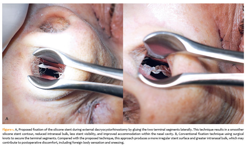
This retrospective, longitudinal study evaluated patients who underwent external dacryocystorhinostomy (DCR) with bicanalicular silicone stent placement. The conventional knotted fixation technique, which is well established among oculoplastic surgeons, was compared with a novel technique using an off-label cyanoacrylate gel adhesive (Loctite Super Bonder cyanoacrylate adhesive, Henkel Corporation, Rocky Hill, Connecticut) that eliminates the need for knots (Figure 1 A,B). All DCR procedures included bicanalicular silicone stent placement and were performed by the same surgeon (MRLS) at a single center (Ver Hospital de Olhos, Goiânia, Brazil) under sedation and local anesthesia.
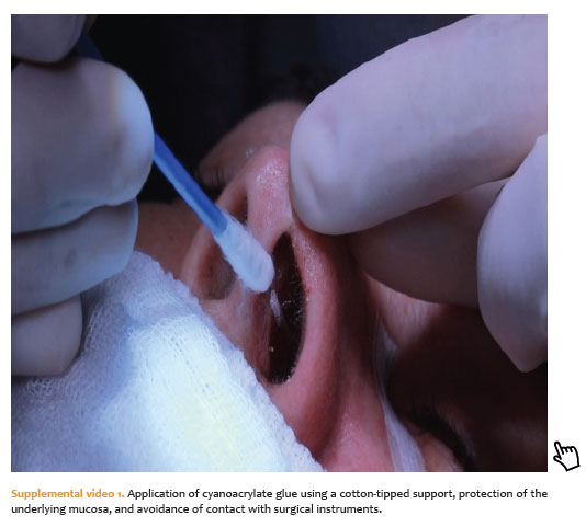
At the time of stent fixation, the nasal and periorbital mucosa were first protected. The anterior mucosa was protected with two to three cotton-tipped bulbs, and the posterior mucosa was protected with a surgical paper strip. Cyanoacrylate adhesive was then applied to all surfaces of the stent using a bare, cotton-free applicator tip. The adhesive was allowed to set for 30 s to ensure adequate consolidation and polymerization. The nasal protectors were subsequently removed, and the stent was trimmed with a No. 15 blade. Correct positioning within the nasal cavity was then confirmed (Supplemental video 1).
Postoperative care was standardized across all cases and included topical ophthalmic antibiotic ointment applied locally three times daily for 1 week, along with combined steroid-antibiotic eye drops administered four times daily for 2 weeks. When intraoperative findings indicated local infection (dacryocystitis), patients were additionally prescribed oral cephalexin at a dose of 500 mg four times daily for 7 days. The study was approved by the Institutional Review Board of the Federal University of Goiás (Approval No. 90857225.4.0000.5078/7.778.206), and informed consent was obtained from all patients or their legal representatives.
Patients were eligible if they had congenital or acquired lacrimal obstruction and a minimum postoperative follow-up of 6 months. Exclusion criteria included incomplete medical records or a follow-up period of less than 6 months. The assessed outcomes included symptoms indicative of tolerability (foreign body sensation, sneezing, and nasal secretion) and surgical complications (glue-induced toxicity, infection, stent extrusion, mobilization, release, and pyogenic granuloma)(12,15,16). Tolerability data were obtained from medical records documenting patient complaints reported during clinical visits. Additional demographic and clinical variables included age, sex, laterality, and etiology of stenosis. Symptoms and complications were documented until silicone stent removal, which typically occurred at 6 weeks(17), or until the last available follow-up visit.
The control group was selected from a historical institutional cohort and frequency-matched by age. Statistical analyses were performed using SPSS (version 29.0.2.0; IBM Corp., Armonk, New York). Continuous variables were analyzed using the Mann–Whitney U test, whereas categorical variables were analyzed using Fisher’s exact test because of the nonparametric nature of the data. Statistical significance was defined as a two-tailed p<0.05. Multivariable comparisons and p-value adjustments were not performed because of the expected small sample size.
RESULTS
A total of 70 patients who underwent external DCR with bicanalicular silicone stent intubation were included in the study: 40 patients underwent cyanoacrylate glue fixation, and 30 controls underwent conventional knot fixation. The overall median age was 69.0 years (range, 3–90 years; interquartile range [IQR], 20 years), with no statistically significant difference between the groups (71.5 years [IQR, 19 years] in the study group vs. 67.0 years [IQR, 22 years] in the control group; p=0.264; Table 1). Overall, 81.4% of patients (n=57) were female, and 18.6% (n=13) were male. The proportion of women was higher in the study group (87.5%) than in the control group (73.3%); however, this difference was not statistically significant (p=0.213).
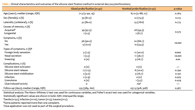
Unilateral obstruction was the predominant presentation (78.6%, n=55), with similar proportions between the groups (80.0% in the study group vs. 78.6% in the control group; p=0.775). Most cases of obstruction were noncongenital (95.7%, n=67), with a comparable distribution between the groups (97.5% vs. 93.3%; p=0.573). Postoperative symptoms were reported by 34.3% of patients (n=24) and were significantly more frequent in the control group (33.3%) than in the study group (10.0%; p<0.001). Among all patients, foreign body sensation was reported by 21.4% (n=15), sneezing by 7.1% (n=5), and nasal secretion by 17.1% (n=12). Each of these symptoms was significantly more common in the control group (p=0.002, p=0.012, and p<0.001, respectively).
Adverse events were generally uncommon. No cases of silicone stent extrusion occurred in either group. Stent release was observed in 17.5% of patients in the study group (n=7) and 20.0% of patients in the control group (n=6; p=0.790), whereas stent mobilization occurred in 5.0% of patients in the study group (n=2) and 16.7% of patients in the control group (n=5; p=0.130). Postoperative infection, characterized by local hyperemia and edema, occurred only in the study group (2.9%, n=2), with no cases reported in the control group; however, this difference was not statistically significant (p=0.503). In these cases, treatment with oral cephalexin (500 mg four times daily for 7 days) resulted in complete resolution of symptoms.
One patient in the control group developed a small granuloma (3.3%, p=0.429), which was managed with oral antibiotics combined with steroid-antibiotic eye drops administered three times daily for 7 days. No cases of glue-induced toxicity were reported. Overall, there were no statistically significant differences in complication rates between the groups.
The overall median follow-up duration was 240 days (range, 918 days; IQR, 183 days) and was similar between the groups (p=0.367).
DISCUSSION
This case series describes a novel application of cyanoacrylate glue for silicone stent fixation during external DCR as an alternative to conventional knotted fixation. Our findings suggest that cyanoacrylate fixation is associated with comparable complication rates and improved postoperative tolerability. Although external DCR with stent fixation is widely used for acquired or congenital nasolacrimal duct obstruction(1, 2), knot tightening remains a debatable surgical step. Several authors have considered it technically dispensable and, in some cases, challenging, particularly in patients with narrow nasal cavities or limited endoscopic visualization(10,11,13,18). Nevertheless, other studies have suggested that knot fixation may help prevent stent slippage, premature extrusion, or migration during postoperative healing(3,7,9,19).
Over the years, several modifications to stent fixation techniques have been proposed to address these challenges. Jones first described silicone stent intubation with intranasal knotting using a monocanalicular probe in 1962(20). Crawford subsequently popularized the use of bicanalicular silicone stents tied intranasally with simple or double knots(5). Subsequent alternatives have included silicone clips, absorbable sutures, and self-retaining stents (e.g., Monoka and Ritleng), all of which were developed to simplify stent handling and reduce complications(8, 21).
Despite these innovations, mechanical irritation of the mucosa, accumulation of nasal secretions, and foreign body sensation remain common postoperative complaints, some of which may be related to the presence and bulk of intranasal knots(1,2,12). In our series, these symptoms were frequently reported during routine follow-up and appeared to affect postoperative comfort, consistent with previous reports(1,12).
The fixation method described here eliminates the need for knot tying and was adopted as a simpler and potentially faster alternative to the standard technique. At the time of stent removal, our routine for both the glued and knotted techniques involved cutting the external loop of the stent at the lacrimal punctum, after which the tube was naturally expelled with sneezing. In our series, postoperative symptoms were significantly less frequent in the glued-fixation group than in the conventional knotted-fixation group (p<0.001). Rates of stent release, mobilization, and infection were comparable between the groups (p=0.130–0.790), whereas discomfort-related symptoms were significantly less frequent in the glued-fixation group (p<0.001–0.012). One possible explanation is that eliminating intranasal knots reduces mucosal friction and irritation during healing. However, the potential for chemical injury associated with glue application remains an important consideration.
Cyanoacrylates are fast-polymerizing adhesives used in various medical fields to promote tissue stabilization, achieve hemostasis, and secure materials through rapid exothermic polymerization(15,22). In ophthalmology, they have been used for corneal perforations, amniotic membrane fixation, conjunctival closure, and minor eyelid procedures, with generally acceptable safety and tolerability(14,15,23). Within oculoplastic surgery, their use has also been described for skin closure after blepharoplasty, punctal occlusion, and eyelid margin repair, generally with a controlled inflammatory response(15,24-26).
The two most common medical-grade forms of cyanoacrylate are n-butyl and 2-octyl cyanoacrylate, both of which have longer polymer chains that reduce the intensity of the exothermic reaction and associated tissue toxicity(27). Ethyl cyanoacrylate, a third type found in commercial adhesives (e.g., Super Bonder), contains shorter polymer chains that result in faster polymerization and greater brittleness, potentially increasing tissue irritation and the inflammatory response(28). For this reason, butyl-based formulations are generally preferred for direct application to human tissues in surgical settings(24,27).
In this study, an ethyl-based cyanoacrylate glue was selected because of its accessibility, low cost, and ease of controlled application along the silicone stent while the surrounding mucosa was protected during polymerization. In our preliminary unpublished experience, 2-octyl cyanoacrylate appeared to provide weaker adhesion to synthetic materials, which further supported our selection of the ethyl-based adhesive for silicone stent fixation. In addition, the gel formulation provided greater intraoperative control and minimized unintended dripping because of its viscous consistency. As some patients transitioned from sedation to an alert state, they briefly reported a mild odor from the glue that resolved within seconds and was not associated with intolerance. During follow-up, no clinically apparent external chemical toxicity, granulomatous reaction, or local erosion was observed while the glued stents remained loosely positioned in the nasal cavity until removal at approximately 6 weeks. Postoperative endoscopic examination was not considered necessary in any of the cases.
Several technical considerations should also be emphasized. First, supporting the silicone stent with a cotton-tipped applicator during trimming helps prevent accidental adhesion to surgical instruments. Second, using a minimal volume of glue, which varied among cases in this series, facilitates rapid polymerization while minimizing unintended spread. Third, the transient odor noted during and after the procedure did not appear to affect patient tolerability.
This study has several limitations. The retrospective design may have introduced selection bias, and the relatively small sample size limits the ability to detect rare adverse events. In addition, the absence of posttraumatic obstruction cases may limit the generalizability of the findings, given the potentially higher risk of infection and scarring in this population. Symptom assessment also relied on clinical notes rather than standardized questionnairese. Furthermore, neither endoscopic nasal evaluation nor stent biofilm analysis was performed to further assess the risk of infection(29).
Future prospective comparative studies using biocompatible, low-odor, ophthalmic-grade adhesives are warranted to further evaluate the safety, tolerability, and long-term effectiveness of this fixation technique.
AUTHORS’ CONTRIBUTIONS:
Significant contribution to conception and design: Marlos Rodrigues Lopes e Silva, Diogo Batista de Queiroz Silveira, Patricia Mitiko Santello Akaishi, Leonardo Lando.Data acquisition: Marlos Rodrigues Lopes e Silva. Data Analysis and interpretation: Marlos Rodrigues Lopes e Silva, Michael Knafo, Mostafa Bondok, Anne-Xuan Lan Nguyen, Leonardo Lando. Manuscript drafting: Marlos Rodrigues Lopes e Silva, Michael Knafo, Mostafa Bondok, Anne-Xuan Lan Nguyen, Leonardo Lando. Significant intellectual content revison of the manuscript: Michael Knafo, Mostafa Bondok, Diogo Batista de Queiroz Silveira, Anne-Xuan Lan Nguyen, Patricia Mitiko Santello Akaishi, Leonardo Lando. Final approval of the submitted manuscript: Marlos Rodrigues Lopes e Silva, Michael Knafo, Mostafa Bondok, Diogo Batista de Queiroz Silveira, Anne-Xuan Lan Nguyen, Patricia Mitiko Santello Akaishi, Leonardo Lando. Statistical analysis: Leonardo Lando. Obtaining funding: not applicable. Supervision of administrative, technical, or material support: Marlos Rodrigues Lopes e Silva, Leonardo Lando. Research group leadership:not applicable.
REFERENCES
1. Ullrich K, Malhotra R, Patel BC. Dacryocystorhinostomy. StatPearls. Treasure Island (FL) ineligible companies. Disclosure: Raman Malhotra declares no relevant financial relationships with ineligible companies. Disclosure: Bhupendra Patel declares no relevant financial relationships with ineligible companies.: StatPearls Publishing Copyright © 2025, StatPearls Publishing LLC.; 2025.
2. Perez Y, Patel BC, Mendez MD. Nasolacrimal Duct Obstruction. StatPearls. Treasure Island (FL) ineligible companies. Disclosure: Bhupendra Patel declares no relevant financial relationships with ineligible companies. Disclosure: Magda Mendez declares no relevant financial relationships with ineligible companies.: StatPearls Publishing Copyright © 2025, StatPearls Publishing LLC.; 2025.
3. Merbs SL, Harris LL, Iwamoto MA, Iliff NT. Prevention of prolapsed silicone stents in lacrimal intubation using an intrasac fixation suture. Arch Ophthalmol. 1999;117(8):1092-5.
4. Feng YF, Cai JQ, Zhang JY, Han XH. A meta-analysis of primary dacryocystorhinostomy with and without silicone intubation. Can J Ophthalmol. 2011;46(6):521-7.
5. Crawford JS. Intubation of obstructions in the lacrimal system. Can J Ophthalmol. 1977;12(4):289-92.
6. Crawford JS. Lacrimal intubation set with suture in the lumen. Ophthalmic Plast Reconstr Surg. 1988;4(4):249-50.
7. Crawford JS. Intubation of the lacrimal system. Ophthalmic Plast Reconstr Surg. 1989;5(4):261-5.
8. Segal KL, Van Tassel SH, Kim C, Hsu N, Kacker A, Lelli GJ, Jr. Comparison of the extrusion rate of Crawford tubes. Int J Ophthalmol. 2015;8(4):791-3.
9. Sabermoghaddam AA, Hosseinpoor SS. Preventing silicone tube extrusion after nasolacrimal duct intubation in children. J Ophthalmic Vis Res. 2010;5(4):280-3.
10. Vahdani K, Thaller VT, Albanese G. Easy dacryocystorhinostomy stent fixation and removal: Use of a silicone sleeve and external stent removal from the medial canthus. Orbit. 2017;36(3):175-7.
11. Hopkisson B, Suharwardy J. Sleeves for fixation of Silastic nasolacrimal tubes. Br J Ophthalmol. 1995;79(7):664-6.
12. Anderson RL, Edwards JJ. Indications, complications and results with silicone stents. Ophthalmology. 1979;86(8):1474-87.
13. Ratliff CD, Meyer DR. Silicone Intubation Without Intranasal Fixation for Treatment of Congenital Nasolacrimal Duct Obstruction. Am J Ophthalmol. 1994;118(6):781-5.
14. Sharma A, Sharma N, Basu S, Sharma R, Aggarwal S, Gupta PC, et al. Tissue Adhesives for the Management of Corneal Perforations and Challenging Corneal Conditions. Clin Ophthalmol. 2023;17:209-23.
15. Strempel I. Complications by liquid plastics in ophthalmic surgery. Histopathologic study. Dev Ophthalmol. 1987;13:137-46.
16. Mimura M, Ueki M, Oku H, Sato B, Ikeda T. Evaluation of granulation tissue formation in lacrimal duct post silicone intubation and its successful management by injection of prednisolone acetate ointment into the lacrimal duct. Jpn J Ophthalmol. 2016;60(4):280-5.
17. Migliori ME, Putterman AM. Silicone intubation for the treatment of congenital lacrimal duct obstruction: successful results removing the tubes after six weeks. Ophthalmology. 1988;95(6):792-5.
18. Quickert MH, Dryden RM. Probes for intubation in lacrimal drainage. Trans Am Acad Ophthalmol Otolaryngol. 1970;74(2):431-3.
19. Christmann LM. Crawford tubes--a technique to avoid dislocation. J Pediatr Ophthalmol Strabismus. 1991;28(5):290-1.
20. Jones LT. The cure of epiphora due to canalicular disorders, trauma and surgical failures on the lacrimal passages. Trans Am Acad Ophthalmol Otolaryngol. 1962;66:506-24.
21. Shinder R, Wu A, Mehendale RA, Tang SX, Nasser QJ, Chou E, et al. Lacrimal intubation during dacryocystorhinostomy utilizing the STENTube. Orbit. 2017;36(1):6-12.
22. Borie E, Rosas E, Kuramochi G, Etcheberry S, Olate S, Weber B. Oral Applications of Cyanoacrylate Adhesives: A Literature Review. Biomed Res Int. 2019;2019:8217602.
23. Singh RB, Zhu S, Yung A, Dohlman TH, Dana R, Yin J. Efficacy of cyanoacrylate tissue adhesive in the management of corneal thinning and perforation due to microbial keratitis. Ocul Surf. 2020;18(4):795-800.
24. Rutherford S, Crawford JS, Hurwitz JJ. Silicone Tubing Used in Intubating the Lacrimal System: Joining the Ends for Easy Removal. Ophthalmology. 1984;91(8):963-5.
25. Kouba DJ, Tierney E, Mahmoud BH, Woo D. Optimizing closure materials for upper lid blepharoplasty: a randomized, controlled trial. Dermatol Surg. 2011;37(1):19-30.
26. Bondok MS, Bondok M, Alharbi AD, Arthurs B, El-Hadad C. Cyanoacrylate glue as a novel skin substitute in periocular skin excisions: case series and literature review. Can J Ophthalmol. 2025;60(2):e263-e8.
27. Athavale A, Thao M, Sassaki VS, Lewis M, Chandra V, Fukaya E. Cyanoacrylate glue reactions: A systematic review, cases, and proposed mechanisms. J Vasc Surg Venous Lymphat Disord. 2023;11(4):876-88.e1.
28. Saska S, Gaspar AM, Hochuli-Vieira E. [Cyanoacrylate adhesives for the synthesis of soft tissue]. An Bras Dermatol. 2009;84(6):585-92.
29. Kim DJ, Park J-H, Chang M. Species-specific characteristics of the biofilm generated in silicone tube: an in vitro study. BMC Ophthalmol. 2018;18(1):85.
Submitted for publication:
June 26, 2026.
Accepted for publication:
September 9, 2026.
Approved by the following research ethics committee: Universidade Federal de Goiás (CAAE: 90857225.4.0000.5078).
Data Availability Statement: The datasets generated and/or analyzed during this study are available from the corresponding author upon reasonable request and can be provided to referees upon request.
Edited by
Editor-in-Chief: Newton Kara-Júnior
Associate Editor: Silvana A. Schellin
Funding: This study received no specific financial support.
Disclosure of potential conflicts of interest: The authors declare no potential conflicts of interest.

How to cite this article:
 ABO is licensed under a Creative Commons Attribution-NonComercial 4.0 Internacional.