
Arq. Bras. Oftalmol. 2026; 89 (6): 10.5935/0004-2749.2026-0078
Total: 22
Caio Cezar Toledo De Conti1; Elisabeth Brandão Guimarães1; Marizilda Rita de Andrade1; Renato Giovedi Filho1 , Pablo Felipe Rodrigues2; Bernardo Kaplan Moscovici2,3
DOI: 10.5935/0004-2749.2026-0078
ABSTRACT
PURPOSE: To compare standard single-step transepithelial photorefractive keratectomy, planned using a population-derived epithelial thickness profile, with MS-39 epithelial thickness map-guided single-step transepithelial photorefractive keratectomy in eyes with regular corneas, with a focus on early epithelial healing and short-term visual and refractive outcomes.
METHODS: This prospective, randomized, patient-masked, contralateral-eye clinical trial included 15 patients (30 eyes) with stable myopia, with or without regular astigmatism. Each patient underwent standard transepithelial photorefractive keratectomy in one eye and MS-39 epithelial map-guided transepithelial photorefractive keratectomy in the fellow eye using the same excimer laser platform and single-step transepithelial workflow. Early postoperative outcomes included pain scores and epithelial-defect area on postoperative day 2 and complete reepithelialization on day 7. Visual and refractive outcomes included uncorrected distance visual acuity at 30 and 90 days, corrected distance visual acuity at 90 days, residual spherical equivalent, and residual cylinder magnitude at 90 days. Paired analyses were performed.
RESULTS: All patients completed the 90-day follow-up. Early postoperative recovery did not differ significantly between groups. On day 2, the mean pain score was 2.33±0.82 in guided eyes and 2.40±0.63 in standard eyes, whereas the mean epithelial-defect area was 3.75±5.34 and 2.78±2.86 mm2, respectively. Complete reepithelialization by ay 7 occurred in all eyes. At 90 days, mean uncorrected distance visual acuity was −0.080±0.056 logMAR in guided eyes and −0.073±0.046 logMAR in standard eyes (p=0.564). Mean residual spherical equivalent was −0.03±0.35 D and 0.00±0.41 D, respectively (p=0.831). A total of 93.3% of guided eyes and 86.7% of standard eyes were within ±0.50 D of plano, and all eyes were within ±1.00 D.
CONCLUSIONS: In this randomized contralateral-eye pilot study, MS-39 epithelial map-guided single-step transepithelial photorefractive keratectomy did not demonstrate a statistically detectable short-term clinical advantage over standard population-based planning in eyes with regular corneas. Because the study was not designed as an equivalence or noninferiority trial, these findings should not be interpreted as evidence of equivalence.
Keywords: Photorefractive keratectomy/methods; Cornea/physiopathology; Corneal epithelium/surgery; Myopia/surgery; Astigmatism/surgery; Corneal topography; Refraction, ocular/physiology
INTRODUCTION
Myopia and astigmatism are among the most prevalent refractive errors worldwide and remain major public health concerns. Although spectacles and contact lenses are the standard methods of correction, many patients seek refractive surgery because of lifestyle limitations, intolerance to optical aids, or cosmetic preferences. Over the past several decades, corneal laser refractive surgery has become widely adopted because of its established efficacy, predictability, and safety in appropriately selected patients(1-8).
Photorefractive keratectomy (PRK) was one of the earliest excimer laser refractive procedures and remains clinically relevant, particularly in eyes with thin corneas, suspicious topographic findings, or contraindications to lamellar procedures such as laser-assisted in situ keratomileusis. By avoiding flap creation, PRK preserves corneal biomechanics and eliminates flap-related complications. However, conventional PRK techniques involving mechanical or alcohol-assisted epithelial removal may be associated with delayed epithelial healing, increased postoperative discomfort, and potential toxicity to adjacent tissues. To address these limitations, transepithelial PRK (tPRK) was developed, enabling epithelial removal and stromal ablation in a single laser-assisted step. This no-touch approach reduces surgical manipulation, shortens treatment time, and has been associated with favorable early postoperative comfort in comparative studies(9-13).
In standard single-step tPRK planning, epithelial ablation is based on a population-derived thickness profile, typically assuming an epithelial thickness of approximately 55 µm centrally and 65 µm peripherally. Although this simplified model has proven clinically effective in many routine cases, it does not account for interindividual variability or localized epithelial remodeling. Corneal epithelial thickness varies substantially among individuals and may compensate for underlying stromal irregularities, potentially influencing the effective stromal ablation delivered during transepithelial treatments(14-17). Advances in anterior segment imaging, particularly the combination of spectral-domain optical coherence tomography and Placido-disc topography, now permit detailed in vivo mapping of epithelial thickness. The MS-39 tomographer (CSO, Florence, Italy) provides high-resolution epithelial maps that can be incorporated into ablation planning to individualize epithelial removal. Theoretically, such customization may improve the accuracy of stromal reshaping, reduce the induction of higher-order aberrations, and optimize postoperative optical quality(18-22).
Despite this biological rationale, the clinical relevance of epithelial thickness-guided tPRK in eyes with regular corneas remains uncertain. Individualized epithelial planning may be more relevant in eyes with irregular corneas, high ametropia, or postsurgical irregularities. However, several randomized and contralateral-eye studies have not demonstrated a clear clinical advantage over standard tPRK when conventional visual and refractive endpoints are evaluated in routine cases(4,5,9,10). More recent prospective studies specifically evaluating MS-39-guided epithelial customization in myopic eyes have likewise reported broadly similar visual and refractive outcomes between customized and standard planning, suggesting that any measurable benefit in regular corneas, if present, may be small or limited to specific subgroups(9,10).
Taken together, the available evidence suggests that epithelial thickness customization is biologically plausible, but its incremental clinical value in routine eyes with regular corneas remains incompletely defined. The most directly comparable published studies have reported no clear short-term advantage of epithelial map-guided tPRK over standard tPRK in regular myopic eyes, while also suggesting that the effect of customization may depend on platform-specific nomograms and refractive targets(9,10). The present study was designed as an incremental and confirmatory evaluation. Its randomized contralateral-eye design controls for patient-level healing and refractive factors, while its assessment of early postoperative pain and epithelial closure complements previous parallel-group evidence. Therefore, this study aimed to compare standard single-step tPRK, planned using a population-derived epithelial thickness profile, with MS-39 epithelial thickness map-guided single-step tPRK in patients with regular corneas, focusing on early postoperative healing and short-term visual and refractive outcomes.
METHODS
Study design and patients
This prospective, randomized, patient-masked, contralateral-eye clinical trial was conducted at the Department of Refractive Surgery, Santa Casa de Misericórdia de São Paulo, São Paulo, Brazil. The study adhered to the principles of the Declaration of Helsinki. All participants provided written informed consent before enrollment, and the study protocol was approved by the local Research Ethics Committee (CEP/Plataforma Brasil; CAAE 85597424.9.0000.5479).
Eligible patients were 22-40 years of age and had stable myopia of up to −7.00 diopters (D), with or without regular astigmatism of up to −5.00 D, and a central corneal thickness of at least 500 µm. Exclusion criteria were autoimmune disease, diabetes mellitus, previous ocular surgery, a history of corneal infection, clinical or tomographic evidence of keratoconus, and use of systemic medications known to impair ocular surface healing.
Sample size calculation
An a priori sample size calculation was performed to estimate the minimum number of eyes required to detect a clinically relevant difference of 0.1 logMAR in uncorrected distance visual acuity (UDVA) on postoperative day 90. Assuming a standard deviation of 0.1 logMAR, a two-sided α level of 0.05, and 80% power, 13 eyes per group were required. To account for potential losses to follow-up, 15 patients (30 eyes) were enrolled. No equivalence or noninferiority margin was prespecified.
Randomization and masking
Randomization was performed immediately before surgery using simple allocation by coin toss to determine which eye would undergo standard single-step transepithelial photorefractive keratectomy (tPRK) and which fellow eye would undergo epithelial thickness map-guided tPRK. Thus, each patient served as their own control. Patients were masked to treatment allocation. Surgeon masking was not feasible because treatment planning differed between the two techniques.
Preoperative evaluation
All patients underwent a comprehensive ophthalmic examination, including UDVA, corrected distance visual acuity (CDVA), manifest and cycloplegic refraction, slit-lamp biomicroscopy, Goldmann applanation tonometry, and dilated fundus examination. Corneal tomography was performed using the Pentacam HR (Oculus, Wetzlar, Germany) to confirm corneal regularity and exclude ectatic disease.
In eyes randomized to epithelial map-guided treatment, epithelial thickness mapping was additionally performed preoperatively using the MS-39 tomographer (CSO, Florence, Italy), which integrates spectral-domain optical coherence tomography with Placido-disc corneal topography. MS-39 imaging was used exclusively for preoperative customization of the epithelial profile; postoperative MS-39 imaging and corneal wavefront analysis were not included in the study protocol.
Surgical procedure
All procedures were performed by a single experienced refractive surgeon using the SCHWIND AMARIS 1050RS excimer laser platform (SCHWIND eye-tech-solutions, Kleinostheim, Germany). Transepithelial ablation was performed in a single-step reverse mode in all eyes.
In the standard tPRK group, epithelial removal was planned using a fixed, population-derived epithelial thickness profile of 55 µm centrally and 65 µm peripherally. In the epithelial map-guided group, ablation was modulated according to the individual epithelial thickness profile obtained using the MS-39 device.
Immediately after laser ablation, mitomycin C 0.02% was applied to the stromal surface for 40 seconds in all eyes, followed by irrigation with 20 mL of balanced saline solution. At the end of surgery, one drop of moxifloxacin 0.5% combined with dexamethasone 0.1% (Vigadexa; Alcon) was instilled, and a therapeutic bandage contact lens was placed.
Postoperative regimen
Postoperatively, patients were instructed to instill topical moxifloxacin 0.5% combined with dexamethasone 0.1% four times daily for 1 week, ketorolac tromethamine 0.4% three times daily for 3 days, and preservative-free lubricant eye drops (Hyabak; Théa) four times daily for 30 days. After removal of the bandage contact lens on postoperative day 7, prednisolone acetate 0.12% was prescribed for an additional 3 weeks. Oral analgesics (paracetamol 500 mg plus codeine 30mg) were permitted as needed for up to 5 days.
Follow-up and outcome measures
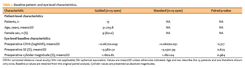
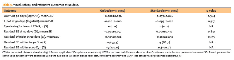
Patients were examined postoperatively on days 2, 7, 30, and 90. At each visit, UDVA, CDVA, and manifest refraction were recorded. Spherical equivalent (SE) and refractive cylinder were calculated from manifest refraction. Preoperative and 90-day baseline characteristics are summarized in table 1, early postoperative healing outcomes are presented in table 2, and visual, safety, and refractive outcomes at 90 days are presented in table 3.

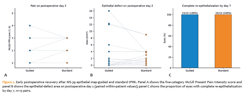
On postoperative day 2, the epithelial-defect area was measured using a millimeter caliper, and postoperative discomfort was graded using the five-category McGill Present Pain Intensity scale (1 = mild, 2 = discomforting, 3 = distressing, 4 = horrible, and 5 = excruciating). Complete reepithelialization was assessed on postoperative day 7. The exact time to epithelial closure was not recorded daily for each eye.
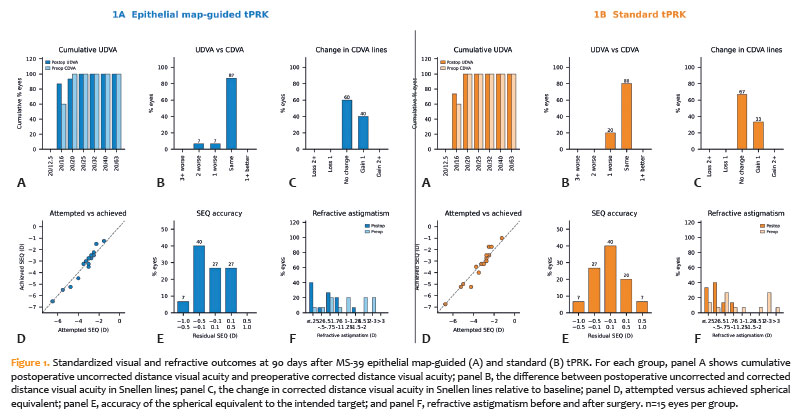
The primary outcome was UDVA on postoperative day 90. Secondary outcomes included CDVA at day 90, UDVA at day 30, residual SE and refractive cylinder at day 90, postoperative pain score and epithelial-defect area at day 2, and complete reepithelialization at day 7. Standardized refractive outcomes at 3 months are illustrated in figure 1, and early postoperative recovery is summarized in figure 2.
Statistical analysis
Statistical analyses were performed using SPSS (version 26.0; IBM Corp., Armonk, New York) and Minitab (version 21.2; Minitab LLC, State College, Pennsylvania). Given the contralateral-eye design, paired comparisons between-treatment groups were performed using the Wilcoxon signed-rank test. Within-group comparisons of preoperative and postoperative measurements were also performed using the Wilcoxon signed-rank test.
Associations between preoperative parameters and patient age were assessed using Spearman rank correlation coefficients. Continuous variables are presented as mean± standard deviation, and categorical variables are presented as counts and percentages. A p-value <0.05 was considered statistically significant. Statistical nonsignificance was not interpreted as evidence of equivalence.
RESULTS
Fifteen patients (30 eyes) completed the study and the 90-day follow-up. Baseline demographic and ocular characteristics were comparable between the two treatment groups, with no statistically significant differences in preoperative CDVA, SE, or refractive cylinder magnitude (Table 1 and Figure 1).
Early postoperative healing
No statistically significant differences in early postoperative recovery were observed between the epithelial map-guided and standard groups (Table 2 and Figure 2). On postoperative day 2, the mean pain score was 2.33±0.82 in the guided group and 2.40±0.63 in the standard group (p=0.564). The mean epithelial-defect area was 3.75±5.34 and 2.78±2.86 mm2, respectively (p=0.440). By postoperative day 7, complete reepithelialization had occurred in all eyes in both groups (100.0%).
At postoperative day 30, mean UDVA was 0.007±0.110 logMAR in the guided group and 0.000±0.093 logMAR in the standard group, with no significant between-group difference (p=0.803; Table 2).
Visual and refractive outcomes at 90 days
At postoperative day 90, both treatment approaches demonstrated favorable visual and refractive outcomes (Table 3; Figure 1). Mean UDVA was −0.080±0.056 logMAR in the epithelial map-guided group and −0.073±0.046 logMAR in the standard group; the between-group difference was not statistically significant (p=0.564). Mean CDVA was −0.100±0.000 logMAR in the guided group and −0.093±0.026 logMAR in the standard group (p=0.317).
No eye in either treatment group lost two or more lines of CDVA during follow-up.
Refractive accuracy at 90 days was high in both groups. The mean residual SE was −0.03±0.35 D in guided eyes and 0.00±0.41 D in standard eyes (p=0.831). The corresponding mean residual refractive cylinder was −0.38±0.39 D and −0.27±0.26 D, respectively (p=0.135). Fourteen of 15 guided eyes (93.3%) and 13 of 15 standard eyes (86.7%) achieved a residual SE within ±0.50 D of plano. All eyes in both groups were within ±1.00 D of plano.
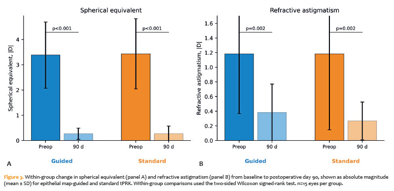
Within each treatment group, SE and refractive astigmatism decreased significantly from baseline to postoperative day 90 (Figure 3). Standardized refractive surgery outcome graphs are presented in figure 1.
DISCUSSION
In this prospective, randomized, contralateral-eye study, no statistically significant differences were observed between standard single-step tPRK planned using a population-derived epithelial profile and MS-39 epithelial thickness map-guided single-step tPRK with respect to early epithelial healing, visual outcomes, or refractive outcomes in eyes with regular corneas. Complete epithelial closure was achieved by postoperative day 7 in all eyes, and no eye lost two or more lines of CDVA. These findings do not demonstrate superiority of either treatment strategy and should not be interpreted as evidence of equivalence.
Our findings suggest that, in routine myopic eyes with regular corneas, individualization of the epithelial component of single-step tPRK using preoperative MS-39 epithelial mapping does not provide a detectable short-term clinical benefit when conventional postoperative endpoints are assessed. This finding is biologically plausible. Although epithelial thickness varies among individuals and may partially compensate for subtle stromal contour variations, standard population-based epithelial profiles appear to provide sufficiently accurate treatment planning in many eyes with regular corneas, particularly when procedures are performed using a modern, high-speed excimer laser platform and standardized surgical nomograms. Under these conditions, any incremental benefit of epithelial customization may be too small to be detected using early postoperative healing measures, conventional visual outcomes, or routine refractive endpoints.
The present findings are consistent with the most directly comparable evidence. Aramberri et al. compared standard and MS-39-customized StreamLight tPRK in 108 randomized eyes and reported no significant differences in visual, refractive, or optical quality outcomes through 6 months(9). Niknam et al.(10), using a map-adjusted approach on a different laser platform, reported a postoperative hyperopic tendency and highlighted the potential need for adjustments to treatment targets or nomograms. The present study extends this evidence by evaluating a different excimer laser platform in a contralateral-eye design and by including early postoperative pain, epithelial-defect area, and epithelial closure by day 7 as additional outcomes. Earlier trials comparing transepithelial with conventional PRK addressed the related but distinct question of epithelial removal technique rather than the potential benefit of epithelial map-guided treatment planning(2,5,18-24).
An important strength of this study is its contralateral-eye design. By assigning one eye of each patient to each treatment strategy, the influence of interindividual variability in corneal wound healing, epithelial behavior, tear-film status, pain perception, and refractive response was substantially reduced. Furthermore, all procedures were performed by a single experienced surgeon using the same excimer laser platform, the same single-step reverse-mode transepithelial technique, and the same postoperative regimen. These methodological features enhanced internal consistency and permitted a more direct comparison of the treatment-planning strategies.
The study also provides useful information regarding early postoperative recovery. Because transepithelial approaches are often associated with improved comfort and faster epithelial recovery, the absence of a measurable difference in postoperative pain or epithelial-defect area between the two groups is noteworthy. These findings suggest that, in regular corneas, the early postoperative experience may be influenced more by the use of the no-touch transepithelial technique itself than by whether epithelial removal is planned using a fixed or individualized thickness profile. Postoperative epithelial remodeling and its platform- and technique-dependent patterns have also been described following tPRK(25-29). Similarly, the finding that all eyes achieved complete reepithelialization by postoperative day 7 supports the favorable early safety profile of both approaches in this population.
At 90 days, both treatment groups demonstrated favorable visual acuity and refractive predictability. Mean residual SE was close to plano, and all eyes were within ±1.00 D of the intended correction. Although 93.3% of guided eyes and 86.7% of standard eyes were within ±0.50 D of plano, these descriptive differences cannot establish superior or equivalent refractive accuracy. In particular, the study was small and no equivalence margin was prespecified.
The findings should also be interpreted in the context of the study population. The trial included eyes with regular corneas and moderate myopia, a population in which standard transepithelial nomograms already provide favorable outcomes. Epithelial thickness customization may be more relevant in eyes exhibiting irregular epithelial compensation, higher refractive errors, asymmetric epithelial profiles, subtle corneal surface irregularities, or postsurgical remodeling. Epithelial mapping may be particularly valuable for identifying such compensatory patterns(27,28). Therefore, the absence of a detectable treatment advantage in the present cohort should not be generalized to all patients undergoing surface ablation.
This study has several limitations. First, the small sample size may have limited the ability to detect modest between-treatment differences, and no equivalence or noninferiority margin was prespecified. Second, the follow-up period was limited to 90 days and therefore did not permit assessment of longer-term epithelial remodeling or refractive stability. Third, postoperative MS-39 imaging, corneal wavefront analysis, contrast sensitivity testing, and validated patient-reported measures of optical quality were not included. Finally, the exact time to complete epithelial closure was not recorded on a daily basis. Consequently, the nonsignificant findings observed in this pilot study should not be interpreted as evidence that the two treatment-planning strategies are equivalent.
Future studies should include larger cohorts, longer follow-up periods, and more comprehensive assessments of epithelial and optical changes. In particular, postoperative epithelial mapping, corneal aberrometry, contrast sensitivity testing, and validated patient-reported outcome measures may help determine whether specific patient subgroups derive a clinically meaningful benefit from individualized epithelial thickness-guided transepithelial ablation. Studies stratifying patients according to epithelial thickness profiles, degree of myopia, corneal morphology, and epithelial-stromal compensation may also help identify populations in which customized planning is most likely to provide an advantage.
Overall, in eyes with regular corneas undergoing single-step transepithelial PRK, MS-39 epithelial thickness map-guided customization did not demonstrate a statistically detectable short-term clinical advantage over standard population-based planning. Given the pilot nature of the study, small sample size, short follow-up, and absence of a prespecified equivalence or noninferiority framework, these findings should be interpreted as an absence of demonstrated superiority rather than evidence of equivalence.
ACKNOWLEDGMENTS
The authors acknowledge Unidade de Cirurgia Ocular (UNICO) for providing access to the excimer laser used in this study.
AUTHORS’ CONTRIBUTIONS:
Significant contribution to conception and design: Caio Cezar Toledo De Conti, Pablo Felipe Rodrigues, Bernardo Kaplan Moscovici. Data acquisition: Caio Cezar Toledo De Conti, Elisabeth Brandão Guimarães. Data Analysis and interpretation: Caio Cezar Toledo De Conti, Pablo Felipe Rodrigues, Bernardo Kaplan Moscovici. Manuscript drafting: Caio Cezar Toledo De Conti, Bernardo Kaplan Moscovici. Significant intellectual content revision of the manuscript: Elisabeth Brandão Guimarães, Marizilda Rita de Andrade, Renato Giovedi Filho, Pablo Felipe Rodrigues, Bernardo Kaplan Moscovici. Final approval of the submitted manuscript: Caio Cezar Toledo De Conti, Elisabeth Brandão Guimarães, Marizilda Rita de Andrade, Renato Giovedi Filho, Pablo Felipe Rodrigues, Bernardo Kaplan Moscovici. Statistical analysis: Pablo Felipe Rodrigues, Bernardo Kaplan Moscovici. Obtaining funding: not applicable. Supervision of administrative, technical, or material support: Marizilda Rita de Andrade, Renato Giovedi Filho, Bernardo Kaplan Moscovici. Research group leadership:Bernardo Kaplan Moscovici.
REFERENCES
1. Yildirim Y, Olcucu O, Alagoz N, Agca A, Karakucuk Y, Demirok A. Comparison of visual and refractive results after transepithelial and mechanical photorefractive keratectomy in myopia. Int Ophthalmol. 2018;38(2):627-33.
2. Kaluzny BJ, Cieślinska I, Mosquera SA, Verma S. Single-step transepithelial PRK versus alcohol-assisted PRK in myopia and compound myopic astigmatism correction. Medicine (Baltimore). 2016;95(6):e1993.
3. Fattah MA, Antonios R, Arba Mosquera S, Abiad B, Awwad ST. Epithelial erosions and refractive results after single-step transepithelial photorefractive keratectomy and alcohol-assisted photorefractive keratectomy in myopic eyes: a comparative evaluation over 12 months. Cornea. 2018;37(1):45-52.
4. Buzzonetti L, Petrocelli G, Laborante A. A new transepithelial phototherapeutic keratectomy mode using the NIDEK CXIII excimer laser. J Refract Surg. 2009;25(1 Suppl):S122-4.
5. Aslanides IM, Selimis VD, Bessis NV, Georgoudis PN. A pharmacological modification of pain and epithelial healing in contemporary transepithelial all-surface laser ablation (ASLA). Clin Ophthalmol. 2015;9:685-90.
6. Arba-Mosquera S, Awwad ST. Theoretical analyses of the refractive implications of transepithelial PRK ablations. Br J Ophthalmol. 2013;97(7): 905-11.
7. Kaluzny BJ, Szkulmowski M, Bukowska DM, Wojtkowski M. Spectral OCT with speckle contrast reduction for evaluation of the healing process after PRK and transepithelial PRK. Biomed Opt Express. 2014;5(4):1089-98.
8. Rymer P, Moscovici BK, Freitas MM, Schor P, Campos M. Comparative outcomes of mechanical and transepithelial PRK on the same excimer laser: a contralateral eye study. Eur J Ophthalmol. 2025;35(3):893-901.
9. Aramberri J, Lauzirika G, Illarramendi I, Mendicute J. A 6-month follow-up comparative study of single-step transepithelial photorefractive keratectomy (Trans-PRK) using the StreamLight software with and without epithelial thickness customization. Clin Ophthalmol. 2024;18:2831-41.
10. Niknam S, Niknam J, Salouti R, Nowroozzadeh MH. Epithelial thickness map-adjusted transepithelial photorefractive keratectomy for treatment of myopic astigmatism: 12-month results. Int Ophthalmol. 2024;44(1):252.
11. Moscovici BK, Rodrigues PF, Dantas MP, Okimoto JT, Naves OS, Taguchi FM, et al. Ten-year follow-up of corneal cross-linking and refractive surface ablation in patients with asymmetric corneal topography. Indian J Ophthalmol. 2023; 71(9):3210-8.
12. Rodrigues PF, Moscovici BK, Lamazales L, Freitas MM, Gomes JÁ, Nosé W, et al. Measurement of the visual axis through two different methods: quantification and differences for measuring chord μ. Arq Bras Oftalmol. 2023;87(4):e2022-0035.
13. Colombo-Barboza GN, Colombo-Barboza MN, Colombo-Barboza LR, Colombo-Barboza MM, Colombo-Barboza FD, Lui-Netto A, et al. Vision quality questionnaire assessment in patients after topography-guided photorefractive keratectomy for irregular astigmatism secondary to radial keratotomy. Clin Ophthalmol. 2022;16:3491-3501.
14. Rios LC, Silva PG, Canamary Junior AM, Rodrigues P, Regatieri CV, Campos M, et al. Centration in refractive surgery. Arq Bras Oftalmol. 2020;83(1):76-81.
15. Rymer P, Moscovici BK, Gomes R, Couto B, Schor P, Campos M. Pain response and symptoms in photorefractive keratectomy: mechanical de-epithelization compared with transepithelial ablation. Arq Bras Oftalmol. 2022;85(2):152-7.
16. Dutra BA, Villabona-Martinez V, Wilson SE. The influence of the epithelium removal method on the outcomes of photorefractive keratectomy. J Refract Surg. 2025;41(5):e510-8.
17. Torricelli AA, Giglio VB, Garcia R, Santhiago MR, Bechara SJ, Wilson SE, et al. Photorefractive keratectomy: technical evolution, refractive outcomes, corneal wound healing response, and complications. J Refract Surg. 2024; 40(10):e754-e767.
18. Adib-Moghaddam S, Soleyman-Jahi S, Sanjari Moghaddam A, Hoorshad N, Tefagh G, Haydar AA, et al. Efficacy and safety of transepithelial photorefractive keratectomy. J Cataract Refract Surg. 2018;44(10):1267-79.
19. Ho T. Clinical outcomes of transepithelial photorefractive keratectomy performed with smart pulse technology for the correction of moderate to high myopia. J Clin Med. 2024;13(11):3058.
20. Abdel-Radi M, Shehata M, Mostafa MM, Aly MO. Transepithelial photorefractive keratectomy: a prospective randomized comparative study between the two-step and the single-step techniques. Eye (Lond). 2023;37(8):1545-52.
21. Sabhapandit S, Abdussamad A, Shaik TA, Perumalla SR. Transepithelial photorefractive keratectomy for hyperopia correction: an uncharted territory. Clin Ophthalmol. 2023;17:1497-1504.
22. Sabau A, Daas L, Behkit A, Wagenpfeil S, Langenbucher A, Ardjomand N, et al. Efficacy, safety, and predictability of transepithelial photorefractive keratectomy: meta-analysis. J Cataract Refract Surg. 2021;47(5):634-40.
23. Vakalopoulos DG, Chatzea MS, Chronopoulou K, Katsimpras M, Tsopouridou RD, Kymionis GD, et al. Corneal irregularity induced by transepithelial photorefractive keratectomy in a patient with partial limbal stem cell deficiency. Cornea. 2024;43(2):249-52.
24. Hashemi H, Alvani A, Aghamirsalim M, Miraftab M, Asgari S. Comparison of transepithelial and conventional photorefractive keratectomy in myopic and myopic astigmatism patients: a randomized contralateral trial. BMC Ophthalmol. 2022;22(1):68.
25. Weng TH, Chang YM, Lin FH, Weng ZX, Wang TW, Chen YH, et al. Investigation of corneal epithelial thickness and irregularity by optical coherence tomography after transepithelial photorefractive keratectomy. Clin Exp Optom. 2024;107(1):23-31.
26. Alshami SAJH, Elmassry AA, Elhennawy HM, Osman IM. Evaluation of corneal epithelial remodeling after conventional versus trans-epithelial photorefractive keratectomy for myopia. Alex J Med. 2024;60(1):162-70.
27. Reinstein DZ, Archer TJ, Vida RS. Epithelial thickness mapping for corneal refractive surgery. Curr Opin Ophthalmol. 2022;33(4):258-68.
28. Asroui L, Dupps WJ Jr, Randleman JB. Determining the utility of epithelial thickness mapping in refractive surgery evaluations. Am J Ophthalmol. 2022; 240:125-34.
29. Shetty R, Narasimhan R, Dadachanji Z, Patel P, Maheshwari S, Chabra A, et al. Early corneal and epithelial remodeling differences identified by OCT imaging and artificial intelligence between two transepithelial PRK platforms. J Refract Surg. 2020;36(10):678-86.
Submitted for publication:
March 16, 2026.
Accepted for publication:
August 12, 2026.
Approved by the following research ethics committee: Santa Casa de Misericórdia de São Paulo – SCMSP (CAAE: 85597424.9.0000.5479).
Data Availability Statement: The datasets generated and/or analyzed during the current study are available from the corresponding author upon reasonable request, subject to justified conditions.
Edited by
Editor-in-Chief: Newton Kara-Júnior Associate
Editor: Camila Koch
Funding: This study received no specific financial support.
Disclosure of potential conflicts of interest: The authors declare no potential conflicts of interest.

How to cite this article:
 ABO is licensed under a Creative Commons Attribution-NonComercial 4.0 Internacional.