

Arq. Bras. Oftalmol. 2026; 89 (5): 10.5935/0004-2749.2025-0332
Total: 441
Camila Matsuura Endo1; Luciene Barbosa de Sousa1; Rodrigo Antônio Brant Fernandes1,2; Mirella Millena Carmo de Andrade1; Christiane Rolim-de-Moura3
DOI: 10.5935/0004-2749.2025-0332
ABSTRACT
PURPOSE:To evaluate the short-term safety and efficacy of bovine pericardium as a patch graft in glaucoma drainage implant surgery.
METHODS: We included 10 eyes from 10 patients with glaucoma. All patients underwent glaucoma drainage implant surgery using a bovine pericardial patch graft. The pericardial membrane was trimmed to 5 × 7 mm and used to cover the tube’s scleral track. Intraocular pressure, best-corrected visual acuity, and the number of antiglaucoma medications were assessed preoperatively and postoperatively on Days 1, 7, 14, 28, 45, 60, 75, and 90. Conjunctival hyperemia and intraocular inflammation were clinically graded postoperatively. Symptoms, including foreign body sensation, itching, and tearing, as well as postoperative complications, were recorded. Surgical failure was defined as tube exposure resulting from erosion of the overlying patch graft and conjunctiva.
RESULTS: The mean patient age was 63.4±15.6 years (range, 45–87 years). The most common postoperative symptom was foreign body sensation. During follow-up, three eyes developed asymptomatic thinning of the pericardial graft overlying the tube; in one of these eyes,
the thinning progressed to tube exposure (10.0%; 95% CI, 0.3–44.5). No cases of endophthalmitis were observed.
CONCLUSIONS: In this short-term follow-up study, bovine pericardium may be a viable alternative for coverage of glaucoma drainage implants. Larger studies with longer follow-up periods are warranted to confirm these findings.
Keywords: Glaucoma; Drainage; Glaucoma drainage implants; Foreign body; Visual acuity; Intraocular pressure; Antiglaucoma agents; Conjunctiva; Pericardium; Bovine
INTRODUCTION
Glaucoma is a chronic, progressive optic neuropathy and the leading cause of irreversible blindness worldwide. It currently affects more than 70 million people globally, approximately 10% of whom are bilaterally blind. With the increasing number and proportion of older adults worldwide, the number of people with glaucoma is projected to reach 111.8 million by 2040. Glaucoma is characterized by optic disc cupping and loss of retinal ganglion cell axons, resulting in progressive visual dysfunction(1). Because elevated intraocular pressure (IOP) is the only known modifiable risk factor, treatment aims to reduce intraocular pressure through medical, laser, or surgical interventions. Despite advances in surgical management, trabeculectomy remains the gold standard for glaucoma surgery. However, it is associated with several short- and long-term complications(2,3).
Glaucoma drainage implants (GDIs) are increasingly used to manage complex cases of glaucoma in adults and children, including neovascular, aphakic, traumatic, and refractory glaucoma that is inadequately controlled with other treatment modalities(4). These devices consist of a plate connected to a tube, which may be inserted into the anterior chamber or through the pars plana in eyes undergoing vitrectomy. Currently available implants include the Ahmed glaucoma valve (New World Medical, Rancho Cucamonga, California, USA), Baerveldt glaucoma implant (Advanced Medical Optics, Santa Ana, California, USA), Molteno implant (Molteno Ophthalmic Limited, Dunedin, New Zealand), Susanna Glaucoma Drainage Device, and Paul Glaucoma Implant (Advanced Ophthalmic Innovations, Singapore, Republic of Singapore)(5,6).
Glaucoma drainage implant surgery is associated with several complications, including acute postoperative hypotony, corneal decompensation, excessive scarring of the capsule surrounding the plate that may reduce its permeability, tube or plate erosion, strabismus, and infection(4,7).
One of the most common delayed complications specific to glaucoma drainage implant surgery is tube exposure through erosion of the overlying conjunctiva. The incidence of this complication varies according to glaucoma etiology, surgical indication, and duration of follow-up. Reported rates range from 1% after primary filtering surgery at 3 years of follow-up to 5% within the first 6 months in refractory cases(8,9). Tube exposure repair was the most common intervention unrelated to IOP control among patients who received Ahmed implants during the first year of follow-up, accounting for 57.7% of surgical interventions(10).
Conjunctival erosion with tube exposure increases the risk of endophthalmitis, a potentially blinding condition, by providing a pathway for microorganisms to migrate from the ocular surface and conjunctiva into the eye(11,12). In a series of 542 eyes that received an Ahmed glaucoma valve, nine eyes (1.7%) developed endophthalmitis. Conjunctival erosion over the Ahmed glaucoma valve was present in six of these eyes(13). Several surgical approaches have been proposed to reduce the risk of tube exposure, including creation of a long scleral tunnel, use of scleral flaps, advancement of Tenon’s tissue, and placement of patch grafts over the anterior aspect of the tube shunt(9,14).
The first material used as a patch graft was glycerin-preserved donor sclera, which yielded successful results and continues to be used in some settings(15-17). To improve the safety and efficacy of tube surgery and reduce the risk of endophthalmitis, various graft materials have been proposed for placement between the tube and conjunctiva. These include autologous sclera, human donor sclera, pericardium, dura mater, cornea, human cadaveric fascia lata, buccal mucosa, amniotic membrane, autologous Tenon’s tissue, expanded polytetrafluoroethylene, and porcine small intestinal submucosa. Each patch graft has advantages and disadvantages related to surgical handling, thickness, cost, availability, and risk of exposure. The ideal material for tube coverage should be immunologically safe, biocompatible, cost-effective, readily available, and easy to handle(13,18-22).
In Brazil, the most commonly used patch grafts for tube coverage are autologous and human donor sclera. Human donor sclera has limited availability in some regions, and its use depends on eye bank availability, which may be particularly problematic in emergency situations.
Bovine pericardium has been used worldwide in cardiovascular surgery for decades, with a low incidence of complications and high durability. It has also been used in ophthalmic surgery as a safe wrapping material for hydroxyapatite orbital implants in patients undergoing enucleation for uveal melanoma(23).
Recently, a study conducted at the Department of Ophthalmology and Visual Sciences of Universidade Federal de São Paulo evaluated the use of bovine pericardium for primary pterygium treatment and reported successful results.
Processed bovine pericardium is commercially available as Ocuguard (Ocuguard Supplies, Bio-Vascular Inc., St. Paul, Minnesota, USA) and Tutoplast (Tutogen Medical GmbH, Germany; marketed in the United Kingdom by Wescott Medical Limited, Durham, UK).
Processed pericardial tissue (Tutoplast Pericardium, IOP Inc., Costa Mesa, California, USA) is commonly used as a patch graft because of its accessibility and long shelf life compared with human donor sclera(17,22).
The advantages of bovine pericardium include processing methods that enhance immunologic safety and reduce the risk of viral transmission, including HIV, as well as prion diseases. Other advantages include uniform dimensions, ease of handling, and commercial availability without dependence on an eye bank.
In Brazil, bovine pericardium is not commercially available for ophthalmic use. Although human sclera and cornea are available and donors undergo epidemiologic and serologic screening, there remains a risk of contamination because of the immunologic window period, unidentified pathogens, and reduced test sensitivity associated with postmortem blood samples. Therefore, this study aimed to evaluate the short-term safety and efficacy of bovine pericardium prepared using a novel preservation technology developed in Brazil for use in glaucoma drainage implant surgery.
METHODS
This prospective study included 10 eyes of 10 patients from the Glaucoma Section, Department of Ophthalmology and Visual Sciences, Universidade Federal de São Paulo, and Hospital Ophthal. Patients aged 18 years or older with a diagnosis of glaucoma and an indication for glaucoma drainage implant surgery were enrolled. All eyes had an IOP >21 mmHg despite receiving maximally tolerated topical medications. The surgical technique was standardized among the surgeons and is described in detail below.
The study was conducted in accordance with the tenets of the Declaration of Helsinki, and the study protocol was approved by the Ethics Committee of the university hospital. Written informed consent was obtained from all participants.
Exclusion criteria were previous glaucoma drainage implant surgery, active uveitis, and a history of intraocular tumor. Sex and ethnicity were not considered exclusion criteria. If a patient had an indication for surgery in both eyes, only the first eye undergoing surgery was included.
Preoperative data included sex, age, race, history of hypertension, diabetes, and other chronic diseases; glaucoma type; topical and oral medications; and the type and number of previous intraocular surgeries. Additional data included the type and position of the tube, preoperative and postoperative visual acuity, and IOP. At each follow-up visit (days 1, 7, 14, 28, 45, 60, 75, and 90 after surgery), signs of inflammation and ocular surface symptoms, including foreign body sensation, itching, and tearing, were recorded.
Treatment failure was defined as extrusion of the tube, specifically the segment extending between the anterior chamber and the plate.
Intraoperative and postoperative complications were recorded, including conjunctival retraction, leakage through the conjunctival incisions, postoperative alteration of the implant tract, and patch graft thinning. Reinterventions for uncontrolled IOP or other causes unrelated to tube extrusion were not considered treatment failures.
Patch graft description
The patch graft used in this study was bovine L-hydro pericardium (Figure 1). L-hydro is a processing method in which bovine pericardium is treated with polyethylene glycol and ethanol to achieve controlled extraction of antigenic substances and enhance biocompatibility. This process also reduces the inflammatory response by incorporating the nonsteroidal anti-inflammatory agent indomethacin. Its thrombogenicity is reduced by heparin, thereby contributing to the long-term durability of the pericardial tissue. The description of the L-hydro processing method is based on the manufacturer’s specifications.
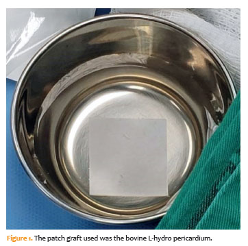
The bovine pericardium was preserved in 500 mL of physiologic saline solution containing heparin. During the perioperative period, the patch was transferred to balanced salt solution (BSS) and maintained in a hydrated state in a sterile surgical bowl for at least 5 minutes before use.
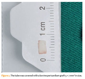
The tube was covered with a 5 × 7-mm bovine pericardial graft (Figure 2), which was secured to the sclera using a 10-0 nylon suture. Any remaining unused graft material was appropriately discarded.
Surgical technique
Under local anesthesia with sedation, a clear corneal traction suture was placed near the limbus to rotate the globe and achieve maximal exposure of the selected quadrant. A fornix-based conjunctival incision was then made to provide adequate exposure of the surgical site. When necessary, cautery was used to control bleeding.
Flow-restricted implants require priming to ensure adequate valve function. This was performed by injecting BSS through the lumen of the glaucoma drainage implant. Nonvalved GDIs require an additional procedure to restrict aqueous humor flow until fibrous encapsulation of the plate occurs. This may be achieved by placing an absorbable suture around the proximal portion of the tube.
The plate was positioned in the selected quadrant beneath the conjunctiva and secured 8–10 mm posterior to the surgical limbus with nonabsorbable sutures placed through the sclera.
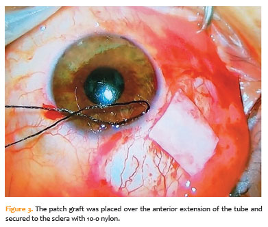
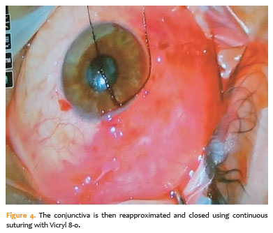
Subsequently, the tube was inserted into the anterior chamber through a scleral track created 2–3 mm posterior to the limbus. The tube was positioned either above or below the iris and secured to the sclera with 10-0 nylon sutures for additional fixation. The patch graft was then positioned over the anterior portion of the tube and secured to the sclera with 10-0 nylon sutures (Figure 3). The conjunctiva was subsequently reapproximated and closed with continuous 8-0 Vicryl sutures (Figure 4).
At the end of the procedure, subconjunctival injections of gentamicin and dexamethasone were administered in the quadrant opposite the surgical site, after which the eye was patched.
Postoperative treatment and follow-up
The postoperative regimen consisted of topical moxifloxacin eye drops for 1 week and topical prednisolone acetate 1% eye drops, which were progressively tapered over 6 weeks. Antiglaucoma medications were added as necessary according to IOP measurements. Follow-up assessments were performed on days 1, 7, 14, 30, 45, 60, 75, and 90 after surgery.
Signs of inflammation, including conjunctival hyperemia and the presence of cells and flare in the anterior chamber, as well as ocular surface symptoms such as foreign body sensation, itching, and tearing, were assessed at each visit.
At each follow-up visit, the patch graft was examined for evidence of graft melting, tube erosion, graft-related infection, or other associated intraocular complications.
Statistical analyses were descriptive because of the small sample size and exploratory nature of this case series. Continuous variables are presented as mean ± standard deviation and range, whereas categorical variables are presented as frequencies and percentages. Exact 95% confidence intervals (95% CIs) were calculated for the proportions of implant exposure and graft thinning using the Clopper–Pearson method. Given the limited sample size, formal hypothesis testing was not performed, and the findings should therefore be interpreted as exploratory
RESULTS
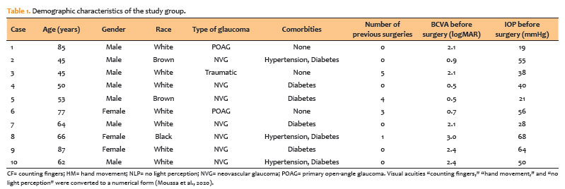
The mean age was 63.4 ± 15.6 years (range, 45–87 years). The average visual acuity was 1.61 ± 0.87 logMAR (range, 0.50–2.90). Neovascular glaucoma was the most common diagnosis, followed by primary open-angle glaucoma. The average preoperative IOP was 43.9 ± 17.4 mmHg (range, 19–68 mmHg). No patient was lost to follow-up during the study. Demographic characteristics are summarized in table 1.
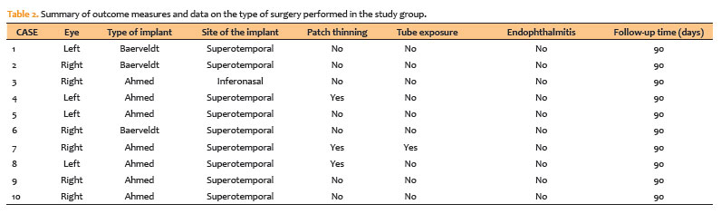
Three patients received the Baerveldt implant, and seven received the Ahmed®-PF7 valve implant. The GDI was positioned in the superotemporal or inferonasal quadrant, depending on conjunctival mobility and the presence of previous surgical conjunctival scarring. The surgical procedure was similar for the superior and inferior valves. The plate was implanted in the superotemporal quadrant in nine eyes and in the inferonasal quadrant in one eye. No intraoperative complications occurred. Data on the type of surgery performed in the study group and outcome measures are summarized in table 2.
Expected immediate postoperative findings included conjunctival hyperemia and intraocular inflammation (anterior chamber cell count/flare). Two eyes showed significant redness, and three eyes presented with mild-to-severe inflammation. Both conditions improved within 2 weeks after surgery.
The most common postoperative symptom was foreign body sensation. Two patients reported foreign body sensation during the second and third weeks after surgery.
Three patients presented with postoperative complications: one had hyphema, and two had patch thinning, with the shunt tube clearly visible through the conjunctiva but without evidence of conjunctival erosion.
One case presented with tube exposure and was classified as a failure (10.0%; 95% CI, 0.3–44.5). The time from surgery to detection of the exposure was 3 months. The patient did not have any illness at the time of exposure detection. Urgent surgical revision was performed, and a human donor scleral patch was used to cover the tube. The post-treatment course was favorable, with stable visual acuity and controlled IOP. Asymptomatic graft thinning occurred in two of 10 eyes (20.0%; 95% CI, 2.5–55.6).
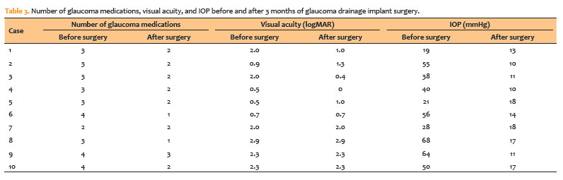
Table 3 summarizes the number of glaucoma medications, visual acuity, and IOP before and 90 days after surgery. These outcomes reflect the performance of the glaucoma drainage device. None of the patients had graft infection or developed endophthalmitis.
DISCUSSION
In our study, three patients had patch thinning, and one of them had tube exposure. All eyes evaluated with patch thinning had comorbidities and neovascular glaucoma. The patient with tube exposure was pseudophakic and had a central vein occlusion episode. There were no signs of inflammation before tube exposure. The other two patients had asymptomatic patch graft thinning. One of them had a rebound inflammatory response after discontinuation of corticosteroid treatment.
Progressive thinning of the pericardial patch graft and tube exposure in our patients may be related to systemic factors and predisposing ocular conditions, such as uveitis and previous surgery. A history of neovascular glaucoma and inflammation before tube exposure has been shown to be a risk factor for GDD exposure in a case-control study(24).
We observed one case of tube exposure in our study, corresponding to 10.0%, with a wide confidence interval (0.3–44.5). The number of tube exposures in our bovine pericardium study was higher than that reported in the literature; however, the small sample size emphasizes that this result may not be clinically relevant. Previous studies have reported tube exposure rates ranging from 2.0% to 8.5%(22). The high proportion of patients with neovascular glaucoma, a high-risk group for tube exposure, limits the generalizability of our findings.
There are many mechanisms related to tube erosion, such as mechanical forces caused by micromotion of the tube during ocular movements and blinking, tension on the conjunctiva and overlying graft, immune-mediated inflammatory processes that lead to melting of the patch graft, and ischemic damage to the conjunctiva due to compression of its small vessels(13,15,18,19,25).
Most studies suggest that Hispanic race, female sex, younger age, neovascular glaucoma, concomitant surgery, ocular surface inflammation, and topical glaucoma medications are associated with tube erosion(18,24). Some authors have shown that smoking, pseudoexfoliation glaucoma, and a history of dry eye syndrome are risk factors for tube exposure(21). Huddleston et al.(7) analyzed different risk factors for recurrent erosion after repair and identified Afro-descendence, diabetes mellitus, a high number of glaucoma medications before shunt implantation, a history of multiple glaucoma laser procedures, and a combination of initial aqueous shunt implantation with another surgery as risk factors.
Glaucoma drainage implant surgery is typically performed in the superotemporal quadrant because there is more space for the GDI and an absence of oblique muscle fibers, which minimizes the risk of diplopia(5). One patient had the GDI placed in the inferonasal quadrant because of silicone oil in the posterior segment. This patient had a good result, with no complications and well-controlled IOP 3 months after surgery.
There is some evidence relating the position of the implant to tube exposure. Some authors have postulated that inferior quadrant implantation leads to greater tube exposure and erosion over the plate, which may be related to limited space within the inferior fornix and less conjunctiva available to cover the implant, thereby possibly increasing the rate of wound dehiscence(26).
We did not observe an association between inferior placement of the tube and exposure; thus, only one patient had a GDI in the inferior quadrant.
GDIs are an important surgical option for the treatment of refractory glaucoma and complex cases in which other procedures are not possible(27).
Bovine pericardium offers several advantages, including a processing method that enhances immunologic safety and reduces the risk of viral transmission, such as HIV, and prion diseases, eliminating the need for an eye bank.
Until now, bovine pericardium has not been available for use in ophthalmic surgery in Brazil.
Limitations of the present study include the small sample size and limited follow-up. The findings are not statistically representative of the entire population; however, given the small number of patients, the use of bovine pericardium could be considered preliminary evidence that it may be an alternative to other graft materials.
This is the first study of bovine pericardium in GDI surgery in Brazil and represents an initial step toward its use in glaucoma surgery. Bovine pericardium can already be produced in various thicknesses and sizes. Using a thicker bovine pericardium patch in GDI surgery might better protect the conjunctiva from erosion, and surgeons might individualize the thickness or number of patch layers according to the patient’s risk factors at the time of surgery.
A study by Lankaranian et al.(28) suggested that double-thickness processed pericardium results in lower rates of erosion than single-thickness grafts, suggesting that this approach could be investigated in further studies in this area.
In conclusion, processed bovine pericardium used in GDI surgery showed encouraging preliminary results in this small case series. Larger studies with longer follow-up periods and comparative designs are required to better establish its safety and effectiveness.
ACKNOWLEDGMENTS
This study was supported by Labcor, which supplied the bovine pericardium used in the study.
AUTHORS’ CONTRIBUTIONS:
Significant contribution to conception and design: Camila Matsuura Endo, Luciene Barbosa Sousa, Christiane Rolim de Moura. Data Acquisition: Camila Matsuura Endo, Luciene Barbosa Sousa, Rodrigo Antonio Brant Fernandes, Mirella Millena Carmo de Andrade, Christiane Rolim de Moura. Data Analysis and interpretation: Camila Matsuura Endo, Luciene Barbosa Sousa, Christiane Rolim de Moura. Manuscript Drafting: Camila Matsuura Endo, Luciene Barbosa Sousa, Rodrigo Antonio Brant Fernandes, Mirella Millena Carmo de Andrade, Christiane Rolim de Moura. Significant intellectual content revision of the manuscript: Camila Matsuura Endo, Luciene Barbosa Sousa, Christiane Rolim de Moura. Final approval of the submitted manuscript: Camila Matsuura Endo, Luciene Barbosa Sousa, Rodrigo Antonio Brant Fernandes, Mirella Millena Carmo de Andrade, Christiane Rolim de Moura. Statistical analysis: Camila Matsuura Endo, Christiane Rolim de Moura. Obtaining funding: not applicable. Administrative, technical, or material support supervision: Camila Matsuura Endo, Luciene Barbosa Sousa, Rodrigo Antonio Brant Fernandes, Mirella Millena Carmo de Andrade, Christiane Rolim de Moura. Research group leadership: Luciene Barbosa Sousa, Christiane Rolim de Moura.
REFERENCES
1. Kang JM, Tanna AP. Glaucoma. Med Clin North Am. 2021;105(3):493-510.
2. Morgan WH, Yu DY. Surgical management of glaucoma: a review. Clin Exp Ophthalmol. 2012;40(4):388-99.
3. Lim R. The surgical management of glaucoma: a review. Clin Exp Ophthalmol. 2022;50(2):213-31.
4. Patel S, Pasquale LR. Glaucoma drainage devices: a review of the past, present, and future. Semin Ophthalmol. 2010;25(5-6):265-70.
5. Schwartz KS, Lee RK, Gedde SJ. Glaucoma drainage implants: a critical comparison of types. Curr Opin Ophthalmol. 2006;17(2):181-9.
6. Moschos MM, Nitoda E, Gouliopoulos N, Damaskos C, Laios K, Bagkli E, et al. The choice of drainage device in complicated glaucomas: comparing Ahmed and Baerveldt implants. In Vivo. 2019;33(3):911-6.
7. Giovingo M. Complications of glaucoma drainage device surgery: a review. Semin Ophthalmol. 2014;29(5-6):397-402.
8. Gedde SJ, Feuer WJ, Lim KS, Barton K, Goyal S, Ahmed IIK, et al.; Primary Tube Versus Trabeculectomy Study Group. Treatment outcomes in the primary tube versus trabeculectomy study after 3 years of follow-up. Ophthalmology. 2020;127(3):333-45.
9. Sheha H, Tello C, Al-Aswad LA, Sayed MS, Lee RK. Outcomes of the shunt tube exposure prevention study: a randomized clinical trial. Ophthalmol Glaucoma. 2019;2(6):392-401.
10. Kirmaci Kabakci A, Solmaz B, Basarir B, Kemer Atik B, Yasar T. Non-intraocular pressure-related revision surgeries after Ahmed glaucoma valve (AGV) implantation in refractory glaucoma. Int Ophthalmol. 2021;41(10):3533-8.
11. Islam YE, Blake CR, Gibran SK. Management of endophthalmitis related to glaucoma drainage devices: review of the literature and our experience. Eye (Lond). 2021;35(7):1850-8.
12. Wentzloff JN, Grosskreutz CL, Pasquale LR, Walton DS, Chen TC. Endophthalmitis after glaucoma drainage implant surgery. Int Ophthalmol Clin. 2007;47(2):109-15.
13. Nardi M, Maglionico MN, Posarelli C, Figus M. Managing Ahmed glaucoma valve tube exposure: surgical technique. Eur J Ophthalmol. 2021;31(2):778-81.
14. Singh M, Chew PT, Tan D. Corneal patch graft repair of exposed glaucoma drainage implants. Cornea. 2008;27(10):1171-3.
15. Tsoukanas D, Xanthopoulou P, Charonis AC, Theodossiadis P, Kopsinis G, Filippopoulos T. Heterologous, fresh, human donor sclera as patch graft material in glaucoma drainage device surgery. J Glaucoma. 2016;25(7):558-64.
16. Smith MF, Doyle JW, Ticrney JW, Jr. A comparison of glaucoma drainage implant tube coverage. J Glaucoma. 2002;11(2):143-7.
17. Wigton E, C Swanner J, Joiner W, Feldman A, McGwin G Jr, Huisingh C, et al. Outcomes of shunt tube coverage with glycerol preserved cornea versus pericardium. J Glaucoma. 2014;23(4):258-61.
18. Bains U, Hoguet A. Aqueous drainage device erosion: a review of rates, risks, prevention, and repair. Semin Ophthalmol. 2018;33(1):1-10.
19. Oana S, Vila J. Tube exposure repair. J Curr Glaucoma Pract. 2012;6(3):139-42.
20. Lind JT, Shute TS, Sheybani A. Patch graft materials for glaucoma tube implants. Curr Opin Ophthalmol. 2017;28(2):194-8.
21. Thakur S, Ichhpujani P, Kumar S. Grafts in glaucoma surgery: a review of the literature. Asia Pac J Ophthalmol (Phila). 2017;6(5):469-76.
22. van Hoefen Wijsard M, Haan M, Rietveld E, van Rijn LJ. Donor sclera versus bovine pericardium as patch graft material in glaucoma implant surgery and the impact of a drainage suture. Acta Ophthalmol. 2018;96(7):692-8.
23. Gupta M, Lyon F, Singh AD, Rundle PA, Rennie IG. Bovine pericardium (Tutopatch) wrap for hydroxyapatite implants. Eye (Lond). 2007;21(4):476-9.
24. Thompson AC, Manjunath V, Muir KW. Risk factors for earlier reexposure of glaucoma drainage devices. J Glaucoma. 2017;26(12):1155-60.
25. Huddleston SM, Feldman RM, Budenz DL, Bell NP, Lee DA, Chuang AZ, et al. Aqueous shunt exposure: a retrospective review of repair outcome. J Glaucoma. 2013;22(6):433-8.
26. Trubnik V, Zangalli C, Moster MR, Chia T, Ali M, Martinez P, et al. Evaluation of risk factors for glaucoma drainage device-related erosions: a retrospective case-control study. J Glaucoma. 2015;24(7):498-502.
27. Mercieca K. Comparison of glaucoma drainage implants. Ophthalmologie. 2023;120(4):372-7.
28. Lankaranian D, Reis R, Henderer JD, Choe S, Moster MR. Comparison of single thickness and double thickness processed pericardium patch graft in glaucoma drainage device surgery: a single surgeon comparison of outcome. J Glaucoma. 2008;17(1):48-51.
Submitted for publication:
February 13, 2026.
Accepted for publication:
August 12, 2026.
Approved by the following research ethics committee: Universidade Federal de São Paulo (CAAE: 26118019.0.0000.5505).
Data Availability Statement: The datasets generated and/or analyzed during the current study are included in the manuscript.
Edited by
Editor-in-Chief: Newton Kara-Júnior
Associate Editor: Heloisa Russ
Disclosure of potential conflicts of interest: The authors declare no potential conflicts of interest.

How to cite this article:
 ABO is licensed under a Creative Commons Attribution-NonComercial 4.0 Internacional.

