

Arq. Bras. Oftalmol. 2026; 89 (5): 10.5935/0004-2749.2025-0385
Total: 416
Pablo Felipe Rodrigues1; Felipe A. Casseb dos Santos2; Bernardo Kaplan Moscovici1,3,4; Renato Ambrósio Jr.5; Mauro Campos1; Walton Nose1
DOI: 10.5935/0004-2749.2025-0385
ABSTRACT
PURPOSE:To investigate tomographic and biomechanical screening artifacts associated with primary pterygium that may mimic keratoconus-like patterns before and after surgical excision.
METHODS: Twenty-five eyes with primary pterygium underwent Scheimpflug corneal tomography and dynamic corneal response analysis before surgery and 3 months after excision. Tomographic and biomechanical (parameters were compared using paired analyses. The proportions of eyes exceeding established screening thresholds (Tomographic Biomechanical Index ≥0.29; Corvis Biomechanical Index ≥0.5) were compared using McNemar’s test.
RESULTS: BAD-D decreased significantly after surgery (1.69±1.22 vs. 1.36±0.90; Wilcoxon, p=0.027). Pentacam irregularity indices also improved significantly, including ISV (51.16±44.21 vs. 36.88±42.94; p<0.001) and IVA (0.39±0.29 vs. 0.30±0.32; p=0.006). No significant changes were observed in maximum keratometry, corneal astigmatism. The mean CBI decreased significantly (0.494±0.353 vs. 0.384±0.298; p=0.048), and the proportion of CBI-positive eyes (≥0.5) declined from 48% (12/25) to 28% (7/25). In contrast, the mean tomographic Biomechanical Index did not change remained unchanged (0.519±0.313 vs. 0.534±0.323; p=0.909), and the proportion of Tomographic Biomechanical Index-positive eyes (≥0.29) remained 76% (19/25). The number of topographic keratoconus classification positive eyes decreased from 8 (32%) to 3 (12%). Integrated Radius showed no significant postoperative change (p=0.131).
CONCLUSIONS: Primary pterygium was associated with tomographic and biomechanical screening abnormalities that mimicked keratoconus-like patterns. Following surgical excision, anterior tomographic parameters improved, and the proportion of Corvis biomechanical index-positive eyes decreased. In contrast, Tomographic Biomechanical Index remained elevated in most eyes, and stiffness-related biomechanical parameters showed no significant change. These findings demonstrate a differential postoperative response among screening indices and suggest that pterygium-induced anterior corneal surface distortion may increase false-positive keratoconus screening rates, particularly for Tomographic Biomechanical Index, which may remain elevated even after lesion removal.
Keywords: Pterygium; Cornea; Corneal topography; Biomechanical phenomena; Tomography; Keratoconus; Refractive surgical procedures
INTRODUCTION
Pterygium is a fibrovascular growth of the conjunctiva that spreads to the cornea, usually from the nasal limbus. Pathogenesis is characterized by complex interactions between genetic, inflammatory, and environmental factors, with chronic ultraviolet (UV) exposure playing a prominent role(1,2). Although pterygium is typically asymptomatic in its early stages, it can cause a variety of visual disturbances, particularly as it progresses into the central cornea. These include irregular astigmatism, decreased visual acuity, and ocular surface instability, which frequently necessitate surgical excision(3-5).
Topographic changes caused by pterygium, such as flattening of the horizontal meridian and increased corneal asymmetry, have been well documented and tend to resolve after surgery(6,7). However, its influence on corneal biomechanics has received relatively little attention. Corneal biomechanics refers to the tissue’s intrinsic viscoelastic properties, which contribute to overall ocular structural integrity and are required to maintain corneal shape and optical quality. With the introduction of non-invasive biomechanical devices like the Corvis ST (Oculus GmbH, Wetzlar, Germany), it is now possible to assess the cornea’s biomechanical behavior in vivo using a variety of dynamic deformation parameters. This is especially important because integrated tomographic biomechanical indices are increasingly being used for ectasia screening and may be prone to surface-driven artifacts from ocular surface lesions(8).
Among the indices measured by this technology, the Corvis Biomechanical Index (CBI) and the Tomographic Biomechanical Index (TBI) are widely used for early detection of ectatic disease, particularly keratoconus. TBI combines tomographic data from Scheimpflug imaging with biomechanical response metrics to produce a sensitive indicator for detecting subclinical keratoconus(9-11). However, both indices may be influenced by factors other than true biomechanical instability, such as anterior surface irregularities caused by ocular surface lesions(12,13).
Previous research has shown that pterygium can resemble ectatic patterns on corneal topography and tomography, potentially leading to false-positive screening results and misinterpretation of preoperative measurements(14-16). More recent evidence shows that pterygium excision improves optical quality metrics, such as higher-order aberrations on both the anterior and posterior corneal surfaces, after pterygium excision(17-24). Furthermore, prospective data show that pterygium excision can affect keratometry and other measurements over time, emphasizing the importance of postoperative reassessment(16). However, it is unclear whether such imaging artifacts also apply to biomechanical screening indices and to what extent these changes revert after lesion excision(17-23).
The purpose of this study was to determine whether tomographic and biomechanical screening abnormalities associated with primary pterygium decrease after surgical excision as well as and whether different categories of indices (anterior surface tomography, composite AI-based screening indices, and stiffness-related parameters) exhibit distinct postoperative response patterns. We hypothesized that pterygium-related abnormalities in keratoconus screening indices are primarily caused by anterior surface distortion rather than by intrinsic stromal biomechanical weakness. A better understanding of this dynamic is critical for increasing diagnostic accuracy and avoiding misinterpretations of keratoconus screening tools in eyes with anterior segment pathology.
METHODS
Study design and ethical considerations
This was a prospective, single-center, observational study carried out at a tertiary ophthalmology hospital. The study followed the Declaration of Helsinki. This study was approved by the Research Ethics Committee (Approval No. 7.791.010; CAAE: 87411125.7.0000.5479). All participants provided written informed consent before enrollment.
Patient selection
Patients with primary nasal pterygium were enrolled sequentially. The inclusion criteria were age 18 or older, unilateral pterygium extending at least 2 mm onto the corneal surface, and no history of ocular surgery or ocular surface disease. Patients were excluded if they had experienced ocular trauma, a keratoconus diagnosis, active inflammation, dry eye syndrome, or used contact lenses within 15 days of the evaluation. The study included 25 eyes from 25 eligible patients. All patients underwent a comprehensive ophthalmologic examination, which included best corrected visual acuity, slit-lamp biomicroscopy, and intraocular pressure measurements. The horizontal extension of the pterygium onto the cornea was measured under the slit lamp with a caliper aligned with the lesion’s limbus and apex.
Imaging protocols
All patients underwent corneal imaging with the Pentacam HR and Corvis ST before and three months after surgery. Tomographic data were collected with the Pentacam HR, a rotating Scheimpflug camera system that measures anterior and posterior elevation, corneal curvature, pachymetric progression, and risk indices. Three scans were taken per eye, and only measurements with an “OK” quality index were considered. The highest-quality scan from each patient was used for analysis. The parameters recorded were flat and steep simulated keratometry (K1 and K2), maximum keratometry (Kmax), anterior and posterior elevation maps, corneal astigmatism, thinnest pachymetric point, BAD-D, and TKC.
Biomechanical measurements were taken with the Corvis ST, which employs a high-speed Scheimpflug camera that records corneal deformation in response to a calibrated air pulse at 4,300 frames per second. The deformation amplitude, peak distance, applanation times and lengths, radius at highest concavity, and composite indices such as the CBI and TBI were all investigated. The study also included five advanced Corvis parameters: DA Ratio, integrated Radius Ambrosio Relational ARTh, SP-A1, and SSI. Each patient underwent three consecutive scans under consistent illumination, with the highest-quality scan being used for analysis.
Surgical technique
All procedures were carried out by a single experienced surgeon using the Moscovici dissection technique. A 30-gauge needle was used to inject a subepithelial air bubble into the superior bulbar conjunctiva while under topical anesthesia, allowing for the atraumatic separation of the autograft. The pterygium’s body and head were removed as well as the Tenon’s capsule beneath them. Following hemostasis, the conjunctival graft was harvested, oriented correctly, and secured to the scleral bed with fibrin glue (Tisseel, Baxter). No sutures used(2). For 4 weeks following surgery, patients were given topical moxifloxacin and dexamethasone four times per day.
Follow-up and outcome measures
At 3 months, all patients returned for their postoperative evaluations. The 3-month time point was chosen because keratometric and topographic parameters have been shown to stabilize in most eyes after pterygium excision with conjunctival autografting at this time(4,16). The primary outcome was a change in corneal tomographic and biomechanical indices from baseline. Secondary analyses included a comparison of the proportion of eyes that exceeded established TBI and CBI screening thresholds prior to and following surgery.
Statistical analysis
The statistical analyses were done with SPSS version 26.0 (IBM Corp., Armonk, NY, USA) and Minitab version 21.2 (Minitab, LLC, State College, PA, USA). The Kolmogorov-Smirnov test was used to determine whether continuous variables were normal. For paired comparisons, the Wilcoxon signed-rank test was used as the primary analysis, with the paired Student’s t-test included for transparency. In addition to paired comparisons of continuous variables, the proportions of eyes that exceeded established keratoconus screening thresholds were determined before and after surgery. Eyes were classified as TBI-positive when TBI ≥0.29 and as CBI-positive when CBI ≥0.5, according to previously established cutoffs(13). The proportions at each time point were compared with McNemar’s test. The proportion of eyes with topographic keratoconus classification (TKC) other than 0 was also compared at different time points. Statistical significance was defined as p<0.05.
Sample size estimation
Using a paired pre-post design, a two-sided α of 0.05, and 80% power, we estimated that 22 eyes are needed to detect a moderate within-eye effect (Cohen’s dz ≈ 0.65) in key screening metrics (e.g., BAD-D and TBI). To account for potential losses and poor image quality, we intended to include 25 eyes. This sample size was deemed appropriate for an exploratory prospective study focusing on changes within the eye following pterygium excision.
RESULTS
Preoperative characteristics
The study included 25 eyes from 25 patients (14 females and 11 males) diagnosed with primary nasal pterygium. The average patient age was 54.8±9.1 yr (range: 41-70 yr). Clinical measurements showed that all pterygia extended at least 2.0mm onto the cornea. There were no cases of reports of recurring pterygium, severe dry eye disease, or previous ocular surgery.
Baseline corneal tomography frequently revealed localized anterior surface distortion, near the pterygium, often accompanied by asymmetric curvature patterns. Several eyes had tomographic profiles similar to early keratoconus, including elevated BAD-D and increased keratoconus screening indices (particularly TBI). However, no participant met the clinical criteria for definitive keratoconus, which were defined in this study as the presence of slit-lamp biomicroscopic signs (such as Vogt striae, Fleischer ring, or apical scarring)(11-14).
Tomographic and biomechanical changes after surgery
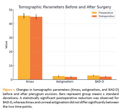
Three months after pterygium removal, anterior corneal tomography parameters showed different responses. BAD-D levels decreased significantly from 1.69±1.22 to 1.36±0.90 (Wilcoxon p=0.027). Pentacam improved irregularity indices, with ISV decreasing from 51.16±44.21 to 36.88±42.94 (p<0.001) and IVA from 0.39±0.29 to 0.30±0.32 (p=0.006). In contrast, Kmax (46.02±2.13 to 46.10±1.93 D; p=0.565) and corneal astigmatism (2.66±4.17 to 2.53±4.59 D; p=0.821) remained stable. In terms of topographic keratoconus classification, 8 of 25 eyes (32%) were TKC-positive (any category other than zero) preoperatively, compared to 3 of 25 (12%) postoperatively (McNemar test, p=0.125; Table 1 and Figure 1).
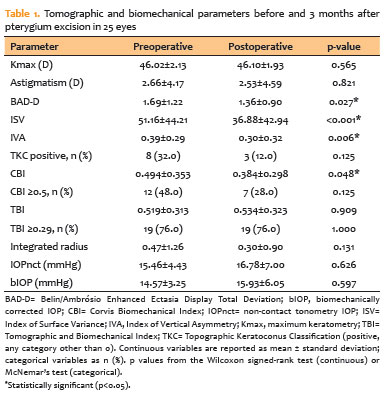
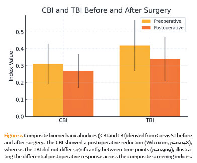
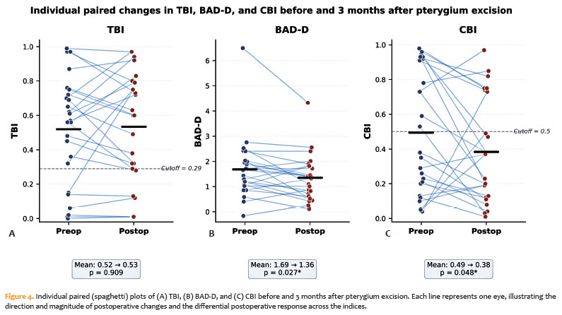
Corvis ST composite screening indices exhibited a differential response pattern. The mean CBI decreased from 0.494±0.353 to 0.384±0.298 (Wilcoxon p=0.048). The proportion of eyes classified as CBI-positive (≥0.5) decreased from 48.0% (12/25) preoperatively to 28.0% (7/25) after surgery (McNemar test, p=0.125). In contrast, mean TBI did not change significantly (0.519±0.313 to 0.534±0.323; p=0.909), and the proportion of eyes classified as TBI-positive (≥0.29) remained at 76% (19/25) before and after surgery (McNemar test, p=1.000; Table 1 and Figures 2-4). Of the 19 eyes classified as TBI-positive preoperatively, 18 (94.7%) remained TBI-positive postoperatively (“persistent TBI elevation”); only one eye normalized, and one more eye that was TBI-negative preoperatively became TBI-positive postoperatively. Figure 4 shows individual paired data for these indices as well as BAD-D.
Parameters intended to reflect intrinsic corneal stiffness remained statistically unchanged following excision. Integrated Radius did not change significantly between time points (0.47±1.26 to 0.30±0.90; Wilcoxon p=0.131). Tonometric measurements remained stable, with IOPnct (15.46±4.43 to 16.78±7.00 mmHg; p=0.626) and bIOP (14.57±3.25 to 15.93±6.05 mmHg; p=0.597) not changing significantly. Other stiffness-related parameters (DA Ratio, ARTh, SP-A1, and SSI) had no significant postoperative changes (Table 1).
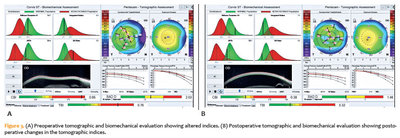
Figure 3 shows a representative case before and after pterygium excision.
Subgroup analysis
Because horizontal pterygium extension in millimeters was not consistently registered in the standardized study database in a way that would allow reliable retrospective extraction for all 25 eyes, planned correlation analyses between pterygium extension and tomographic/biomechanical changes were unable to be performed, which was acknowledged as a study limitation. Eyes that remained TBI-positive postoperatively (n=18) had a mean preoperative TBI of 0.68±0.20, compared to 0.16±0.14 in eyes that remained TBI-negative (n=5); the single eye that normalized had a preoperative TBI of 0.48, while the single eye that became newly TBI-positive had a preoperative TBI of 0.15.
Complications
There were no complications either during or after the procedure. At the 3-month visit, all conjunctival grafts remained stable with no recurrences, graft retraction, or epithelial defects. There was no additional surgical or medical intervention required for any of the patients.
DISCUSSION
This prospective study demonstrates that primary pterygium can cause significant changes in corneal imaging and biomechanical screening results, mimicking patterns commonly associated with ectatic disease(5,8). Importantly, the current data show a differential postoperative response among screening indices: anterior tomographic metrics such as BAD-D and Pentacam irregularity indices, as well as the proportion of eyes classified as CBI-positive decreased after surgical excision, whereas TBI remained elevated in the majority of eyes. This pattern emphasizes the importance of careful diagnostic timing and interpretation when keratoconus screening is performed in the presence of ocular surface lesions as well as the fact that postoperative reassessment does not always reclassify previously screening-positive eyes(3,15,20).
In our cohort, pterygium removal resulted in significant postoperative reductions in BAD-D and Pentacam irregularity indices (ISV and IVA) as well as a numerical decrease in the proportion of eyes classified as TKC-positive (from 32% to 12%). However, due to the limited sample size(1,4-6), this did not reach statistical significance. Mean Kmax and corneal astigmatism did not differ between time points, which is likely due to the wide preoperative variability in these parameters in eyes with pterygium and the small sample size, rather than the lack of clinical change. These results are consistent with previous research showing that pterygium distorts anterior corneal curvature and causes irregular astigmatism(1,4,6). The current study provides a clinically relevant biomechanical perspective by investigating how surface-driven distortion affects Corvis-derived keratoconus screening indices in vivo. Our findings support the hypothesis that anterior surface distortion contributes to apparent abnormalities in keratoconus screening indices in eyes with pterygium, but they also show that this contribution varies across indices(5,9).
One of the most clinically relevant findings was the difference in postoperative behavior between the two AI-derived composite screening indices. Using the previously proposed positivity threshold of CBI ≥0.5(13), the proportion of eyes classified as CBI-positive decreased from 48% preoperatively to 28% postoperatively, and the mean CBI decreased significantly (Wilcoxon signed-rank test, p=0.048). In contrast, the TBI showed no change in its mean value or the proportion of eyes above the established positivity threshold of TBI ≥0.29: 19 of 25 eyes (76%) were classified as TBI-positive both before and after surgery, and 18 of 19 preoperatively TBI-positive eyes remained TBI-positive at 3 months(11-13). This pattern suggests that the TBI, which integrates tomographic and biomechanical data using artificial intelligence algorithms, may be especially sensitive to abnormalities that cannot be fully resolved by removing the visible pterygium itself. While this high sensitivity is useful for detecting early ectatic disease, it may also increase the likelihood of false-positive keratoconus screening rates in eyes with a history of nonectatic anterior segment abnormalities, such as pterygium, even after lesion removal(5,11,12). The CBI, which becomes dynamic deformation response metrics with corneal thickness profiles, may be more sensitive to postoperative reductions in anterior surface distortion than the TBI. However, in this small sample(13,18) the McNemar comparison of CBI-positivity rates failed to reach statistical significance (p=0.125).
Our findings also show that the deeper corneal stroma is stable(8,22). No significant postoperative changes were observed in parameters such as DA ratio, integrated radius, ARTh, SP-A1, or SSI, which reflect intrinsic biomechanical properties and may be less affected by surface shape or epithelial remodeling(14-16). In particular, SSI, which is derived from finite element modeling and is intended to be less dependent on corneal geometry, did not differ between time points, supporting the hypothesis that the lesion does not impair intrinsic stromal stiffness(10,14). Taken together, the postoperative reduction in anterior surface tomographic abnormalities, the partial decrease in CBI-positivity, the persistent elevation of TBI in most eyes, and the stability of stiffness-related parameters point to a heterogeneous origin of the keratoconus-like screening pattern in eyes with pterygium: a component related to anterior surface and geometric irregularity, which is more readily attenuated after surgery, and a residual component captured by the TBI that may persist after lesion removal(14-16).
These findings have significant implications for clinical decision-making(15,20). Clinicians should be aware that anterior surface lesions such as pterygium can produce tomographic and biomechanical screening profiles similar to early keratoconus, potentially leading to diagnostic uncertainty or unnecessary exclusion from corneal procedures based solely on keratoconus screening(11-13). Our data show that postoperative reductions in composite screening indices do not always imply complete normalization in every eye: 18 of 19 TBI-positive eyes (94.7%) remained TBI-positive at 3 months. Postoperative TBI and CBI values should not be interpreted in isolation, but rather in conjunction with slit-lamp findings, full tomographic context, epithelial remodeling (if available), and long-term stability. In eyes with residual borderline values, a follow-up visit may be necessary before using keratoconus screening results to guide refractive or cataract surgery planning(19,20). It is unclear whether a longer follow-up (for example, 6 months) would result in a further reduction in screening indices, and future research is needed. Our protocol included a reassessment at 3 months, which is when most eyes’ keratometric and topographic parameters are reported to be stable following pterygium excision(16).
While our study provides new evidence in this area, there are some limitations to consider. First, the lack of pre-pterygium measurements is a significant methodological limitation. Because corneal imaging before the development of pterygium was not available, the postoperative changes observed in this study should not be interpreted as definitive evidence of true reversibility or return to a predisease baseline. Rather, they indicate that pterygium-associated tomographic and biomechanical screening abnormalities decrease slightly after surgical excision, with a variable pattern across indices. Second, horizontal pterygium extension in millimeters was not consistently recorded in the standardized study database in a way that would allow reliable retrospective extraction for all 25 eyes, preventing planned correlation analyses between pterygium size and the magnitude of tomographic or biomechanical changes; this analysis will be included prospectively in future studies on this topic. Third, while an a priori sample size estimation suggested including 25 eyes for paired pre-post comparisons, the sample size was small, limiting subgroup analyses and the statistical power for binary comparisons (e.g., McNemar comparisons of CBI-positivity and TKC reclassification rates); thus, findings with marginal p-values should be interpreted with caution. A postoperative reassessment was only performed after 3 months; longer follow-up would be beneficial to confirm whether residual TBI elevation persists or decreases over time. The lack of epithelial thickness mapping (e.g., OCT epithelial maps) limits our ability to distinguish between epithelial compensation from stromal effects and better understand the mechanisms underlying the partial tomographic improvement. Furthermore, the contralateral eyes were not used as internal controls(4,5,7,19,25).
Despite these limitations, the convergence of a postoperative reduction in anterior tomographic indices with the stability of deeper biomechanical parameters lends credence to the hypothesis that pterygium can mimic keratoconus-like screening patterns, at least in part, by distorting the anterior surface without compromising stromal material stiffness(14-16). At the same time, the persistence of TBI elevation in the majority of preoperatively positive eyes suggests that not all of the screening abnormalities seen in these patients are due to a directly reversible surface artifact. This distinction is clinically significant: keratoconus screening results in eyes with pterygium should be interpreted in context, and a positive screening result after lesion removal should not be interpreted solely as residual artifact(11-13). Future studies that include epithelial thickness mapping, fellow-eye controls, larger cohorts stratified by pterygium size, and longer follow-up are needed to refine the response patterns described here and their clinical implications.
Primary pterygium has been linked to tomographic and biomechanical screening abnormalities that may resemble keratoconus-like patterns, contributing to false-positive keratoconus screening rates in eyes with this lesion(5,11-13). Following surgical excision, anterior tomographic parameters (BAD-D, ISV, IVA) and the proportion of CBI-positive eyes decreased. In contrast, TBI levels remained elevated in 18 of 19 preoperative TBI-positive eyes, and stiffness-related parameters (DA ratio, integrated radius, ARTh, SP-A1, and SSI) did not change significantly(14-16). These findings show a difference in postoperative response among screening indices and suggest that pterygium-related anterior surface distortion can contribute to keratoconus-like screening patterns without necessarily reflecting intrinsic stromal biomechanical weakening, and that postoperative TBI values should not be interpreted in isolation. Post-excisional reassessment may improve diagnostic interpretation in this setting, while also acknowledge that persistent TBI elevation can occur, necessitating an integrated clinical and tomographic context(19,20,26-30).
AUTHORS’ CONTRIBUTIONS:
Significant contribution to conception and design: Pablo Felipe Rodrigues; Bernardo Kaplan Moscovici; Renato Ambrósio Jr.; Mauro Campos; Walton Nose. Data acquisition: Pablo Felipe Rodrigues; Felipe A. Casseb dos Santos. Data analysis and interpretation: Pablo Felipe Rodrigues; Renato Ambrósio Jr.; Bernardo Kaplan Moscovici. Manuscript drafting: Pablo Felipe Rodrigues; Felipe A. Casseb dos Santos; Bernardo Kaplan Moscovici. Significant intellectual content revision of the manuscript: Renato Ambrósio Jr.; Mauro Campos; Walton Nose; Bernardo Kaplan Moscovici. Final approval of the submitted manuscript: Pablo Felipe Rodrigues; Felipe A. Casseb dos Santos; Bernardo Kaplan Moscovici; Renato Ambrósio Jr.; Mauro Campos; Walton Nose. Statistical analysis: Pablo Felipe Rodrigues; Renato Ambrósio Jr. Obtaining funding: not applicable. Supervision of administrative, technical, or material support: Mauro Campos; Walton Nose; Bernardo Kaplan Moscovici. Research group leadership: Bernardo Kaplan Moscovici.
REFERENCES
1. Maheshwari S. Pterygium-induced corneal refractive changes. Indian J Ophthalmol. 2007;55(5):383-6.
2. Moscovici BK, Rodrigues PF, Freitas MM, E Silva FB, Felberg S, Dantas PE. Donor graft harvesting with air in pterygium surgery. Eur J Ophthalmol. 2022;32(5):3103-9.
3. Tang Y, Qian D, Wei L, Du Y, Qiu X, Lu Y, et al. Influences of the three-dimensional parameters of pterygium on corneal astigmatism and the intraocular lens power calculation. Sci Rep. 2020;10(1):5017.
4. Kam KW, Kuan TA, Belin MW, Young AL. Long-term stability of keratometry, Scheimpflug-derived true net power, and total corneal refractive power after primary pterygium excision. Cornea. 2017;36(11):1358-63.
5. Vanathi M, Goel S, Ganger A, Agarwal T, Dada T, Khokhar S. Corneal tomography and biomechanics in primary pterygium. Int Ophthalmol. 2018;38(2):663-71.
6. Yoon CH, Seol BR, Choi HJ. Effect of pterygium on corneal astigmatism, irregularity and higher-order aberrations: a comparative study with normal fellow eyes. Sci Rep. 2023;13(1):7328.
7. Wiącek MP, Kuśmierz-Wojtasik M, Kowalska B, Machalińska A. Effect of pterygium removal combined with conjunctival autograft on corneal parameters in swept-source imaging. J Clin Med. 2022;11(2):329.
8. Öner V, Taş M, Özkaya E, Bulut A. Influence of pterygium on corneal biomechanical properties. Curr Eye Res. 2016;41(7):913-6.
9. Esporcatte LP, Salomão MQ, Lopes BT, Vinciguerra P, Vinciguerra R, Roberts C, et al. Biomechanical diagnostics of the cornea. Eye Vis (Lond). 2020;7(1):9.
10. Liu Y, Pang C, Ming S, Fan Q. Effect of myopia and astigmatism deepening on the corneal biomechanical parameter stress-strain index in individuals of Chinese ethnicity. Front Bioeng Biotechnol. 2022;10:1018653.
11. Ambrósio R Jr, Lopes BT, Faria-Correia F, Salomão MQ, Bühren J, Roberts CJ, et al. Integration of Scheimpflug-based corneal tomography and biomechanical assessments for enhancing ectasia detection. J Refract Surg. 2017;33(7):434-43.
12. Dupps WJ Jr. Biomechanical modeling of corneal ectasia. J Refract Surg. 2005;21(2):186-90.
13. Vinciguerra R, Ambrósio R Jr, Elsheikh A, Roberts CJ, Lopes B, Morenghi E, et al. Detection of keratoconus with a new biomechanical index. J Refract Surg. 2016;32(12):803-10.
14. Salomão MQ, Hofling-Lima AL, Faria-Correia F, Lopes BT, Rodrigues-Barros S, Roberts CJ, et al. Dynamic corneal deformation response and integrated corneal tomography. Indian J Ophthalmol. 2018;66(3):373-82.
15. Dogan E, Cakir B, Aksoy NO, Alagöz G. The effect of pterygium surgery on intraocular lens power and ocular biometric parameters. Ir J Med Sci. 2022;191(5):2399-403.
16. Anutarapongpan O, Sintopachakul M, Petpansri C, Thanathanee O, Luanratanakorn P, Suwan-Apichon O, et al. Intraocular Lens Power and Corneal Topographic Change After Pterygium Surgery. Am J Ophthalmol. 2025;269:409-18.
17. Eliasy A, Chen KJ, Vinciguerra R, Maklad O, Vinciguerra P, Ambrósio R Jr, et al. Ex-vivo experimental validation of biomechanically-corrected intraocular pressure measurements on human eyes using the CorVis ST. Exp Eye Res. 2018;175:98-102.
18. Roberts CJ, Mahmoud AM, Bons JP, Hossain A, Elsheikh A, Vinciguerra R, et al. Introduction of two novel stiffness parameters and interpretation of air puff-induced biomechanical deformation parameters with a dynamic Scheimpflug analyzer. J Refract Surg. 2017;33(4):266-73.
19. Aguilar-González M, España-Gregori E, Pascual-Camps I, Pinazo-Durán MD, Peris-Martínez C. Association between tomographic characteristics of pterygium and preoperative anterior and posterior topography measured by anterior segment optical coherence tomography. Life (Basel). 2024;14(10):1245.
20. Fernandes M, Sangwan VS, Bansal AK, Gangopadhyay N, Sridhar MS, Garg P, et al. Outcome of pterygium surgery: analysis over 14 years. Eye (Lond). 2005;19(11):1182-90.
21. Lopes BT, Ramos IC, Salomão MQ, Guerra FP, Schallhorn SC, Schallhorn JM, et al. Enhanced Tomographic assessment to detect corneal ectasia based on artificial intelligence. Am J Ophthalmol. 2018 Nov:195:223-232
22. Roberts CJ, Dupps WJ Jr. Biomechanics of corneal ectasia and biomechanical treatments. J Cataract Refract Surg. 2014;40(6):991-8.
23. Feltrin de Barros G, Susanna BN, Brito L, Lima VL, Moscovici BK. Results of fibrin glue applied over the corneal surface immediately after pterygium surgery: a novel pain relief technique. Cornea. 2023;42(10):1327-31.
24. Igarashi A, Tsuchiya A, Shimizu T, Yuda K, Kato N, Yamagami S, et al. Importance of Higher-Order Aberrations on Both Anterior and Posterior Surfaces After Pterygium Surgery. Cornea. 2025;44(2):180-8.
25. De Stefano VS, Ford MR, Seven I, Dupps WJ Jr. Depth-dependent corneal biomechanical properties in normal and keratoconic subjects by optical coherence elastography. Transl Vis Sci Technol. 2020;9(7):4.
26. Herber R, Wittig D, Lochmann F, Pillunat LE, Raiskup F. The increase in corneal stiffness after accelerated corneal cross-linking in progressive keratoconus using different methods of epithelial debridement. Transl Vis Sci Technol. 2024;13(10):38.
27. Miao YY, Ma XM, Qu ZX, Eliasy A, Wu BW, Xu H, et al. Performance of Corvis ST parameters including updated stress-strain index in differentiating between normal, forme-fruste, subclinical, and clinical keratoconic eyes. Am J Ophthalmol. 2024 Feb;258:196-207.
28. Juliani MG, Santis LA, Oliveira SB, Wilbert DD, Rodrigues PF, Felberg S, et al. Different therapeutic preferences for recurrent pterygia. Questionnaire in recurrent pterygia. Rev Bras Oftalmol. 2025;84:e0031.
29. Carvalho KA, Oliveira NA, Melice RS, Rocha RH, Rodrigues PF, Moscovici BK. Comparison of Pentacam data in patients with diagnostic criteria for keratoconus in only one eye. Rev Bras Oftalmol. 2024;83:e0047.
30. Moreno CB, Cruz JC, Soares PV, Barboza GN, Barboza LR, Barboza MN, et al. Analysis of the thickness of conjunctival autograft in pterygium surgery with the Moscovici dissection technique compared with manual dissection. Rev Bras Oftalmol. 2023;82:e0042.
Submitted for publication:
January 8, 2026.
Accepted for publication:
July 18, 2026.
Approved by the following research ethics committee: Santa Casa de Misericórdia de São Paulo - SCMSP (CAAE: 87411125.7.0000.5479).
Data Availability Statement: The datasets generated and/or analyzed during the current study are available from the corresponding author upon reasonable request, subject to justified conditions.
Edited by
Editor-in-Chief: Newton Kara-Júnior
Associate Editor: Richard Y. Hida
Funding: This study received no specific financial support.
Disclosure of potential conflicts of interest: The authors declare no potential conflicts of interest.

How to cite this article:
 ABO is licensed under a Creative Commons Attribution-NonComercial 4.0 Internacional.

