

Arq. Bras. Oftalmol. 2026; 89 (5): 10.5935/0004-2749.2026-0157
Total: 522
Rakesh Kumar Jha1; Sankalp Seth2
DOI: 10.5935/0004-2749.2026-0157
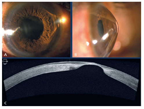
A 24-year-old male presented with progressive, painless visual decline in the right eye, with a best-corrected visual acuity of 20/80. Slit-lamp photographs obtained with temporal illumination demonstrated a sharply demarcated conical light reflex on the nasal corneal surface (Figure 1A, B), consistent with Rizzuti's sign(1). Anterior segment optical coherence tomography (Figure 1C) revealed apical corneal thinning, with a minimum corneal thickness of 422 μm. Rizzuti's sign results from localized corneal steepening that focuses incident light onto the opposite limbal region and is a classic clinical sign of advanced keratoconus.
REFERENCE
1. Rabinowitz YS. Keratoconus. Surv Ophthalmol. 1998;42(4):297-319.
Submitted for publication:
May 27, 2026.
Accepted for publication:
June 23, 2026.
Data Availability Statement: The datasets generated and/or analyzed during the current study are included in the manuscript.
Edited by
Editor-in-Chief: Newton Kara-Júnior
Associate Editor: Carlos Augusto Moreira Neto
Funding: This study received no specific financial support.
Disclosure of potential conflicts of interest: The authors declare no potential conflicts of interest.

How to cite this article:
 ABO is licensed under a Creative Commons Attribution-NonComercial 4.0 Internacional.

