

Arq. Bras. Oftalmol. 2026; 89 (5): 10.5935/0004-2749.2025-0395
Total: 500
Alper Can Yilmaz; Onder Ayyildiz
DOI: 10.5935/0004-2749.2025-0395
ABSTRACT
PURPOSE: This study aimed to evaluate the agreement between the internal astigmatism axis obtained using the optical path difference-Scan III (NIDEK Co., Japan) and conventional slit-lamp biomicroscopic measurements for determining the toric intraocular lens axis in non-dilated eyes.
METHODS: This retrospective study included 56 eyes that underwent toric intraocular lens implantation and were evaluated at postoperative week 6. The toric intraocular lens axis was measured using the optical path difference-derived internal astigmatism axis under mesopic conditions without pharmacologic pupil dilation and compared with slit-lamp biomicroscopic measurements obtained after pharmacologic dilation. Agreement between the two methods was assessed using Bland–Altman analysis, the intraclass correlation coefficient, angular error analysis, and the distribution of clinically relevant deviations. Subgroup analyses were performed for plate-haptic and C-loop haptic intraocular lens.
RESULTS: The mean signed angular difference between the two methods was 0.00º (95% CI:, −0.41º to +0.41º), with 95% limits of agreement ranging from −2.99º to +2.99º. No systematic or proportional bias was observed. The mean absolute angular error was 1.21º±0.91º, and no eye demonstrated an intermethod discrepancy exceeding 5º. The difference between the two methods was ≤1º in 73.2% of eyes, ≤3º in 98.2%, and ≤5º in all eyes. Excellent agreement was observed between the two techniques (intraclass correlation coefficient >0.99, p<0.001). Comparable agreement was also observed in both the plate-haptic and C-loop haptic intraocular lens subgroups.
CONCLUSIONS: Optical path difference-derived internal astigmatism axis measurements demonstrated excellent agreement with conventional slit-lamp biomicroscopic assessment for postoperative evaluation of toric intraocular lens alignment. The low angular error, narrow limits of agreement, and absence of clinically meaningful discrepancies suggest that this technique may serve as a reliable alternative for determining the toric intraocular lens axis in non-dilated eyes with adequate pupil size.
Keywords: Internal astigmatism; OPD scan III; Rotation; Toric intraocular lens axis; Haptic design
INTRODUCTION
Refractive assessment has become increasingly important in cataract surgery. With the advent of modern surgical techniques and advances in intraocular lens (IOL) technology, refractive cataract surgery has emerged as a key concept(1). In this context, the intraoperative management of astigmatism represents a critical aspect of cataract surgery. If corneal astigmatism is left uncorrected during surgery, it can compromise postoperative visual outcomes, necessitate spectacle correction, and negatively affect patients' quality of life(2). Astigmatism of 1 diopter (D) or less generally does not cause significant visual impairment; however, among patients with cataracts, the prevalence of corneal astigmatism exceeding 1.00 D, 1.50 D, and 2.00 D has been reported to be 32.5%-45.5%, 21.0%-26.2%, and 8%, respectively(3-6). Astigmatism at these levels often persists after surgery, resulting in residual postoperative astigmatism and the subsequent need for corrective lenses(2,7).
Limbal relaxing incisions, opposite clear corneal incisions, and toric IOLs are commonly used to correct corneal astigmatism(8-10). Studies have demonstrated that toric IOLs effectively correct astigmatism of 1 D or greater and may be considered a first-line treatment option(11,12). Furthermore, toric IOLs provide more predictable outcomes than other techniques and, unlike some surgical approaches, can be repositioned if necessary(2,13). The key to successful toric IOL implantation is rotational stability. Each degree of IOL rotation reduces astigmatic correction by approximately 3% and may increase higher-order aberrations(14,15). When IOL rotation is sufficient to compromise the effectiveness of the toric IOL, early postoperative repositioning surgery can realign the IOL with the intended axis(16).
Therefore, accurate assessment of IOL rotation is essential. Traditionally, IOL rotation has been measured using slit-lamp biomicroscopy, whereas more standardized methods, such as retroillumination photography and aberrometry, are now commonly employed(17,18). In this study, we aimed to evaluate whether the internal astigmatism axis, measured under mesopic conditions in non-dilated eyes using the OPD-Scan III (Optical Path Difference; NIDEK Co., Ltd., Japan), can be used to determine the toric IOL axis and assess IOL rotation.
METHODS
In this retrospective comparative study, data from 56 eyes that underwent cataract surgery with toric IOL implantation for astigmatism correction at the Department of Ophthalmology, Gulhane Faculty of Medicine, between October 2024 and March 2025 were reviewed. Patients with ocular surface disorders, keratoconus, or posterior capsule opacification were excluded. The study was conducted in accordance with the ethical principles of the Declaration of Helsinki and was approved by the Scientific Research Ethics Committee of the University of Health Sciences (Approval No.s 2025-412).
Preoperative refractive errors were assessed using an autorefractometer (Tonoref III, NIDEK Co., Ltd., Aichi, Japan). Best-corrected visual acuity was evaluated using the logarithm of the minimum angle of resolution (logMAR) chart. Following anterior segment examination by slit-lamp biomicroscopy, pupil dilation was induced using tropicamide eye drops (Tropamid, 0.5% tropicamide, 5 mL; Bilim Pharmaceutical, Turkey), and the retina and optic nerve head were examined using a +90 D lens (Volk Optical, Ohio, USA). In patients with advanced cataracts that precluded retinal examination, B-mode ocular ultrasonography was performed. Intraocular pressure was measured using a pneumotonometer (NT-530, NIDEK Co., Ltd., Aichi, Japan) and/or Goldmann applanation tonometry. The IOLMaster 500 (Carl Zeiss Meditec AG, Germany) was used to measure axial length, keratometry, and anterior chamber depth and to calculate IOL power. The SRK/T formula was routinely used for IOL power calculation, whereas the Hoffer Q formula was used for eyes with an axial length of less than 22 mm. Before surgery, all patients received detailed information regarding the procedure and its associated risks, and written informed consent was obtained.
Keratometry readings, corneal axis values, axial length, anticipated surgically induced astigmatism (0.50 D), and the planned incision site (90º-100º) were used to calculate the toric IOL power and implantation axis. The IOL model predicted to result in the lowest residual astigmatism was selected from the preoperative calculations. After topical anesthesia, the 0º and 180º corneal meridians were marked preoperatively using slit-lamp biomicroscopy. Immediately before surgery, after the patient was positioned on the operating table, a Mendez corneal marker was aligned with the previously marked horizontal meridians. The main incision site and intended IOL axis were then marked on the cornea. During surgery, 28 patients received a plate-haptic toric IOL (Acriva BB T UDM 611, VSY Biotechnology, Amsterdam, Netherlands), whereas the remaining 28 patients received a C-loop haptic toric IOL (Eyecryl, Biotech Vision Care, India).
Postoperative assessments were performed at 6 weeks. Internal astigmatism and its axis were measured under mesopic conditions (room illumination: 10-15 lx) using the OPD-Scan III. Pupil dilation was then induced with tropicamide eye drops, and the toric IOL axis was determined by slit-lamp biomicroscopy. OPD measurements were performed by an experienced technician, whereas slit-lamp assessments were conducted by an independent ophthalmologist who had not participated in the surgical procedures. Agreement between the two methods was primarily evaluated using Bland-Altman analysis. Pearson correlation, linear regression, and intraclass correlation coefficient (ICC) analyses were performed as supportive analyses.
Statistical analysis
Statistical analyses were performed using SPSS Statistics version 30.0 (IBM Corp., Armonk, New York). Continuous variables were expressed as mean±standard deviation (SD), and categorical variables were presented as frequencies and percentages. Data normality was evaluated using the Shapiro-Wilk test, along with skewness and kurtosis values.
Because toric IOL axis measurements represent circular data within a 0º-180º range, differences between measurement methods were calculated using a circular angular difference formula rather than simple arithmetic subtraction. Signed angular differences were normalized to the interval of −90º to +90º, whereas absolute angular errors were expressed as values ranging from 0º to 90º.
Agreement between the two measurement methods was primarily assessed using Bland-Altman analysis. The mean difference (bias), SD of the differences, and 95% limits of agreement (LoA; bias±1.96 × SD) were calculated. A one-sample t-test was performed to determine whether the mean bias differed significantly from 0. Proportional bias was assessed by linear regression analysis of the Bland-Altman difference-versus-mean plots.
As supplementary analyses, intraclass correlation coefficients (two-way random-effects model, absolute agreement, single measurement [ICC 2,1]), Pearson correlation coefficients, and simple linear regression analyses were calculated. The absolute angular error between methods was determined for each eye, and the frequency of clinically relevant deviations (≤1º, ≤3º, ≤5º, >5º, and >10º) was evaluated.
Comparisons between the plate-haptic and C-loop haptic IOL groups were performed using the independent-samples t-test or the Mann-Whitney U test, as appropriate. Categorical variables were compared using the chi-square test or Fisher's exact test when the expected cell count was less than five. All statistical tests were two-sided, and a p value of <0.05 was considered statistically significant.
RESULTS
A total of 56 eyes from 56 patients were included in the analysis. Among the study participants, 31 (55.4%) were female. The mean age was 65.46±10.76 years (range, 43-85 years). All patients had a mesopic pupil diameter greater than 3 mm, with a mean diameter of 5.29±0.64 mm (range, 4.08-6.81 mm). The demographic and clinical characteristics of the study population are summarized in Table 1.
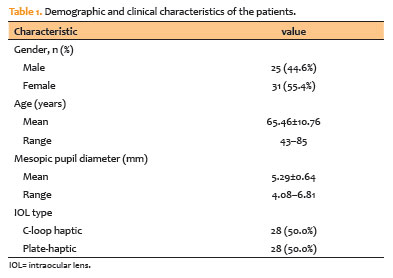
No significant differences were observed between the plate-haptic and C-loop haptic IOL groups with respect to age, sex distribution, mesopic pupil diameter, or ocular biometric parameters (all p>0.05), indicating that the two groups were well matched at baseline.
Preoperative and postoperative outcomes
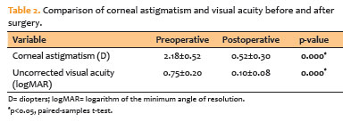
Mean corneal astigmatism was significantly reduced from 2.18±0.52 D before surgery to 0.52±0.30 D at 6 weeks postoperatively (p<0.001). Likewise, uncorrected visual acuity improved significantly, decreasing from 0.75±0.20 logMAR preoperatively to 0.10±0.08 logMAR postoperatively (p<0.001; Table 2).
Agreement between OPD and slit-lamp biomicroscopic measurements
Bland-Altman analysis demonstrated excellent agreement between the OPD-derived internal astigmatism axis and the toric IOL axis measured by slit-lamp biomicroscopy (Figure 1). The mean signed angular difference (bias) between the two methods was 0.00º (95% confidence interval [95% CI], −0.41º to +0.41º), indicating the absence of systematic measurement error. The 95% LoA ranged from −2.99º to +2.99º, demonstrating that measurements obtained with the two techniques differed by no more than approximately ±3º in 95% of eyes.
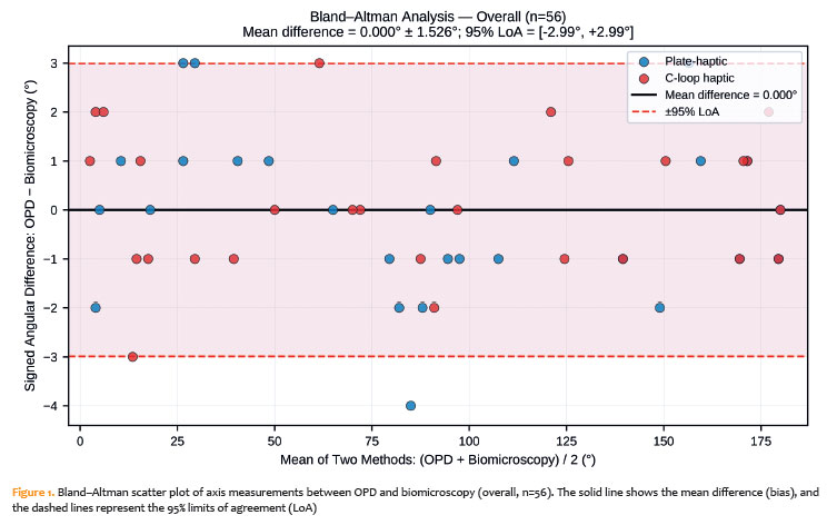
Regression analysis of the Bland-Altman plots revealed no evidence of proportional bias (slope=−0.00, p=0.642). The mean absolute angular error between the two methods was 1.21º±0.91º (range, 0º-4º). Agreement was excellent across the study population, with intermethod differences of ≤1º in 41 eyes (73.2%), ≤3º in 55 eyes (98.2%), and ≤5º in all 56 eyes (100%). No eye exhibited a discrepancy greater than 5º. Intraclass correlation coefficient analysis further confirmed excellent agreement between the two measurement techniques (ICC [2,1] >0.99, p<0.001).
Agreement according to the IOL design
Subgroup analysis by IOL design showed a similarly high level of agreement between the two measurement methods. The mean bias was −0.14º (95% LoA, −3.38º to +3.09º) in the plate-haptic group and +0.14º (95% LoA, −2.61º to +2.90º) in the C-loop haptic group (Figure 2). No significant proportional bias was observed in either subgroup.
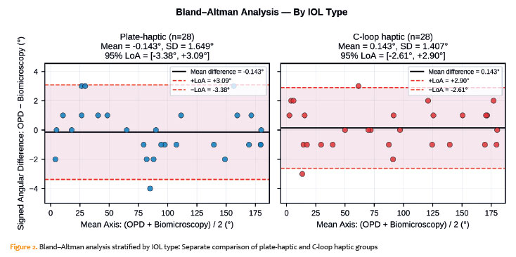
The mean absolute angular error was 1.29º±1.01º in the plate-haptic group and 1.14º±0.80º in the C-loop haptic group. This small difference was not statistically significant (p=0.745), indicating comparable measurement performance regardless of IOL design.
Correlation and regression analyses
As supportive analyses, both the biomicroscopically measured IOL axis and the OPD-derived internal astigmatism axis showed a strong correlation with the target axis (r=0.998 for both methods, p<0.001). Linear regression analysis demonstrated a strong relationship between each measurement method and the target axis (biomicroscopic: R2=0.996, p<0.001; OPD: R2=0.995, p<0.001). Direct comparison of the two measurement methods also revealed an almost perfect linear relationship (r=0.999, p<0.001; R2=0.999, p<0.001). Similar findings were observed in both the plate-haptic and C-loop haptic IOL subgroups.
Degree of IOL rotation
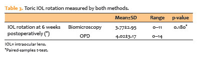
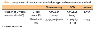
The degree of IOL rotation ranged from 0º to 11º (mean±SD: 3.77º±2.95º) when measured using the conventional biomicroscopic method, and from 0º to 14º (4.02º±3.17º) when assessed by OPD. No statistically significant difference was observed between the rotation degrees measured by the two methods (p=0.180). The mean values were similar and did not demonstrate a clinically significant difference (Table 3). Comparison between the plate-haptic and C-loop haptic IOL groups revealed no significant differences in mean rotation degree measured by either method (p=0.055 and p=0.999, respectively; Table 4).
Distribution of rotation degrees
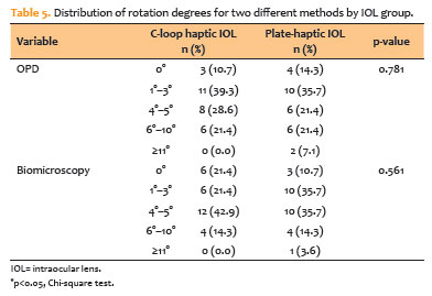
Based on OPD measurements, the proportion of IOLs demonstrating a rotation degree within 0º-5º was 78.6% in the C-loop haptic IOL group and 71.4% in the plate-haptic IOL group. In contrast, corresponding proportions for the conventional biomicroscopic method were 85.7% and 82.1%, respectively. A detailed comparison of rotation degree distributions by IOL type and measurement method is presented in Table 5.
DISCUSSION
In the present study, agreement between toric IOL axis measurements obtained using the OPD-derived internal astigmatism axis and conventional slit-lamp biomicroscopy was evaluated using Bland-Altman analysis. Bland-Altman analysis demonstrated minimal bias (0.00º) and narrow 95% LoA (−2.99º to +2.99º) between the two methods, with no evidence of systematic or proportional bias. Furthermore, the mean absolute angular error was only 1.21º, and no eye showed an intermethod discrepancy greater than 5º. These findings indicate that OPD-derived measurements provide clinically comparable results to conventional slit-lamp biomicroscopy for toric IOL axis determination.
Toric IOL implantation is an effective and safe surgical method for correcting astigmatism in eyes with cataracts and regular corneal astigmatism. However, achieving optimal outcomes requires careful preoperative planning, precise intraoperative alignment, and thorough postoperative evaluation(19). Accurate preoperative calculation of keratometric values, corneal axis, surgically induced astigmatism, axial length, and toric IOL power, together with precise alignment of the IOL along the intended axis during surgery, is crucial for successful outcomes(20). Preoperative corneal marking or the use of eye-tracking systems is intended to ensure accurate positioning of the IOL along the target axis. However, even when the IOL is correctly aligned, postoperative rotation may occur before complete stabilization of the capsular bag and IOL(21). Postoperative rotation of toric IOLs occurs most frequently within the first postoperative day, and the maximum rotation between postoperative day 1 and month 3 has been reported to be no more than 2º(22).
Each degree of toric IOL rotation results in approximately a 3% reduction in astigmatic correction; therefore, a rotation of 30º completely negates the astigmatic correction provided by the lens(14). Accordingly, repositioning surgery is generally recommended when rotation exceeds 10º(23). Therefore, postoperative assessment of the toric IOL axis and the degree of rotation is important for optimizing visual outcomes. In the present study, two eyes implanted with plate-haptic IOLs exhibited rotation greater than 10º. However, the small number of such cases limited our ability to identify potential risk factors or perform meaningful subgroup analyses. Nevertheless, agreement between OPD-derived and biomicroscopic measurements remained high, and no clinically relevant discrepancies were observed in these eyes.
Postoperative toric IOL alignment is conventionally assessed by slit-lamp biomicroscopy after pharmacological pupil dilation. However, the measurements may be influenced by patient-related factors, such as head position and cooperation during the examination, as well as examiner-related factors. In addition, poor pupil dilation and capsular fibrosis may hinder visualization of the toric IOL alignment marks. Photographic and camera-based systems can also be used to determine the IOL axis, but, similar to the conventional method, they require pupil dilation to visualize the toric IOL reference marks(17,18). Several techniques have been developed to reduce patient- and examiner-related errors and provide more objective measurements. Viestenz et al.(17) measured IOL rotation using anterior segment landmarks and reported an average measurement difference of 2.5º. In another study, the baseline IOL position was determined from intraoperative video recordings, and IOL orientation was subsequently evaluated at 2 weeks and 6 months postoperatively using digital retroillumination images. Measurement variability ranged from 2.3º to 3.1º(24).
Previous studies evaluating OPD-derived internal astigmatism measurements for toric IOL axis assessment have primarily reported strong correlations between OPD and conventional measurement techniques. Carey et al.(25) reported a mean intermethod difference of 3.27º±2.98º, with 86.3% of eyes demonstrating a discrepancy within 5º, supporting the validity of the OPD-based approach. Similarly, Gurdal et al.(26) demonstrated a high degree of agreement between OPD and slit-lamp measurements at 1 and 6 months postoperatively. In the present study, agreement was evaluated using Bland-Altman analysis, which is considered the preferred method for assessing agreement between measurement techniques. The observed bias of 0.00º, LoA within approximately ±3º, and absence of proportional bias provide further evidence that OPD-derived internal astigmatism axis measurements may serve as a clinically reliable alternative to conventional slit-lamp biomicroscopy.
The observed distribution of intermethod differences further supports the clinical applicability of the OPD-based approach. In the present study, 73.2% of eyes showed an intermethod difference of ≤1º, 98.2% showed a difference of ≤3º, and all eyes demonstrated a difference within 5º. Given that each degree of toric IOL misalignment reduces astigmatic correction by approximately 3%, these findings suggest that the differences between OPD-derived and biomicroscopic measurements are unlikely to have a clinically meaningful impact on decision-making or postoperative refractive outcomes.
An additional strength of the present study is the separate evaluation of plate-haptic and C-loop haptic toric IOLs. Excellent agreement was observed in both subgroups, and neither the mean bias nor the absolute angular error differed significantly according to IOL design. These findings suggest that the applicability of OPD-derived internal astigmatism axis measurements is not limited to a specific toric IOL platform and that comparable results can be obtained with different haptic designs.
The OPD-based measurement of the internal astigmatism axis has several limitations. First, OPD measurements require a minimum pupil diameter of 3 mm. In patients with smaller pupils, the iris may partially obstruct the projected light beam, potentially reducing measurement accuracy. Therefore, OPD scans should be performed under mesopic conditions, and pharmacological pupil dilation should be considered when the mesopic pupil diameter is less than 3 mm. Second, although two different toric IOL designs were evaluated, the study was not designed or statistically powered to compare their rotational stability. Therefore, any observations regarding differences between plate-haptic and C-loop haptic IOLs should be interpreted with caution.
The present study should be considered a clinical validation study that further supports the use of the OPD-derived internal astigmatism axis for toric IOL assessment. Previous studies have already demonstrated the feasibility of this approach(25,26). Accordingly, the primary contribution of our study is not the introduction of a new measurement concept but the confirmation of its applicability in an independent patient cohort and across toric IOLs with different haptic designs. Further prospective studies with larger sample sizes and additional IOL platforms are warranted to confirm the generalizability of these findings.
Early postoperative evaluation may provide valuable information regarding initial toric IOL rotational changes. However, measurements were obtained at 6 weeks postoperatively to minimize the potential effects of residual corneal edema and incomplete capsular bag-IOL stabilization on OPD-derived assessments. Another limitation is that slit-lamp biomicroscopy, an observer-dependent technique, was used as the reference method rather than digital photographic or image-based rotational tracking systems. Consequently, some degree of examiner-related measurement variability cannot be excluded. In addition, residual postoperative astigmatism may be influenced by factors other than toric IOL rotation, including IOL tilt, decentration, and posterior corneal astigmatism. Therefore, changes in the internal astigmatism axis should not be interpreted as evidence of IOL rotation alone.
In conclusion, the excellent agreement demonstrated by Bland-Altman analysis, together with the low absolute angular error and the absence of clinically relevant intermethod discrepancies, suggests that OPD-derived internal astigmatism axis measurements may serve as a reliable and practical alternative to conventional slit-lamp biomicroscopy for postoperative toric IOL axis assessment. Furthermore, in eyes with an adequate pupil diameter, this method enables axis assessment without pharmacological pupil dilation, potentially simplifying postoperative evaluation in routine clinical practice.
AUTHORS' CONTRIBUTIONS:
Significant contribution to conception and design: Alper Can Yilmaz, Onder Ayyildiz.
Data Acquisition: Alper Can Yilmaz.
Data Analysis and Interpretation: Alper Can Yilmaz.
Manuscript Drafting: Alper Can Yilmaz.
Significant intellectual content revision of the manuscript: Alper Can Yilmaz, Onder Ayyildiz.
Final approval of the submitted manuscript: Alper Can Yilmaz, Onder Ayyildiz.
Statistical analysis: Alper Can Yilmaz.
Obtaining funding: not applicable.
Supervision of administrative, technical, or material support: Alper Can Yilmaz, Onder Ayyildiz.
Research group leadership: Onder Ayyildiz
REFERENCES
1. Narang R, Agarwal A. Refractive cataract surgery. Curr Opin Ophthalmol. 2024;35(1):23-7.
2. Kessel L, Andresen J, Tendal B, Erngaard D, Flesner P, Hjortdal J. Toric intraocular lenses in the correction of astigmatism during cataract surgery: a systematic review and meta-analysis. Ophthalmology. 2016;123(2):275-86.
3. Day AC, Dhariwal M, Keith MS, Ender F, Perez Vives C, Miglio C, et al. Distribution of preoperative and postoperative astigmatism in a large population of patients undergoing cataract surgery in the UK. Br J Ophthalmol. 2019;103(7):993-1000.
4. Yu JG, Zhong J, Mei ZM, Zhao F, Tao N, Xiang Y. Evaluation of biometry and corneal astigmatism in cataract surgery patients from Central China. BMC Ophthalmol. 2017;17(1):56-62.
5. Curragh DS, Hassett P. Prevalence of corneal astigmatism in an NHS cataract surgery practice in Northern Ireland. Ulster Med J. 2017;86(1):25-7.
6. Collier Wakefield O, Annoh R, Nanavaty MA. Relationship between age, corneal astigmatism, and ocular dimensions with reference to astigmatism in eyes undergoing routine cataract surgery. Eye (Lond). 2016 ;30(4):562-9.
7. Ferrer-Blasco T, Montés-Micó R, Peixoto-de-Matos SC, González-Méijome JM, Cerviño A. Prevalence of corneal astigmatism before cataract surgery. J Cataract Refract Surg. 2009;35(1):70-5.
8. Abu-Ain MS, Al-Latayfeh MM, Khan MI. Do limbal relaxing incisions during cataract surgery still have a role? BMC Ophthalmol. 2022;22(1):102.
9. Priyamvada PV, Reddy JK, Vijayaraghavan P, Thulasidas M, Sundaram V. Comparison between opposite clear corneal incision versus toric intraocular lens in correcting astigmatism. Indian J Ophthalmol. 2024;72(9):1304-7.
10. Ang RE. Long-term trifocal toric intraocular lens outcomes in Asian eyes after cataract surgery. J Cataract Refract Surg. 2023;49(8):832-9.
11. Rubenstein JB, Raciti M. Approaches to corneal astigmatism in cataract surgery. Curr Opin Ophthalmol. 2013;24(1):30-4.
12. Singh A, Kapoor G, Baranwal VK, Kalra N. Rotational stability of Toric intraocular lenses. Med J Armed Forces India. 2022;78(1):68-73.
13. Tan QQ, Liao X, Lan CJ, Lin J, Tian J. [Comparison of Toric intraocular lenses and corneal incisional procedures for correction of low and moderate astigmatism during cataract surgery: A meta-analysis]. Zhonghua Yan Ke Za Zhi. 2019;55(7):522-30.
14. Ahmed II, Rocha G, Slomovic AR, Climenhaga H, Gohill J, Grégoire A, et al.; Canadian Toric Study Group. Visual function and patient experience after bilateral implantation of toric intraocular lenses. J Cataract Refract Surg. 2010;36(4):609-16.
15. Waltz KL, Featherstone K, Tsai L, Trentacost D. Clinical outcomes of TECNIS toric intraocular lens implantation after cataract removal in patients with corneal astigmatism. Ophthalmology. 2015;122(1):39-47.
16. Oshika T, Inamura M, Inoue Y, Ohashi T, Sugita T, Fujita Y, et al. Incidence and Outcomes of Repositioning Surgery to Correct Misalignment of Toric Intraocular Lenses. Ophthalmology. 2018;125(1):31-5.
17. Viestenz A, Seitz B, Langenbucher A. Evaluating the eye's rotational stability during standard photography: effect on determining the axial orientation of toric intraocular lenses. J Cataract Refract Surg. 2005;31(3):557-61.
18. Elhofi AH, Helaly HA. Comparison Between Digital and Manual Marking for Toric Intraocular Lenses: A Randomized Trial. Medicine (Baltimore). 2015;94(38):e1618.
19. Koshy JJ, Nishi Y, Hirnschall N, Crnej A, Gangwani V, Maurino V, et al. Rotational stability of a single-piece toric acrylic intraocular lens. J Cataract Refract Surg. 2010;36(10):1665-70.
20. Wolffsohn JS, Buckhurst PJ. Objective analysis of toric intraocular lens rotation and centration. J Cataract Refract Surg. 2010;36(5):778-82.
21. Lin X, Ma D, Han X, Hua Z, Shen W, Qiu X, et al. Rotational stability of plate-haptic toric intraocular lenses implantation with a capsular tension ring: prospective randomized paired-eye study. J Cataract Refract Surg. 2025;51(1):31-9.
22. Bachernegg A, Rückl T, Riha W, Grabner G, Dexl AK. Rotational stability and visual outcome after implantation of a new toric intraocular lens for the correction of corneal astigmatism during cataract surgery. J Cataract Refract Surg. 2013;39(9):1390-8.
23. Li H, Sun J, Bai H, Leng L, Dai Y, Wu X. Incidence and Risk Factors of Repositioning Surgery to Correct Misalignment of Toric Intraocular Lenses after Cataract Surgery: A Single-Center Retrospective Observational Study. Ophthalmic Res. 2023;66(1):259-64.
24. Patel CK, Ormonde S, Rosen PH, Bron AJ. Postoperative intraocular lens rotation: a randomized comparison of plate and loop haptic implants. Ophthalmology. 1999;106(11):2190-5.
25. Carey PJ, Leccisotti A, McGilligan VE, Goodall EA, Moore CB. Assessment of toric intraocular lens alignment by a refractive power/corneal analyzer system and slitlamp observation. J Cataract Refract Surg. 2010;36(2):222-9.
26. Gurdal F, Ozge G, Ayyildiz O. Determination of the toric axis by using internal astigmatism axis in non-dilated eyes. Int Ophthalmol. 2024;44(1):46.
Submitted for publication:
January 13, 2026.
Accepted for publication:
June 23, 2026.
Approved by the following research ethics committee: Gulhane Faculty of Medicine, Health Sciences University (approval number 2025-412).
Data Availability Statement: The datasets produced and/or analyzed in this study can be provided to referees upon request.
Edited by
Editor-in-Chief: Newton Kara-Júnior
Associate Editor: André Márcio V. Messias
Funding: This study received no specific financial support.
Disclosure of potential conflicts of interest: The authors declare no potential conflicts of interest.

How to cite this article:
 ABO is licensed under a Creative Commons Attribution-NonComercial 4.0 Internacional.

