

Arq. Bras. Oftalmol. 2026; 89 (5): 10.5935/0004-2749.2025-0367
Total: 634
Marcos Jacob Cohen1,2; Sabrina Veloso Avi Cohen1,2; Luci Meire Pereira da Silva3,4; Ariane Pamela Luttecke Anders3,5; Jacob Moyses Cohen1,2; Rubens Belfort Jr3,4
DOI: 10.5935/0004-2749.2025-0367
ABSTRACT
PURPOSE: To determine the indications and epidemiological characteristics of corneal transplant recipients at a referral center in Amazonas, Brazil, and to evaluate postoperative visual outcomes.
METHODS: This retrospective, descriptive, observational study included adult patients who underwent corneal transplantation performed by a single surgeon at the Instituto de Oftalmologia de Manaus between 2013 and 2023. Demographic characteristics, occupation, comorbidities, surgical indications, graft type, and best-corrected visual acuity at baseline and 12 months postoperatively were analyzed.
RESULTS: A total of 413 corneal transplantations were performed, predominantly in male patients (64.4%) aged 41–70 years (43.3%). Leucoma was the most common indication (26.2%), followed by perforated corneal ulcers (21.1%), bullous keratopathy (17.9%), and keratoconus (10.7%). Penetrating keratoplasty accounted for 94.9% of all procedures. Optical keratoplasty was the most frequently performed procedure (76.3%), followed by tectonic (22.0%) and therapeutic (1.7%) keratoplasty. At 12 months postoperatively, visual improvement was greatest among patients who underwent optical keratoplasty, particularly those with keratoconus, bullous keratopathy, and leucoma. Functional vision (≤0.3 logMAR) was achieved in 32.6% of eyes overall and in 40.3% of eyes that underwent optical keratoplasty. The number of transplantations declined after 2019, coinciding with the COVID-19 pandemic.
CONCLUSIONS: This study represents the largest retrospective series of corneal transplantations in Amazonas and one of the few such studies from Northern Brazil. The predominance of leucoma and infectious corneal perforation reflects regional socioeconomic and occupational vulnerabilities. Although visual outcomes improved significantly after transplantation, visual recovery was limited by the advanced stage of disease at presentation. These findings underscore the need to expand access to early diagnosis, strengthen eye banking and donor tissue procurement systems, and increase the adoption of lamellar keratoplasty techniques to optimize visual outcomes in the Amazon region.
Keywords: Corneal transplantation; Corneal opacity; Eye infections; Keratoplasty; Epidemiology; Visual acuity; Amazonas; Brazil
INTRODUCTION
Corneal transplantation is the most commonly performed form of human tissue transplantation worldwide and is associated with high success rates(1). Corneal diseases remain the second leading cause of reversible blindness globally(2).
In Brazil, corneal transplantation has been the most frequently performed transplant procedure since 1998(3,4), largely owing to advances in eye banking, donor tissue selection, pharmacology, immunology, and microsurgical techniques. These developments have expanded the range of treatable corneal disorders and enabled successful management of cases previously considered inoperable(5,6).
According to data from the Eye Bank Association of America, more than 35,000 corneal transplantations are performed annually in the United States(7). In Brazil, the Brazilian Association of Organ Transplantation reported 15,968 corneal transplant procedures in 2023, including 83 performed in the state of Amazonas. Approximately 30% of these procedures were conducted by teams at the Instituto de Oftalmologia de Manaus (IOM). Since 1989, the IOM has served as a pioneer in corneal transplantation in Northern Brazil. Initially reliant on donor tissue supplied by the Lions Club in the United States, the institution has more recently benefited from the establishment of the Amazonas Eye Bank, which has contributed to reducing the waiting list for corneal transplantation in the region.
Numerous studies have investigated the epidemiological characteristics of corneal transplant recipients and the principal indications for transplantation across different geographic regions(6,8-11). In the state of São Paulo, keratoconus is consistently reported as the leading indication for corneal transplantation(8,10,12). In contrast, Carvalho et al.(13) identified leucoma as the most common indication in Amazonas in 1996, followed by keratoconus.
The present study aimed to identify the primary indications for corneal transplantation at the Instituto de Oftalmologia de Manaus, a referral center in Amazonas, and to characterize the demographic and epidemiological profile of transplant recipients.
METHODS
Data were collected using a structured data extraction form. Information was obtained from medical records and anonymized through the assignment of numerical identifiers. All data were collected manually and independently verified to ensure accuracy and consistency.
The study protocol was approved by the Research Ethics Committee of the Universidade Federal do Amazonas (CAAE No. 86457725.8.0000.5020; Approval No. 7.489.137) and was conducted in accordance with Resolution No. 466/2012 of the Brazilian National Health Council.
This retrospective observational study included patients aged ≥18 years who underwent corneal transplantation performed by a single surgeon at the IOM between January 2013 and December 2023. The variables analyzed included sex, age, ethnicity, place of residence, occupation, educational level, indication for transplantation, laterality, corrected visual acuity before surgery and at 12 months postoperatively, type of transplantation, and systemic comorbidities.
A total of 480 medical records were reviewed. After exclusion of records with incomplete data, 413 patients were included in the final analysis.
Occupations were categorized according to the risk of ocular injury. High-risk occupations included farming, fishing, and metalworking, whereas low-risk occupations included teaching and public service. Transplantations were classified as low risk when no factors associated with reduced graft survival were present and as high risk in cases involving previous transplantation, emergency transplantation, glaucoma, or corneal neovascularization affecting more than two quadrants(14).
Statistical analysis
Categorical variables were summarized as absolute frequencies and percentages and compared using Pearson's chi-square test or Fisher's exact test, as appropriate. The distribution of ordinal variables was assessed using the Shapiro–Wilk test, and nonparametric comparisons were performed using the Mann–Whitney U test.
Visual acuity was assessed using standardized optotype charts and categorized according to the Brazilian Amazon Region Eye Survey (BARES)(15) as follows: (1) normal vision (≥20/32); (2) near-normal vision or mild visual impairment (<20/32 to ≥20/63); (3) visual impairment (<20/63 to ≥20/200); and (4) blindness, including moderate blindness (<20/200 to ≥20/400) and severe blindness (<20/400)(2).
A two-sided p-value <0.05 was considered statistically significant. Statistical analyses were performed using R software (version 4.4.1; R Foundation for Statistical Computing, Vienna, Austria) with RStudio, and graphical representations were generated using the ggplot2 package(16,17).
Visual acuity was also analyzed as a continuous variable using the logarithm of the minimum angle of resolution (logMAR) values. A two-way mixed-effects model with restricted maximum likelihood (REML) estimation was used, with time (preoperative vs. 12 months postoperatively) as the within-subject factor and diagnosis as the between-subject factor. Significant interactions were further evaluated using Šídák-adjusted post hoc comparisons. These analyses were performed using GraphPad Prism 8 (GraphPad Software, San Diego, California).
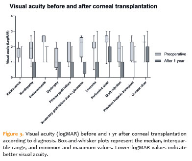
Box-and-whisker plots were used to illustrate the distribution of logMAR visual acuity at baseline and 12 months after surgery. The boxes represent the interquartile range, the central lines indicate the median values, and the whiskers denote the minimum and maximum values. Lower logMAR values correspond to better visual acuity.
RESULTS
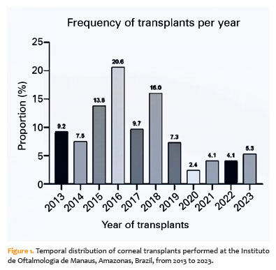
A total of 413 corneal transplantations were performed during the study period. The highest annual volume was recorded in 2016 (20.6%, n=85), followed by 2018 (16.0%, n=66) and 2015 (13.8%, n=57). Beginning in 2019 (7.3%, n=30), the number of procedures declined markedly, reaching its lowest level in 2020 (2.4%, n=10). In the subsequent years, modest fluctuations were observed, with annual proportions ranging from 4.1% to 5.3% between 2021 and 2023 (Figure 1).
Most transplant recipients were aged 41–70 years (43.3%, n=179), followed by those aged 18–40 years (35.6%, n=147) and >70 years (21.1%, n=87). The cohort was predominantly male (64.4%, n=266) and self-identified as brown (99.3%, n=410); only two patients identified as White (0.5%) and one as Black (0.2%). Corneal transplantation was performed in 202 right eyes (49%) and 211 left eyes (51.0%).
Regarding educational attainment, most patients had completed secondary education (51.3%, n=212), followed by primary education (28.6%, n=118). Smaller proportions had no formal education (13.6%, n=56) or had completed higher education (6.5%, n=27).
Most patients resided in Manaus (65.9%, n=272), whereas 22.5% (n=93) were from municipalities in the interior of Amazonas and 11.6% (n=48) originated from other locations.
Based on occupational classification, 18.2% (n=75) of patients were engaged in high-risk occupations, whereas 81.8% (n=338) were classified as having low-risk occupations.
Among systemic and ocular comorbidities, glaucoma was present in 25.2% (n=104) of patients, diabetes mellitus in 5.6% (n=23), and retinopathy in 1.0% (n=4).
Most operated eyes were phakic (63.0%, n=260), followed by pseudophakic (32.7%, n=135) and aphakic eyes (4.4%, n=18).
Overall, 57.1% (n=236) of transplantations were classified as low risk and 42.9% (n=177) as high risk. Penetrating keratoplasty accounted for 94.9% (n=392) of procedures, whereas lamellar keratoplasty represented 5.1% (n=21), including deep anterior lamellar keratoplasty (DALK; 76.2%, n=16) and Descemet membrane endothelial keratoplasty (DMEK; 23.8%, n=5). Postoperative graft rejection occurred in 2,5% (n=8/315) of optical keratoplasties.
Table 1 presents the distribution of demographic and socioeconomic variables according to occupational risk classification. A significant association was observed between age group and occupational risk (p=0.003). In the high-risk group, 54.7% (n=41) of patients were aged 41–70 years compared with 40.8% (n=138) in the low-risk group. Conversely, patients older than 70 years were more frequently represented in the low-risk group (24.3%, n=82).
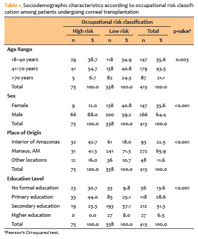
Male patients predominated in the high-risk occupational category (88.0%, n=66). Patient origin was also significantly associated with occupational risk (p<0.001), with residents of the interior of Amazonas more commonly represented in the high-risk group (42.7%, n=32).
Educational level was significantly associated with occupational risk classification (p<0.001). Patients in the high-risk group more frequently had no formal education (30.7%, n=23) or only primary education (44.0%, n=33), whereas those in the low-risk group predominantly had secondary education (57.1%, n=193) or higher education (8.0%, n=27). Notably, no patient in the high-risk group had completed university education.
The geographic distribution of transplant recipients demonstrated that most patients originated from Amazonas (n=368). Additional patients came from Pará (n=27), Roraima (n=6), Acre (n=3), Amapá (n=3), Maranhão (n=3), and Mato Grosso (n=2), and one patient was referred from Venezuela.
Table 2 summarizes the distribution of surgical indications according to age group. Keratoconus predominated among patients aged 18–40 years (26.5%, n=39), whereas its frequency was substantially lower among those aged 41–70 years (2.8%, n=5), and no cases were observed in patients older than 70 years.
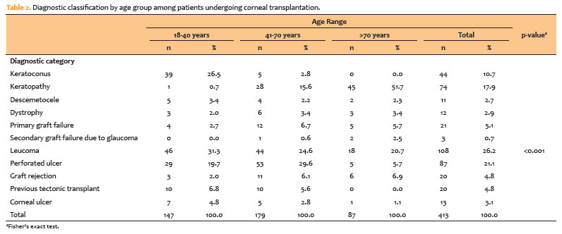
Bullous keratopathy showed the opposite pattern, with the highest prevalence among patients aged >70 years (51.7%, n=45), all of whom had previously undergone cataract surgery. The prevalence was lower in patients aged 41–70 years (15.6%, n=28), most of whom also had a history of cataract surgery, with one case associated with post-uveitic bullous keratopathy. Only one case (0.7%) was identified in the 18–40-year age group and was secondary to ocular trauma.
Leucoma was among the most common indications across all age groups, accounting for 31.3% (n=46) of cases in patients aged 18–40 years, 24.6% (n=44) in those aged 41–70 years, and 20.7% (n=18) in those older than 70 years. This distribution differed significantly among age groups (p<0.001).
Overall, leucoma was the most frequent indication for transplantation (26.2%, n=108), followed by perforated corneal ulcer (21.1%, n=87), bullous keratopathy (17.9%, n=74), and keratoconus (10.7%, n=44). Less common indications included primary graft failure (5.1%, n=21), previous graft rejection (4.8%, n=20), previous tectonic graft (4.8%, n=20), corneal ulcer (3.1%, n=13), corneal dystrophy (2.9%, n=12), descemetocele (2.7%, n=11), and graft failure secondary to glaucoma (0.7%, n=3).
Table 3 presents the distribution of indications according to occupational risk classification. A significant association was observed between occupational risk and transplantation indication (p<0.001).
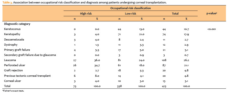
Leucoma was the most common indication in both groups but was more prevalent among high-risk workers (36.0%, n=27) than among low-risk workers (24.0%, n=81). Similarly, perforated corneal ulcer occurred more frequently in the high-risk group (34.7%, n=26) than in the low-risk group (18.0%, n=61). Conversely, all cases of keratoconus occurred among patients in the low-risk occupational category (13%, n=44).
A significant association was found between transplant type and waiting time for surgery (p<0.001). Among optical transplantations, 54% (n=170) were performed within 6 months of listing, 19% (n=60) between >6 and 12 months, and 27% (n=85) after more than 12 months (Table 4).
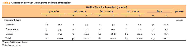
Table 5 shows the distribution of patients according to place of origin and selected sociodemographic and clinical variables.
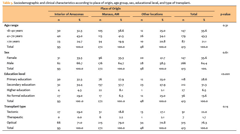
Educational level differed significantly according to place of origin (p<0.001). In Manaus, most patients had completed secondary education (57.7%, n=157), whereas patients from the interior of Amazonas more frequently had no formal education (29%, n=27). Among patients from other locations, secondary education (47.9%, n=23) and absence of formal education (25%, n=12) were both common, highlighting regional disparities in educational attainment.
No significant association was found between transplant type and place of origin (p=0.14). Optical transplantation predominated across all geographic regions, particularly among patients from Manaus (79.0%, n=215). Tectonic transplantation accounted for 22.0% (n=91) of all procedures and was more frequent among patients from the interior of Amazonas (29.0%, n=27). Therapeutic transplantation was uncommon and occurred sporadically across all regions.
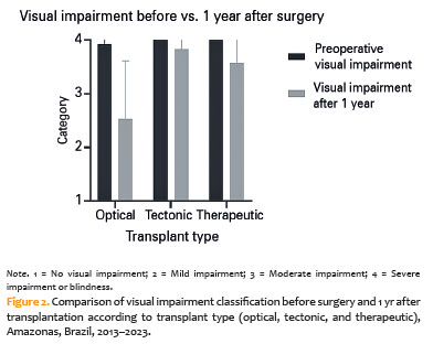
Comparison of visual status before surgery and at 1 year postoperatively demonstrated a significant improvement in visual function. The mean visual impairment score decreased from 3.94 preoperatively to 2.79 at 1 year (p<0.001), indicating substantial functional recovery following transplantation.
When stratified by transplant type, all groups exhibited severe visual impairment preoperatively, with mean scores approaching the maximum value of the scale. Patients undergoing tectonic and therapeutic transplantation both had a mean baseline score of 4.0, whereas the optical transplantation group had a mean score of 3.92. After 1 year, no significant improvement was observed in the tectonic (3.835; p=0.384) or therapeutic (3.571; p=0.550) groups. In contrast, the optical transplantation group showed a significant reduction in mean visual impairment score to 2.53 (p<0.001), indicating improvement from severe visual impairment to moderate or mild impairment in a proportion of patients. These findings suggest that meaningful visual recovery was primarily associated with optical and low-risk transplantations. The distribution of visual status over time is shown in figure 2.
At 1 year, functional vision (≤0.3 logMAR) was achieved in 32.6% (128/393) of eyes overall and in 40.3% (127/315) of eyes that underwent optical keratoplasty. Of the 413 transplanted eyes, 393 (95.2%) completed the 1-year follow-up, whereas 20 eyes required retransplantation before this time point. Among eyes that underwent penetrating keratoplasty, 29.7% (112/377) achieved functional vision, whereas all eyes that underwent lamellar keratoplasty (16/16, 100%) achieved this outcome.
Analysis of visual acuity as a continuous variable (logMAR) using a two-way mixed-effects model revealed a significant effect of time (F(1,382)=295.5, p<0.0001), indicating overall visual improvement from baseline to 1 year after surgery. Significant effects were also observed for diagnosis (F(10,402)=21.23, p<0.0001) and for the time × diagnosis interaction (F(10,382)=7.756, p<0.0001), demonstrating that the magnitude of visual improvement varied according to the underlying diagnosis.
Estimated marginal means showed an improvement in visual acuity from 1.890 logMAR preoperatively to 0.909 logMAR at 1 year, corresponding to a mean reduction of 0.981 logMAR units (95% confidence interval [CI], 0.868–1.093).
Šídák-adjusted post hoc analyses demonstrated significant improvements in visual acuity for most diagnostic categories. The greatest improvements were observed in keratoconus (mean difference, 1.609 logMAR; p<0.0001), bullous keratopathy (1.219 logMAR; p<0.0001), and leucoma (1.149 logMAR; p<0.0001). Significant improvements were also observed in graft rejection (1.025 logMAR; p<0.0001), corneal dystrophy (1.003 logMAR; p=0.0011), graft failure secondary to glaucoma (1.325 logMAR; p=0.0032), previous tectonic transplantation (0.855 logMAR; p<0.0001), corneal ulcer (0.813 logMAR; p=0.0011), primary graft failure (0.669 logMAR; p=0.0007), and perforated corneal ulcer (0.612 logMAR; p<0.0001). No significant improvement was observed among patients with descemetocele (mean difference, 0.508 logMAR; p=0.1595) (Figure 3).
DISCUSSION
This study represents the largest retrospective series of corneal transplantation in Amazonas and one of the few from Northern Brazil, helping to address a regional epidemiological gap. The characterization of demographic and clinical profiles, as well as transplant indications at IOM, provides insight into the specific features of ophthalmic practice in the Amazon region.
Since the establishment of the Banco de Olhos do Amazonas in 2004, corneal transplantation has increased steadily. Between 2013 and 2023, the highest number of procedures was observed in 2016 (20.6%; Figure 1), whereas the lowest rates occurred in 2020, likely because of the COVID-19 pandemic and its impact on corneal procurement and distribution. This pattern mirrors national trends(18), with a marked decline during the pandemic followed by gradual recovery after 2021.
The predominant age group was 41–70 years (43.3%), and the mean age was similar to that reported in national epidemiological studies(4,9,19). A predominance of male patients was also observed (64.4%).
The predominance of individuals with brown skin color (99.3%) reflects the demographic composition of the state of Amazonas(20) and reinforces the need to include ethnic and racial variables in regional ophthalmologic records.
Regarding educational level, a significant association was observed with place of origin (p<0.001). Patients from the interior of the state had a higher proportion of individuals without formal education (29.0%) than residents of Manaus (6.3%). This disparity, also described in the BARES(15), directly affects access to information, treatment adherence, and timely utilization of ophthalmologic services.
Occupational analysis revealed a predominance of low-risk activities (81.8%). However, stratified analysis showed statistically significant associations between occupation and demographic variables. High-risk occupations were more common among individuals younger than 70 years (93.4%; p=0.003), males (88%; p<0.001), and residents of the interior of the state (p<0.001). Workers engaged in high-risk occupations, such as farming, fishing, and metalworking, exhibited a higher frequency of leucomas and perforated ulcers. This pattern is consistent with findings from studies conducted in the North(19,21), Northeast(22), and South(23) regions of Brazil. Similarly, the BARES(15) identified corneal leucoma as one of the causes of blindness and severe visual impairment among adults in the Amazon region, highlighting the persistence of preventable corneal diseases associated with trauma and ocular infections in Northern Brazil.
The most frequent diagnoses were leucoma (26.2%), followed by perforated corneal ulcer (21.1%), bullous keratopathy (17.9%), and keratoconus (10.7%). These findings differ from those reported in large centers in Southeastern Brazil, such as Sorocaba(10) and Minas Gerais(4), where keratoconus is the leading indication for transplantation. However, they are consistent with previous findings from Amazonas(13) and the Northeast region(22,24), where infectious diseases and leucomas remain predominant.
Optical transplantation was the most common procedure (76.3%), followed by tectonic (22.0%) and therapeutic (1.7%) transplantation, findings similar to those reported in other studies(5,19,22).
Optical transplants were more frequent among patients with longer waiting times, although most procedures (54%) were still performed within 6 months. In contrast, tectonic and therapeutic transplants were predominantly performed in emergency situations, with waiting times of less than 3 months.
The predominance of penetrating keratoplasty (94.9%) over lamellar keratoplasty (5.1%) is consistent with the national pattern. However, centers in Southeastern Brazil and other countries have demonstrated increasing adoption of selective lamellar techniques, with rates increasing from 4.8% in 2010 to 34.3% in 2020, while penetrating keratoplasty decreased from 95.2% to 65.7% during the same period (p<0.001)(11).
Functional analysis revealed a significant improvement in visual acuity 1 year after transplantation, with a reduction in the mean visual impairment scale from 3.94 to 2.79 (p<0.001). This improvement was particularly evident among patients undergoing optical transplantation, for whom the postoperative mean score was 2.53 (p<0.001), indicating a transition from severe to mild or moderate visual impairment. In contrast, tectonic and therapeutic transplants did not show significant improvement, as expected, because of their emergency nature and associated comorbidities. Similar findings have been reported in studies from Saudi Arabia(25) and Pernambuco(24), highlighting the superior functional outcomes of optical keratoplasty when performed under elective, low-risk conditions.
Overall, the combined graft rejection and failure rate was 10.6%, slightly lower than that reported in the literature(5,26,27), possibly due to a uniform surgical technique, standardized follow-up protocols, and appropriate case selection. The predominance of low-risk transplants (57.1%) may also have contributed to this outcome.
Postoperative visual outcomes were influenced by the underlying corneal pathology, with greater functional recovery observed in patients with keratoconus, keratopathy, and leucoma, and more limited improvement in conditions associated with ocular comorbidities or urgent surgical indications.
Among patients who underwent optical keratoplasty, the mean postoperative visual acuity was 0.70 logMAR, which was slightly worse than that reported for penetrating keratoplasty in the literature (~0.6 logMAR). This difference likely reflects the higher burden of advanced and infectious diseases in this cohort(28,29). Functional vision (≤0.3 logMAR) was achieved in 32.6% of eyes and in 40.3% of eyes that underwent optical keratoplasty. In contrast, lamellar techniques have consistently demonstrated superior visual outcomes(30), with postoperative visual acuity of approximately 0.3 logMAR and a greater likelihood of achieving functional vision. These findings support the need to expand access to lamellar procedures and promote earlier surgical intervention in the Amazon region.
In conclusion, this study provides the largest epidemiological assessment of corneal transplantation in Amazonas and one of the few robust datasets from Northern Brazil. The findings demonstrate a predominance of leucomas, infectious perforations, and advanced disease associated with socioeconomic and geographic vulnerabilities. Although corneal transplantation resulted in meaningful visual improvement, outcomes were limited by disease severity at presentation and the high burden of preventable conditions. This regional profile contrasts with that of more developed areas and underscores the need to strengthen eye banking systems, expand corneal donation programs, improve access to early diagnosis and timely surgical intervention, and increase the adoption of lamellar techniques to optimize visual outcomes in this population.
AUTHORS' CONTRIBUTIONS:
Significant contribution to conception and design: Marcos Jacob Cohen; Sabrina Veloso Avi Cohen; Jacob Moyses Cohen.
Data Acquisition: Marcos Jacob Cohen; Sabrina Veloso Avi Cohen.
Data Analysis and Interpretation: Marcos Jacob Cohen; Sabrina Veloso Avi Cohen; Ariane Pamela Luttecke Anders.
Manuscript Drafting: Marcos Jacob Cohen; Sabrina Veloso Avi Cohen; Jacob Moyses Cohen; Rubens Belfort Mattos Junior.
Significant intellectual content revision of the manuscript: Marcos Jacob Cohen; Rubens Belfort Mattos Junior; Jacob Moyses Cohen.
Final approval of the submitted manuscript: Marcos Jacob Cohen, Sabrina Veloso Avi Cohen, Luci Meire Pereira da Silva, Ariane Pamela Luttecke Anders, Jacob Moyses Cohen, Rubens Belfort Mattos Junior.
Statistical analysis: Ariane Pamela Luttecke Anders; Sabrina Veloso Avi Cohen.
Obtaining funding: not applicable.
Supervision of administrative, technical, or material support: Marcos Jacob Cohen; Jacob Moyses Cohen, Luci Meire Pereira da Silva, Sabrina Veloso Avi Cohen; Rubens Belfort Mattos Junior.
Research group leadership: Marcos Jacob Cohen.
REFERENCES
1. Gain P, Jullienne R, He Z, et al. Global survey of corneal transplantation and eye banking. JAMA Ophthalmol. 2016;134(2):167-73.
2. World Health Organization (WHO). World report on vision. Geneva: WHO; 2019 [cited 2026 Jun 10]. Available from: https://www.who.int/publications-detail/world-report-on-vision.
3. Associação Brasileira de Transplante de Órgãos (ABTO). Registro Brasileiro de Transplantes (RBT) 2023. São Paulo: ABTO; 2024 [citado 2026 Jun 10]. Disponível em: https://site.abto.org.br/wp-content/uploads/2024/04/rbt2023-restrito.pdf.
4. Neves RC, Boteon JE, Santiago APMS. Indicações de transplante de córnea no Hospital São Geraldo da Universidade Federal de Minas Gerais. Rev Bras Oftalmol. 2010;69(2):84-8.
5. Araújo AA, Melo GB, Silva RL, Neta VM. Perfil epidemiológico dos pacientes na lista de espera para transplante de córnea no Estado de Sergipe. Arq Bras Oftalmol. 2004;67(4):613-6.
6. Lindquist TD, McGlothan JS, Rotkis WM, Chandler JW. Indications for penetrating keratoplasty: 1980-1988. Cornea. 1991;10(3):210-8.
7. Aiken-O'Neill P, Mannis MJ. Summary of corneal transplant activity: Eye Bank Association of America. Cornea. 2002;21(1):1-3.
8. Sano FT, Dantas PE, Silvino WR, Sanchez JZ, Sano RY, Adams F, Nishiwaki-Dantas MC. Tendência de mudança nas indicações de transplante penetrante de córnea. Arq Bras Oftalmol. 2008;71(3):400-4.
9. Netto AA, Schlindwein CA, Felicíssimo LC. Indicações e perfil epidemiológico dos pacientes submetidos à ceratoplastia no estado de Santa Catarina. Rev Bras Oftalmol. 2014;73(3):162-6.
10. Calix Netto MJ, Giustina ED, Ramos GZ, Peccini RF, Sobrinho M, Souza LB. Principais indicações de transplante penetrante de córnea em um serviço de referência no interior de São Paulo (Sorocaba-SP, Brasil). Arq Bras Oftalmol. 2006;69(5):661-4.
11. Flores VG, Dias HL, Castro RS. Indicações para ceratoplastia penetrante no Hospital das Clínicas–UNICAMP. Arq Bras Oftalmol. 2007;70(3):505-8.
12. Moriyama AS, Forseto AD, Pereira NC, Ribeiro AC, Almeida MC, Figueras-Roca M, et al. Trends in corneal transplantation in a tertiary hospital in Brazil. Cornea. 2022;41(7):857-66.
13. Carvalho RC, Moss M, Garrido C, Cohen JM, Chaves C. Indicações de transplante de córnea no Amazonas: experiência de 11 anos no Instituto de Oftalmologia de Manaus. Rev Bras Oftalmol. 1996;55(8):619-22.
14. Porto CS. Transplante de córnea, curva de sobrevida e resultados [dissertação]. Porto Alegre (RS): Universidade Federal do Rio Grande do Sul; 2013.
15. Salomão SR, Mitsuhiro MRKH, Belfort R Jr. The Brazilian Amazon Region Eye Survey: design and methods. Ophthalmic Epidemiol. 2017;24(4):257-64.
16. R Core Team. R: A language and environment for statistical computing. Vienna (Austria): R Foundation for Statistical Computing; 2024 [cited 2026 Jun 10]. Available from: https://www.R-project.org/.
17. Wickham H. Programming with ggplot2. In: ggplot2. Cham: Springer; 2016. p. 241-53. [cited 2026 Jun 10]. Available from: https://cran.microsoft.com/snapshot/ 2015-01-06/web/packages/ggplot2/ggplot2.pdf.
18. Moriyama AS, Pessoa JLE, Bessa TRS, Pereira NC, Mehta JS, Hofling-Lima AL, Forseto ADS. The impact of the COVID-19 pandemic on corneal transplantation in Brazil. Cornea. 2022;41(3):322-7.
19. Araújo AVS, Marinho CG, Rodrigues FSR, Alves MS, Frazão-Neto OC. Perfil socioclínico de pacientes submetidos a transplante de córnea em hospital de referência (Belém-PA). Rev Bras Oftalmol. 2022;81:e0062.
20. Instituto Brasileiro de Geografia e Estatística (IBGE). Censo Demográfico 2022: Identificação étnico-racial da população, por sexo e idade. Rio de Janeiro: IBGE; 2022.
21. Sobrinho EFA, Negrão BC, Almeida HG. Perfil epidemiológico de pacientes na fila de transplante penetrante de córnea no estado do Pará, Brasil. Rev Bras Oftalmol. 2011;70(6):384-90.
22. Tonhá CD, Santos AM, Souza JC, Muniz MC. Estudo retrospectivo dos transplantes de córnea no estado de Alagoas. Braz J Transplant. 2010;13(2):1316-20.
23. Cattani S, Kwitko S, Kroeff MAH, Marinho D, Rymer S, Bocaccio FL. Indicações de transplante de córnea no Hospital de Clínicas de Porto Alegre. Arq Bras Oftalmol. 2002;65(1):95-8.
24. Amaral CSR, Duarte JY, Silva PLS, Valbuena R, Cunha F. Indicações de ceratoplastia penetrante em Pernambuco. Arq Bras Oftalmol. 2005;68(5):635-7.
25. Omar N, Bou Chacra CT, Tabbara KF. Outcome of corneal transplantation in a private institution in Saudi Arabia. Clin Ophthalmol. 2013;7:1311-8.
26. Gurnani B, Czyz CN, Mahabadi N, Havens SJ. Corneal graft rejection. StatPearls [Internet]. Treasure Island (FL): StatPearls Publishing; 2024 [cited 2025 Oct 31]. Available from: https://www.ncbi.nlm.nih.gov/books/NBK519043/.
27. Chalita MR, Diazgranados EBM, Sato EH, Branco BC, Freitas D. Rejeição corneana pós-transplante de córnea: análise de dados do Banco de Olhos do Hospital São Paulo, Escola Paulista de Medicina. Arq Bras Oftalmol. 2000;63(1):29-33.
28. Tan DT, Dart JK, Holland EJ, Kinoshita S. Corneal transplantation. Lancet. 2012;379(9827):1749-61.
29. Wagoner MD. Penetrating keratoplasty: indications and outcomes. Middle East Afr J Ophthalmol. 2009;16(3):143-50.
30. Reinhart WJ, Musch DC, Jacobs DS, Lee WB, Kaufman SC, Shtein RM. Deep anterior lamellar keratoplasty as an alternative to penetrating keratoplasty. Ophthalmology. 2011;118(1):209-18.
Submitted for publication:
December 10, 2025.
Accepted for publication:
June 9, 2026.
Approved by the following research ethics committee: Universidade Federal do Amazonas – UFAM (CAAE: 86457725.8.0000.5020).
Data Availability Statement: The datasets produced and/or analyzed in this study can be provided to referees upon request.
Edited by
Editor-in-Chief: Newton Kara-Júnior
Associate Editor: André M. V. Messias
Funding: This study received no specific financial support.
Disclosure of potential conflicts of interest: The authors declare no potential conflicts of interest.

How to cite this article:
 ABO is licensed under a Creative Commons Attribution-NonComercial 4.0 Internacional.

