

Arq. Bras. Oftalmol. 2026; 89 (5): 10.5935/0004-2749.2025-0337
Total: 612
Augusto Villar1; Heloisa Nascimento2,3; Jorge Kalil1; Thaís Maria da Mata Martins3,4; Luiz Vicente Rizzo5; Gabriel Barbieri3; Pedro Pires1; Lucimeire N. Carvalho2; Larissa R. Rosa3; Rodrigo Moreira2; Carmen Luz Pessuti3; Henrique Ferrer3; José Álvaro Pereira Gomes3; Silvano Raia1; Rubens Belfort Jr2,3
DOI: 10.5935/0004-2749.2025-0337
ABSTRACT
Corneal blindness affects more than 12 million individuals worldwide; however, the availability of human donor corneas meets only a small fraction of the demand. Porcine corneal xenotransplantation represents a promising alternative because of the cornea's immune privilege and the anatomical similarities between human and porcine corneas. However, the galactose-α1,3-galactose antigen expressed on porcine cells induces hyperacute rejection through the activation of human anti-Gal antibodies, complement pathways, and innate immune cell infiltration. This review summarizes the mechanisms underlying corneal immune privilege and contrasts them with galactose-α1,3-galactose-mediated rejection pathways. In addition, it evaluates five major mitigation strategies: GGTA1-knockout pigs, enzymatic α-galactosidase treatment, RNA interference, decellularization, and immunosuppressive approaches, including CD40-CD154 blockade and topical tacrolimus. Preclinical studies have demonstrated that GTKO corneas can maintain transparency for longer than 1 year, whereas decellularized matrices preserve corneal architecture while exhibiting low antigenicity. These findings support the future clinical translation of porcine corneal xenotransplantation.
Keywords: Corneal transplantation; Xenotransplantation; Graft rejection; Galactose; Immunosuppressive agents.
INTRODUCTION
The cornea is a transparent, avascular tissue responsible for approximately two-thirds of the eye's refractive power(1). It consists of five layers—the stratified epithelium, Bowman's membrane, collagen-rich stroma, Descemet's membrane, and a monolayer of nonregenerating endothelial cells—which collectively maintain transparency, protect intraocular structures, and preserve barrier function(2). Loss of corneal clarity or structural integrity resulting from keratoconus, Fuchs dystrophy, infectious keratitis, chemical injury, or corneal scarring can lead to severe visual impairment. In such cases, penetrating or lamellar keratoplasty remains the only effective treatment option(3).
Despite the high survival rates of corneal allografts in ideal avascular host beds, an estimated 12.7 million people worldwide still lack access to donor corneas(4,5). Porcine corneal xenotransplantation may help address this shortage; however, immunological rejection remains a major challenge, primarily because of the galactose-α1,3-galactose (α-Gal) antigen(6,7). This review discusses corneal immune privilege, the biosynthesis and immunogenicity of α-Gal, the mechanisms underlying graft destruction, current mitigation strategies—including genetic ablation, enzymatic removal, decellularization, RNA interference, and immunosuppression—and future directions involving gene editing and bioengineering approaches for long-term graft survival.
Corneal transplantation is the most commonly performed form of human solid tissue transplantation(8). Owing in part to the immune privilege of the cornea, transplantation outcomes are generally favorable, with survival rates reaching 90% at 1 year and 55% at 15 years in avascular, noninflamed host beds(9).
The principal limitation remains the availability of ethically sourced donor tissue. More than half of all countries (53.3%) lack eye-banking services, whereas 35.7% can meet only minimal transplantation demands(5). The burden is greatest in low- and middle-income countries, where approximately 90% of the global visually impaired population resides(10).
The α-Gal antigen is an oligosaccharide expressed in most nonprimate mammals, including pigs and cattle. In humans and higher primates, this antigen is highly immunogenic because these species naturally produce anti-Gal antibodies against the molecule, making α-Gal a central trigger of xenograft hypersensitivity reactions(11-13). Understanding the role of α-Gal in xenotransplant rejection requires an understanding of ocular immunology and corneal immune privilege(14).
Immune privilege in the eye was first recognized through studies involving corneal transplantation(15). This phenomenon is attributed to three major mechanisms: anatomical and molecular barriers, immune tolerance pathways, and an immunosuppressive intraocular microenvironment(16-18). Together, these mechanisms regulate immune activity and protect ocular tissues from excessive inflammatory damage. A comprehensive understanding of these processes is essential for overcoming the challenges associated with xenotransplantation and for developing strategies capable of modulating immune responses to improve graft survival(19,20).
1. OCULAR IMMUNE PRIVILEGES
Immune privilege in the eye enables specific tissues, particularly the cornea, to evade immune rejection more effectively than tissues located at nonprivileged sites. This feature is essential for vision preservation because excessive immune responses can damage irreplaceable structures, such as the nonregenerating corneal endothelium, through scarring and inflammation(21,22). Ocular immune privilege is based on three principal mechanisms: anatomical and molecular barriers, immune tolerance pathways, and an immunosuppressive intraocular microenvironment(18).
Complement regulatory proteins (CRPs), including CD46, CD55, and CD59, are expressed by intraocular cells and are also present in soluble form within the aqueous and vitreous humor(23). These CRPs bind complement fragments, inhibit the formation of C3 and C5 convertases, and prevent deposition of the membrane attack complex, thereby protecting avascular ocular cells from complement-mediated lysis and preserving tissue integrity(24). Studies of human aqueous humor have confirmed that CD46, CD55, and CD59 play key roles in complement regulation, with their soluble forms contributing substantially to the maintenance of immune privilege and corneal transparency under both physiological and inflammatory conditions(24).
Together, soluble and membrane-bound CRPs exemplify the immunosuppressive microenvironment that characterizes ocular immune privilege. The following section discusses the anatomical and molecular barriers that protect the cornea from antigen exposure(23).
1.1 Anatomical and molecular barriers in the eye
The anatomy of the eye plays a central role in immune privilege. Under normal conditions, the cornea is avascular(25,26). This "angiogenic privilege" preserves corneal transparency and minimizes antigen exposure(27). The development of blood or lymphatic vessels in the cornea—caused by injury, infection, or inflammatory disease—increases the risk of graft rejection after corneal transplantation(14,21).
At the molecular level, ocular tissues produce antiangiogenic and anti-inflammatory factors, including thrombospondin-1, VEGFR-3, endostatin, and pigment epithelium-derived factor(20,24,28,29). These molecules inhibit vascular growth into the corneal stroma, thereby preserving immune privilege. Bowman's membrane and the limbal barrier also help prevent vascular invasion from the conjunctiva. When these structures are disrupted by trauma or inflammation, vascular invasion and immune activation increase(30).
1.2 Immune tolerance mechanisms
Conjunctiva-associated lymphoid tissue (CALT), which is part of the mucosa-associated lymphoid system, is a key component of ocular mucosal immunity(31,32). CALT contains intraepithelial lymphocytes (CD8, HML-1), M cells involved in antigen capture, and antimicrobial peptides such as defensins(31-33). The lacrimal gland and tear film further contribute to ocular defense through the secretion of IgA, lysozyme, lactoferrin, and complement proteins, which inhibit pathogen adherence and neutralize microorganisms(33). In addition to protecting the ocular surface, CALT and the lacrimal mucosal system can also induce immune tolerance through mechanisms that contribute to the immune-privileged status of the eye(34).
Anterior chamber-associated immune deviation (ACAID) is one of the best-characterized immune tolerance mechanisms in the eye(35,36). ACAID is essential for preventing immune-mediated damage and depends on interactions between ocular antigen-presenting cells (APCs) and regulatory T cells(36).
When antigens enter the anterior chamber, they are captured by APCs, such as dendritic cells, which subsequently migrate to the spleen(37). In the spleen, these APCs induce selective suppression of delayed-type hypersensitivity and cytotoxic T-cell responses (type III and IV hypersensitivity reactions). This process promotes the expansion of regulatory T cells and the production of non-complement-fixing antibodies(38). Such systemic immune tolerance contributes to corneal graft survival and protects against ocular autoimmunity(39).
1.3 Immunosuppressive intraocular microenvironment
In addition to anatomical barriers and immune tolerance, the eye maintains an immunosuppressive microenvironment that actively limits inflammation(40). Soluble factors present in the aqueous humor—including TGF-β2, α-MSH, VIP, and CGRP—suppress macrophage and T-cell activity(28,35,41). These molecules downregulate inflammatory pathways and help prevent immune-mediated damage to ocular tissues(18,19).
The corneal endothelium and other ocular tissues also express PD-L1 and FasL, which induce apoptosis in activated immune cells(41). Binding of PD-L1 to PD-1 receptors on T cells further suppresses immune-mediated tissue injury(42).
These local immunosuppressive mechanisms are essential for maintaining ocular immune privilege and supporting long-term corneal allograft survival, even when systemic immunosuppression is limited.
Immune responses in the eye are also influenced by neuroimmune interactions, commonly referred to as neuroimmune crosstalk(43). The aqueous humor contains neuropeptides such as VIP and CGRP, which exert anti-inflammatory effects by inhibiting immune cell activity within the eye(44,45). In addition, these neuropeptides contribute to tissue regeneration and wound healing by promoting a tightly regulated inflammatory response(46).
2. WHAT IS THE α-GAL ANTIGEN?
The α-Gal antigen is a carbohydrate epitope found on glycolipids and glycoproteins. It is synthesized by the enzyme α-1,3-galactosyltransferase (GGTA1), which is commonly expressed in nonprimate mammals such as pigs, cattle, and other species considered for xenotransplantation(11,12). The absence of α1,3GT in humans and higher primates results in the production of natural anti-α-Gal antibodies because of continuous immune exposure to gastrointestinal bacteria expressing α-Gal-like antigens(13,47,48). These antibodies trigger strong immune responses when α-Gal-positive tissues are transplanted, leading to hyperacute rejection through complement activation(11,12). α-Gal is abundantly expressed in the porcine corneal endothelium and stroma, making these tissues highly immunogenic(11,13,49).
2.1 Immunological mechanisms of rejection mediated by α-Gal
2.1.1 The primary immune reaction
Rejection of α-Gal-positive xenografts is driven by a rapid immune response. Natural anti-α-Gal antibodies, which account for approximately 1%-3% of human plasma immunoglobulins, bind to transplanted tissues and activate the classical complement pathway(11,47).
This cascade leads to membrane attack complex formation, resulting in cellular lysis and rapid graft destruction(11). Hyperacute rejection occurs within hours and remains a major barrier to successful corneal xenotransplantation(12,13).
In addition to complement activation, xenotransplantation induces a cytokine storm characterized by increased levels of TNF-α, IL-2, and IFN-γ, which promote severe inflammation at the graft site(13,47).
2.1.2 Role of immune cells
In addition to complement activation, innate immune cells such as macrophages and natural killer (NK) cells are recruited by chemokines, including MCP-1, thereby amplifying inflammation and accelerating graft rejection(6,13). Studies involving porcine corneal xenotransplantation have demonstrated marked infiltration of macrophages and NK cells, supporting their important role in acute rejection(11,13). Studies using GGTA1-knockout mice have provided further insight, showing that reduced immune activation is associated with improved graft survival(6,13).
3. IMPACT OF α -GAL ON CORNEAL GRAFT REJECTION
α-Gal epitopes in porcine corneas play a central role in graft rejection. Quantitative studies have reported up to 10¹2 epitopes per milligram of stroma, which is sufficient to induce strong antibody binding and complement activation(11,49). Binding of natural anti-α-Gal antibodies to these epitopes rapidly activates the complement system, followed by recruitment of neutrophils, macrophages, and NK cells, ultimately leading to hyperacute rejection(11,13). Without immunosuppressive intervention, most porcine corneal grafts are rejected within 48 h, as demonstrated in several experimental models(12,13).
4. STRATEGIES TO MITIGATE α-GAL-MEDIATED XENOTRANSPLANT REJECTION
4.1 Use of GGTA1-knockout pigs
The most effective and safest method for eliminating α-Gal is the genetic deletion of GGTA1 in pigs (GTKO)(7,50). These animals do not express α-Gal on their cells, thereby preventing human antibody binding and complement activation(51).
In practice, corneas harvested from GTKO pigs are devoid of α-Gal, preventing hyperacute rejection without the need for additional manipulation. Preclinical studies have shown that GTKO corneas transplanted into rhesus macaques under immunosuppression with prednisolone, basiliximab, tacrolimus, intravenous immunoglobulin, and anti-CD20 antibodies remained transparent and functional for >375 days, surpassing outcomes observed with wild-type donors. Complications were minimal, with no clinically significant neovascularization or evidence of porcine endogenous retrovirus transmission(50).
Yoon et al.(50) reported that nine rhesus macaques underwent full-thickness corneal xenotransplantation using 7.5-mm grafts from α1,3-galactosyltransferase-knockout (GTKO) pigs. Five animals in the control group received systemic steroids, basiliximab, intravenous immunoglobulin, and tacrolimus, whereas four animals in the treatment group additionally received anti-CD20 antibody therapy.
The addition of anti-CD20 significantly prolonged graft survival (p=0.008), with treated grafts surviving from >83 to >375 days compared with 37-165 days in the control group. Treated grafts maintained corneal transparency, showed minimal inflammation, exhibited lower B-cell activation, and demonstrated reduced aqueous humor C3a levels. However, this strategy depends on continuous systemic immunosuppression using multiple agents and is limited by variable tacrolimus dosing, small cohort size, and restricted availability of GTKO donor pigs(50).
4.2 Enzymatic removal of α-Gal using recombinant α-galactosidase
Another experimental strategy involves degradation of α-Gal epitopes through perfusion with recombinant α-galactosidase, such as Endo-β-galactosidase C (EndoGalC). In pig-to-baboon renal xenotransplantation models, this approach removed >98% of α-Gal epitopes without toxicity while preserving renal function. However, the effect was transient, with antigen re-expression occurring within 24 h and requiring repeated enzyme administration(52).
No studies have evaluated this strategy in corneal grafts. Hypothetically, immersion treatment could reduce the α-Gal load before transplantation, but the effects would likely be temporary because keratocytes and endothelial cells would resume antigen synthesis. In addition, uniform enzyme penetration and potential effects on corneal transparency remain untested.
4.3 Ex vivo silencing of GGTA1
RNA interference (RNAi) has been used to suppress α-1,3-galactosyltransferase (α-GalT, encoded by GGTA1) expression in preimplantation porcine embryos cultured in vitro(53). Both cytoplasmic microinjection of dsRNA and pronuclear injection of siRNA expression vectors produced sustained reductions in α-GalT expression through the blastocyst stage, as demonstrated by markedly reduced binding of Bandeiraea simplicifolia isolectin-B4 and RT-PCR evidence of decreased GGTA1 mRNA expression.
In addition, somatic cell nuclear transfer using embryonic fibroblasts stably transfected with siRNA constructs generated cloned blastocysts in which GGTA1 transcripts were undetectable in 100% of cases, compared with approximately 43% spontaneous suppression in controls. However, these findings are limited to an in vitro embryonic model and have not yet been evaluated in multilayered tissues or in terms of graft viability(53).
4.4 Decellularization process
Decellularization of porcine corneas represents a promising strategy because it preserves the native extracellular matrix architecture while removing highly immunogenic cellular components. Osmotic, enzymatic, and irradiation techniques can generate acellular scaffolds capable of supporting host cell adhesion and migration(54).
Lamellar transplantation of a fully decellularized porcine cornea in a single human patient maintained corneal transparency for 2 months, with progressive stromal repopulation by host keratocytes and epithelial regeneration, as demonstrated by invivo confocal microscopy, hematoxylin and eosin staining, and transmission electron microscopy(55). However, interpretation of these findings is limited by the single-case design, short follow-up duration, and lack of evaluation of residual α-Gal antigens or endothelial function(55).
4.5 Isolated potent immunosuppression
The CD40-CD154 interaction provides an essential costimulatory signal that enables APCs to fully activate CD4⁺ T cells and promotes B-cell class switching and high-affinity antibody production. Blockade of CD154 with monoclonal antibodies interrupts this pathway, thereby suppressing Th1 cytokine release, T-cell proliferation, memory formation, and alloantibody generation. Consequently, both cellular and humoral immune responses against porcine corneal xenografts are reduced, substantially prolonging graft survival(56).
A study evaluating blockade of the CD40-CD154 costimulatory pathway demonstrated prolonged survival of wild-type porcine corneal xenografts in rhesus macaques, with grafts remaining functional for >318 days and showing no clinical evidence of cellular rejection or marked inflammation(47).
In Vivo Evidence(47):
• Model: Wild-type porcine corneal grafts under CD40-CD154 blockade in nonhuman primates.
• Results: Graft survival exceeded 318 days with minimal inflammatory infiltration.
• Limitation: Continuous systemic immunosuppressive therapy was required, raising concerns regarding long-term systemic adverse effects.
Because of the limitations associated with prolonged systemic immunosuppression, alternative approaches using local immunosuppression have been investigated to reduce systemic toxicity while preserving graft viability(57).
■ Model: Xenogeneic Descemet's stripping automated endothelial keratoplasty (xeno-DSAEK) using corneas from Wuzhishan miniature pigs transplanted into rhesus monkey eyes under local immunosuppression protocols.
■ Methods:
• Group D1: Received subconjunctival injections of 3.5-mg betamethasone every 10 days for five doses and monthly thereafter, in addition to topical tobramycin/dexamethasone ointment.
• Group D2: Received topical 0.1% tacrolimus (TALYMUS) combined with Pred Forte (0.1% prednisolone acetate) in a tapered regimen over 9 months, together with postoperative tobramycin/dexamethasone eye drops.
■ Results: Graft survival rates of 71.4%-87.5% at 9 months were observed, with preserved endothelial cell density (D1: ~2,716/mm2; D2: ~2,220/mm2). Rejection occurred in 25%-28.6% of grafts.
■ Limitation: No statistically significant difference was observed between the subconjunctival steroid group (D1) and the topical tacrolimus plus steroid group (D2). In addition, some cases developed irreversible rejection or local complications, including anterior synechiae and retrocorneal membranes.
In a nonhuman primate model, Choi et al.(47) demonstrated that systemic CD40-CD154 blockade prolonged graft survival beyond 318 days with minimal inflammatory infiltration; however, this strategy required continuous high-potency immunosuppression and was associated with systemic adverse effects such as weight loss and lymphopenia. In contrast, Li et al.(57) evaluated local immunosuppressive regimens using either subconjunctival betamethasone injections or topical tacrolimus combined with prednisolone. These approaches achieved 9-month graft survival rates of 87.5% and 71.4%, respectively, while preserving endothelial cell density and reducing systemic toxicity. Nevertheless, moderate rejection rates (25%-28.6%) and localized complications, including anterior synechiae, were still observed.
The main strategies investigated to mitigate α-Gal–mediated rejection in corneal xenotransplantation, together with their supporting evidence and principal limitations, are summarized in table 1.
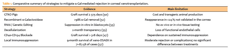
In summary, GTKO pigs remain the most effective strategy for preventing α-Gal-mediated rejection, whereas enzymatic, genetic, and decellularization approaches show promise but still require further validation. Immunosuppressive regimens can prolong graft survival; however, systemic toxicity and local rejection continue to present important challenges.
5. IMMUNOSUPPRESSIVE STRATEGIES AND PHARMACOLOGICAL APPROACHES
Effective pharmacological control of the immune response is essential for the success of corneal xenotransplantation. The combination of cyclosporine and mycophenolate mofetil has been shown to reduce immune responses against α-Gal(49). Cyclosporine inhibits T-lymphocyte activation, whereas mycophenolate suppresses lymphocyte proliferation, thereby reducing the overall adaptive immune response(49). Animal studies involving porcine corneal xenotransplantation have demonstrated that this combination therapy significantly prolongs graft survival(49).
6. PERSPECTIVES
Significant advances in corneal xenotransplantation have been achieved through the development of GTKO pigs and improvements in decellularization techniques. Recent studies suggest that combining GTKO grafts with immunosuppressive therapies can extend graft survival for up to 6 months in nonhuman primate models(13,47). Nevertheless, small residual amounts of α-Gal remain a challenge(11,13).
In addition to α-Gal, non-Gal antigens—including the sialic acid N-glycolylneuraminic acid, the SDa blood-group glycan, and proteins such as GABA type A receptor-associated protein-like 1 and cyclooxygenase-2 as well as cross-reactive swine leukocyte antigen epitopes—have also been identified as contributors to xenograft rejection(58). Even in GGTA1-knockout pigs, immune responses against other xenogeneic antigens persist, indicating that targeting non-Gal epitopes may also be necessary to fully prevent xenograft rejection(12,13,47).
ACKNOWLEDGMENTS
This study was supported by FAPESP (2023/15538-8, 2024/00877-4).
AUTHORS' CONTRIBUTIONS:
Significant contribution to conception and design: Augusto Villar.
Data Acquisition: Heloisa Nascimento, Thaís Maria da Mata Martins, Larissa R. Rosa.
Data Analysis and interpretation: José Álvaro Pereira Gomes.
Manuscript Drafting: Augusto Villar, Pedro Pires, Rodrigo Moreira, Gabriel Barbieri.
Significant intellectual content revision of the manuscript: Jorge Kalil, Heloisa Nascimento, Luiz Vicente Rizzo, Rubens Belfort Jr., Silvano Raia, José Álvaro Pereira Gomes.
Final approval of the submitted manuscript: Augusto Villar, Heloisa Nascimento, Jorge Kalil, Thaís Maria da Mata Martins, Luiz Vicente Rizzo, Gabriel Barbieri, Pedro Pires, Lucimeire N. Carvalho, Larissa R. Rosa, Rodrigo Moreira, Carmen Luz Pessuti, Henrique Ferrer, José Álvaro Pereira Gomes, Silvano Raia, Rubens Belfort Jr.
Statistical analysis: Rodrigo Moreira.
Obtaining funding: Lucimeire N. Carvalho.
Supervision of Administrative, technical, or material support: Carmen Luz Pessuti, Henrique Ferrer, Lucimeire N. Carvalho.
Research group leadership: Heloisa Nascimento, Rubens Belfort Jr., Jorge Kalil, Silvano Raia.
REFERENCES
1. Meek KM, Knupp C, Lewis PN, Morgan SR, Hayes S. Structural control of corneal transparency, refractive power and dynamics. Eye (Lond). 2025;39(4):644-50.
2. Meek KM, Knupp C. Corneal structure and transparency. Prog Retin Eye Res. 2015;49:1-16.
3. Coster DJ, Lowe MT, Keane MC, Williams KA; Australian Corneal Graft Registry Contributors. A comparison of lamellar and penetrating keratoplasty outcomes: a registry study. Ophthalmology. 2014;121(5):979-87.
4. World Health Organization (WHO). Elimination of avoidable blindness. Fifty-Sixth World Health Assembly. Geneva: WHO; 2003.
5. Gain P, Jullienne R, He Z, Aldossary M, Acquart S, Cognasse F, et al. Global survey of corneal transplantation and eye banking. JAMA Ophthalmol. 2016;134(2):167-73.
6. Chen L, Wei L, Shao A, Xu L. Immune risk assessment of residual αGal in xenogeneic decellularized cornea using GTKO mice. Regen Biomater. 2020; 7(4):427-34.
7. Yoon CH, Choi HJ, Kim MK. Corneal xenotransplantation: where are we standing? Prog Retin Eye Res. 2021;80:100876.
8. Niederkorn JY. Corneal transplantation and immune privilege. Int Rev Immunol. 2013;32(1):57-67.
9. Tahvildari M, Amouzegar A, Foulsham W, Dana R. Therapeutic approaches for induction of tolerance and immune quiescence in corneal allotransplantation. Cell Mol Life Sci. 2018;75(9):1509-20.
10. Forrest SL, Mercado CL, Engmann CM, Stacey AW, Hariharan L, Khan S, et al. Does the Current Global Health Agenda Lack Vision? Glob Health Sci Pract. 2023;11(1):e2200091.
11. Lu Y, Shao A, Shan Y, Zhao H, Leiguo M, Zhang Y, et al. A standardized quantitative method for detecting remnant alpha-Gal antigen in animal tissues or animal tissue-derived biomaterials and its application. Sci Rep. 2018;8(1):15424.
12. Lee HI, Kim MK, Oh JY, Ko JH, Lee HJ, Wee WR, et al. Gal α(1-3)Gal expression of the cornea in vitro, in vivo and in xenotransplantation. Xenotransplantation. 2007;14(6):612-8.
13. Kim YG, Oh JY, Gil GC, Kim MK, Ko JH, Lee S, et al. Identification of α-Gal and non-Gal epitopes in pig corneal endothelial cells and keratocytes by using mass spectrometry. Curr Eye Res. 2009;34(10):877-95.
14. Streilein JW. Ocular immune privilege: therapeutic opportunities from an experiment of nature. Nat Rev Immunol. 2003;3(11):879-89.
15. Medawar PB. Immunity to homologous grafted skin: the relationship between the antigens of blood and skin. Br J Exp Pathol. 1946;27(1):15-24.
16. Ferguson TA, Griffith TS. A vision of cell death: insights into immune privilege. Immunol Rev. 1997;156(1):167-84.
17. Notara M, Lentzsch A, Coroneo M, Cursiefen C. The role of limbal epithelial stem cells in regulating corneal (Lymph)angiogenic Privilege and the micromilieu of the limbal niche following UV Exposure. Stem Cells Int. 2018;2018:8620172.
18. Hori J, Vega JL, Masli S. Review of ocular immune privilege in the year 2010: modifying the immune privilege of the eye. Ocul Immunol Inflamm. 2010;18(5):325-33.
19. Hori J, Yamaguchi T, Keino H, Hamrah P, Maruyama K. Immune privilege in corneal transplantation. Prog Retin Eye Res. 2019;72:100758.
20. Shimmura-Tomita M, Wang M, Taniguchi H, Akiba H, Yagita H, Hori J. Galectin-9-mediated protection from allo-specific T cells as a mechanism of immune privilege of corneal allografts. PLoS One. 2013;8(5):e63620.
21. Niederkorn JY. See no evil, hear no evil, do no evil: the lessons of immune privilege. Nat Immunol. 2006;7(4):354-9.
22. Hori J, Streilein JW. Survival in high-risk eyes of epithelium-deprived orthotopic corneal allografts reconstituted in vitro with syngeneic epithelium. Invest Ophthalmol Vis Sci. 2003;44(2):658-64.
23. Ramos MF, Brassard J, Masli S. Immunology and Pathology in Ocular Drug Development. Toxicol Pathol. 2021;49(3):483-504.
24. Sohn JH, Kaplan HJ, Suk HJ, Bora PS, Bora NS. Complement regulatory activity of normal human intraocular fluid is mediated by MCP, DAF, and CD59. Invest Ophthalmol Vis Sci. 2000;41(13):4195-202.
25. Ambati BK, Nozaki M, Singh N, Takeda A, Jani PD, Suthar T, et al. Corneal avascularity is due to soluble VEGF receptor-1. Nature. 2006;443(7114):993-7.
26. Cursiefen C, Chen L, Saint-Geniez M, Hamrah P, Jin Y, Rashid S, et al. Nonvascular VEGF receptor 3 expression by corneal epithelium maintains avascularity and vision. Proc Natl Acad Sci USA. 2006;103(30):11405-10.
27. Bachmann BO, Bock F, Wiegand SJ, Maruyama K, Dana MR, Kruse FE, et al. Promotion of graft survival by vascular endothelial growth factor a neutralization after high-risk corneal transplantation. Arch Ophthalmol. 2008;126(1):71-7.
28. Sugita S, Yamada Y, Horie S, Nakamura O, Ishidoh K, Yamamoto Y, et al. Induction of T regulatory cells by cytotoxic T-lymphocyte antigen-2α on corneal endothelial cells. Invest Ophthalmol Vis Sci. 2011;52(5):2598-605.
29. Sugita S, Streilein JW. Iris pigment epithelium expressing CD86 (B7-2) directly suppresses T cell activation in vitro via binding to cytotoxic T lymphocyte-associated antigen 4. J Exp Med. 2003;198(1):161-71.
30. Nishida T, Saika S, Morishige N. Cornea and sclera: anatomy and physiology. In: Mannis MJ, Holland EJ, editors. Cornea: fundamentals, diagnosis, and management. 5th ed. Amsterdam (NL): Elsevier; 2022.
31. Gallo RL, Hooper LV. Epithelial antimicrobial defence of the skin and intestine. Nat Rev Immunol. 2012;12(7):503-16.
32. Dua HS, Gomes JA, Donoso LA, Laibson PR. The ocular surface as part of the mucosal immune system: conjunctival mucosa-specific lymphocytes in ocular surface pathology. Eye (Lond). 1995;9(Pt 3):261-7.
33. McDermott AM. Antimicrobial compounds in tears. Exp Eye Res. 2013;117:53-61.
34. Knop N, Knop E. Conjunctiva-associated lymphoid tissue in the human eye. Invest Ophthalmol Vis Sci. 2000;41(6):1270-9.
35. Kunishige T, Taniguchi H, Terada M, Akiba H, Yagita H, Abe R, et al. Protective role of ICOS and ICOS ligand in corneal transplantation and in maintenance of immune privilege. Invest Ophthalmol Vis Sci. 2016;57(15):6815-23.
36. Stein-Streilein J, Streilein JW. Anterior chamber associated immune deviation (ACAID): regulation, biological relevance, and implications for therapy. Int Rev Immunol. 2002;21(2-3):123-52.
37. Hara Y, Okamoto S, Rouse B, Streilein JW. Evidence that peritoneal exudate cells cultured with eye-derived fluids are the proximate antigen-presenting cells in immune deviation of the ocular type. J Immunol. 1993;151(10):5162-71.
38. Hori J, Joyce NC, Streilein JW. Immune privilege and immunogenicity reside among different layers of the mouse cornea. Invest Ophthalmol Vis Sci. 2000;41(10):3032-42.
39. Skelsey ME, Mayhew E, Niederkorn JY. Splenic B cells act as antigen presenting cells for the induction of anterior chamber-associated immune deviation. Invest Ophthalmol Vis Sci. 2003;44(12):5242-51.
40. Apte RS, Sinha D, Mayhew E, Wistow GJ, Niederkorn JY. Cutting edge: role of macrophage migration inhibitory factor in inhibiting NK cell activity and preserving immune privilege. J Immunol. 1998;160(12):5693-6.
41. Griffith TS, Brunner T, Fletcher SM, Green DR, Ferguson TA. Fas ligand-induced apoptosis as a mechanism of immune privilege. Science. 1995;270(5239):1189-92.
42. Hori J, Wang M, Miyashita M, Tanemoto K, Takahashi H, Takemori T, et al. B7-H1-induced apoptosis as a mechanism of immune privilege of corneal allografts. J Immunol. 2006;177(9):5928-35.
43. Forrester JV. Privilege revisited: an evaluation of the eye's defence mechanisms. Eye (Lond). 2009;23(4):756-66.
44. Satitpitakul V, Sun Z, Suri K, Amouzegar A, Katikireddy KR, Jurkunas UV, et al. Vasoactive Intestinal Peptide Promotes Corneal Allograft Survival. Am J Pathol. 2018;188(9):2016-24.
45. Paunicka KJ, Mellon J, Robertson D, Petroll M, Brown JR, Niederkorn JY. Severing corneal nerves in one eye induces sympathetic loss of immune privilege and promotes rejection of future corneal allografts placed in either eye. Am J Transplant. 2015;15(6):1490-501.
46. Steinman L. Elaborate interactions between the immune and nervous systems. Nat Immunol. 2004;5(6):575-81.
47. Choi HJ, Lee JJ, Kim DH, Kim MK, Lee HJ, Ko AY, et al. Blockade of CD40-CD154 costimulatory pathway promotes long-term survival of full-thickness porcine corneal grafts in nonhuman primates: clinically applicable xenocorneal transplantation. Am J Transplant. 2015;15(3):628-41.
48. Galili U, Mandrell RE, Hamadeh RM, Shohet SB, Griffiss JM. Interaction between human natural anti-α-galactosyl immunoglobulin G and bacteria of the human flora. Infect Immun. 1988;56(7):1730-7.
49. Lee HI, Kim MK, Oh JY, Ko JH, Lee HJ, Wee WR, et al. The role of cyclosporine and mycophenolate in an orthotopic porcine-to-rat corneal xenotransplantation. J Korean Med Sci. 2008;23(3):492-501.
50. Yoon CH, Choi SH, Choi HJ, Lee HJ, Kang HJ, Kim JM, et al. Long-term survival of full-thickness corneal xenografts from α1,3-galactosyltransferase gene-knockout miniature pigs in non-human primates. Xenotransplantation. 2020;27(1):e12559.
51. Ali A, Kemter E, Wolf E. Advances in Organ and Tissue Xenotransplantation. Annu Rev Anim Biosci. 2024;12(1):369-90.
52. Liu D, Kobayashi T, Yokoyama I, Ogawa H, Nagasaka T, Muramatsu H, et al. Enzymatic removal of alphaGal antigen in pig kidneys by ex vivo and in vivo administration of endo-β-galactosidase C. Xenotransplantation. 2002;9(3):228-36.
53. Chi H, Shinohara M, Yokomine T, Sato M, Takao S, Yoshida M, et al. Successful suppression of endogenous α-1,3-galactosyltransferase expression by RNA interference in pig embryos generated in vitro. J Reprod Dev. 2012;58(1):69-76.
54. Wilson SL, Sidney LE, Dunphy SE, Rose JB, Hopkinson A. Keeping an eye on decellularized corneas: a review of methods, characterization and applications. J Funct Biomater. 2013;4(3):114-61.
55. Shi Y, Bikkuzin T, Song Z, Jin X, Jin H, Li X, et al. Comprehensive evaluation of decellularized porcine corneal after clinical transplantation. Xenotransplantation. 2017;24(6):e12338.
56. Kim J, Kim DH, Choi HJ, Lee HJ, Kang HJ, Park CG, et al. Anti-CD40 antibody-mediated costimulation blockade promotes long-term survival of deep-lamellar porcine corneal grafts in non-human primates. Xenotransplantation. 2017;24(3):e12298.
57. Li X, Huang Y, Liang Q, Li G, Feng S, Song Y, et al. Local Immunosuppression in Wuzhishan Pig to Rhesus Monkey Descemet's Stripping Automated Endothelial Keratoplasty: An Innovative Method to Promote the Survival of Xenografts. Ophthalmic Res. 2022;65(2):196-209.
58. Zhou Q, Li T, Wang K, Zhang Q, Geng Z, Deng S, et al. Current status of xenotransplantation research and the strategies for preventing xenograft rejection. Front Immunol. 2022;13:928173.
Submitted for publication:
April 9, 2026.
Accepted for publication:
May 8, 2026.
Data Availability Statement: The datasets generated and/or analyzed during the current study are already available.
Disclosure of potential conflicts of interest: The authors declare no potential conflicts of interest.

How to cite this article:
 ABO is licensed under a Creative Commons Attribution-NonComercial 4.0 Internacional.

