Arq. Bras. Oftalmol. 2004; 67 (2): 10.1590/S0004-27492004000200005
Total: 2546
Juliana Simões Munhoz; Solange Rios Salomão1; Adriana Berezovsky2; Paula Yuri Sacai3
DOI: 10.1590/S0004-27492004000200005
ABSTRACT
PURPOSE: To determine normative values for EOG in a group of healthy adult volunteers, according to the standard protocol recommended by the International Society for Clinical Eletrophisiology of Vision - ISCEV. METHODS: 33 volunteers aging from 18 to 55 years (mean=26.75±10.50) were included in the study. EOG was recorded by skin electrodes positioned in the external canthi of the eyes, with LED stimulus presented in a Ganzfeld dome. Subject's task was to fix the LED stimulus either straight ahead or with saccadic horizontal movements. The potential difference between the cornea and posterior pole of the eye was recorded each minute. The examination was done in 3 phases: 1- pre-adaptation; 2- dark (scotopic) and 3- light (photopic). The Arden ratio was calculated as the proportion between the maximum peak amplitude in the light phase divided by the minimum peak amplitude in the dark. RESULTS: At the scotopic phase, the minimum peak amplitude occurred between 14 and 21 minutes after the beginning of the stimulation (mean=17.82±2.11 min) and the amplitude ranged from 206 to 635 µV (mean 365.73±122.52 µV). At the photopic phase the maximun peak amplitude occurred between 27 and 34 minutes (mean=30.06±1.80 min); the amplitude ranged from 646 to 1250 µV (mean= 950.70±179.16 µV). Arden ratio ranged from 1.85 to 4.02 with a lower normal limit of 1.92 and the upper limit of 4.00. CONCLUSION: The results obtained in this study are comparable to those previously reported in the literature, with a lower normal limit of 1.92 for Arden ratio. These values will be helpful to evaluate patients with several retinal disorders. Inclusion of other age groups will provide extended normative data.
Keywords: Electrooculography; Reference values; Electrophysiology; Retina; Pigment epithelium of eye
RESUMO
OBJETIVO: Estabelecer valores normativos para o eletro-oculografia (EOG), em grupo de voluntários normais, segundo o protocolo recomendado pela Sociedade Internacional de Eletrofisiologia Visual Clínica (ISCEV). MÉTODO: Participaram 34 sujeitos com idades de 18 a 55 anos (média 26,75 ± 10,50), 9 homens e 24 mulheres. O EOG foi realizado utilizando eletrodos e estímulos luminosos. A tarefa do sujeito foi fixar estímulos luminosos (LEDs) em frente ou realizando movimentos sacádicos horizontais. O exame constou de três fases: 1- Pré-adaptação; 2- Escotópica e 3- Fotópica. O índice de Arden foi determinado como sendo a proporção entre o pico máximo da amplitude na fase fotópica, dividido pelo vale máximo da amplitude na fase escotópica. RESULTADO: Na fase escotópica, o vale máximo ocorreu entre 14 e 21 minutos após o início do exame (média 17,82 ± 2,11 minutos) e a amplitude variou de 206 a 635 µV (média 365,73 ± 122,52 µV). Na fase fotópica, o pico máximo ocorreu entre 27 a 34 minutos (média 30,06 ± 1,80 minuto) e a amplitude variou de 646 a 1249,50µV (média 950,70 ± 179,16). O índice de Arden variou de 1,85 a 4,02 (média 2,79±0.63) sendo o limite inferior de 1,92 e o superior de 4,00. CONCLUSÃO: Os valores para o índice de Arden estão de acordo com valores descritos anteriormente na literatura, sendo o limite inferior de normalidade quando este for maior ou igual a 1,92 e serão úteis para avaliação de pacientes com distúrbios retinianos. A inclusão de outras faixas etárias fornecerá dados normativos mais abrangentes.
Descritores: Eletro-oculografia; Valores de referência; Eletrofisiologia; Retina; Epitélio pigmentado ocular
INTRODUÇÃO
O eletro-oculograma (EOG), corresponde ao potencial de repouso do olho equivalente em média a 6 µV, sendo que para alterações na iluminação retiniana, há uma diferença de potencial entre a córnea, que é eletropositiva e o epitélio pigmentado da retina, que é eletronegativo(1). Essa polarização também é conhecida como diferença de potencial córneo-retiniana.
O termo eletro-oculografia foi introduzido em 1951 por investigadores que mediram a diferença de potencial entre a córnea e o pólo posterior do globo ocular, mas anteriormente, Holmgren demonstrou haver uma alteração na amplitude deste potencial em diferentes níveis de iluminação. Em 1960 utilizou-se o EOG no estudo de movimentos oculares normais e patológicos(2). Em 1962 Arden, Barrada e Kelsey definiram a eletro-oculografia como novo teste da função retiniana. Assim, o potencial de repouso ou potencial córneo-retiniano, ou mesmo ainda corrente de repouso passou a ser uma perspectiva no estado da fisiologia e da fisiopatologia retiniana(3).
O objetivo desta pesquisa consistiu em estabelecer valores normativos para o exame eletrofisiológico visual de eletro-oculografia em uma população de adultos, empregando técnicas recomendadas pela Sociedade Internacional de Eletrofisiologia Visual Clínica – ISCEV. Esta sociedade por sua vez, recomenda que cada laboratório de eletrofisiologia visual obtenha dados normativos próprios(4). Outro motivo importante para a realização deste trabalho é a escassez de dados e estudos relacionados aos valores normais e aos procedimentos que devem ser padronizados para se obter resultados confiáveis.
MÉTODOS
Este estudo foi realizado no período de 06/1999 a 12/2001 no Laboratório de Eletrofisiologia Visual Clínica do Departamento de Oftalmologia da Universidade Federal de São Paulo.
Participaram deste estudo 33 (trinta e três) sujeitos com idades variando entre 18 a 55 anos (média 26,75 ± 10,50), sendo 9 homens e 24 mulheres.
Foram observados os seguintes critérios de inclusão para a pesquisa: ter acuidade visual igual a 20/20 na notação de Snellen ou 0,0 na notação LogMar, com a melhor correção óptica, ausência de distúrbios visuais ou neurológicos, ausência de história familiar positiva para doenças oculares, ausência de queixas visuais.
Todos os sujeitos assinaram o termo de consentimento livre e esclarecido, o qual informava os procedimentos do exame, seus riscos e benefícios. O protocolo foi aprovado pelo comitê de ética em Pesquisa da Universidade Federal de São Paulo.
O EOG foi realizado nos dois olhos sem dilatação pupilar. Após limpeza local da pele dos cantos periorbitários mediais e laterais com álcool 70%, foram aplicados quatro eletrodos de cúpula de ouro, preenchidos com gel condutor eletrolítico. Os eletrodos foram fixados com fita adesiva e conectados ao cabo do sistema de captação de respostas.
O sujeito foi então posicionado na cúpula de Ganzfeld e orientado a olhar fixamente, durante todo o exame, para estímulos luminosos (LEDs vermelhos). A estimulação foi feita inicialmente com um LED central, por 45 segundos, seguido de LEDs laterais, posicionados a 15º do estímulo central, que se iluminavam alternadamente, por um intervalo de 15 segundos. Para o registro do EOG foi utilizado o sistema eletrodiagnóstico UTAS E-3000 (LKC Technologies Inc. Gaithersburg, MD,USA). As amplitudes das respostas foram medidas em microVolts (µV) e o tempo de culminação em milissegundos (ms).
O registro do EOG foi acompanhado em osciloscópio, permitindo o controle da direção do olhar do sujeito, durante o potencial de repouso e na movimentação sacádica para a direita e para esquerda. O exame constou de três fases: Fase 1- Pré-adaptação, com estimulação em condição fotópica com luz de fundo com duração de 6 minutos; Fase 2- Escotópica, com duração de 16 minutos; Fase 3- Fotópica, com a mesma iluminação de fundo da fase 1, com duração de 14 minutos.
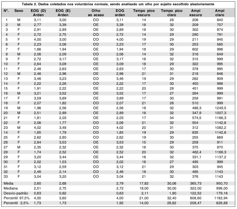
As análises estatísticas foram realizadas através do programa estatístico Jandel Sigmastat - Statistical Software Version 2.0, USA. Foram calculadas a média, o desvio-padrão, a mediana e 2,5 e 97,5 percentis dos parâmetros de índice de Arden, amplitude e tempo tanto para fase escotópica como para fase fotópica(5). Para os cálculos foram utilizados os resultados de apenas um olho por sujeito escolhido aleatoriamente (Tabela 1).
RESULTADOS
O vale na fase escura ocorreu entre 14 e 21 minutos, média 17,82 ± 2,11 minutos, após o início do exame. A amplitude por sua vez variou de 206 a 635 µV sendo a média 365,73 ± 122,52. O pico na fase clara ocorreu entre 27 e 34 minutos, média 30,06 ± 1,80 minuto após o início do exame. Em relação à amplitude, a variação foi de 646 a 1249,50 µV e a média igual a 950,70 ± 179,16. O índice de Arden variou de 1,85 a 4,02; sendo a média igual a 2,79 ± 0,63, o limite inferior (percentil 2,5%) foi igual a 1,92 e o limite superior (percentil 97,5%) igual a 4,00.
A relação interocular do índice de Arden foi calculada usando-se a divisão do Índice de Arden obtido no olho esquerdo pelo olho direito, assim temos que, este valor variou de 0,76 a 1,45 a média encontrada foi de 1,04 ± 0,16. Esta relação foi calculada para verificarmos o quanto o índice de Arden variou de um olho em relação ao outro.
DISCUSSÃO
O eletro-oculograma é importante para auxiliar no diagnóstico de doenças retinianas, em que o epitélio pigmentado da retina se encontra comprometido. Atua como diagnóstico diferencial, juntamente com o eletrorretinograma, na distrofia macular de Best(6).
O EOG possui dois componentes importantes que nos dão respostas referentes aos registros à adaptação ao escuro e ao claro. O componente insensível à luz do EOG - potencial escotópico - depende da integridade do epitélio pigmentado da retina (EPR) e outras estruturas oculares como córnea, cristalino e corpo ciliar, mas não é influenciado pela iluminação retiniana prévia e independe do estado funcional dos fotorreceptores. Já o componente fotossensível do EOG - potencial fotópico - é gerado pela despolarização da membrana basal do epitélio pigmentado da retina e depende da integridade dos fotorreceptores chamados cones e bastonetes e da retina interna(1).
A onda na fase escotópica tende a decrescer devido a uma saturação de íons potássio no epitélio pigmentário da retina, logo após o início da fase fotópica, esta onda tende a aumentar em indivíduos normais, devido a uma despolarização da membrana basal do epitélio pigmentado da retina que não está relacionado diretamente com os níveis de aumento de potássio nas células deste epitélio (Figura 1). Esta despolarização na membrana basal resulta em um aumento do potencial elétrico através das células do epitélio conhecido como potencial trans-epitelial. Para que esta resposta seja gerada, os fotorreceptores devem estar intactos. Além disso, o aumento da onda ocorre apenas quando há um contato entre os fotorreceptores e o epitélio pigmentado da retina(7).
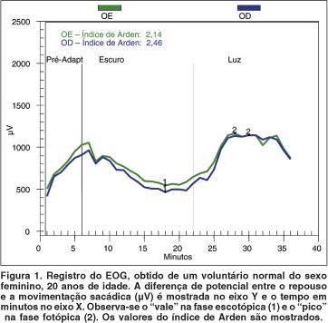
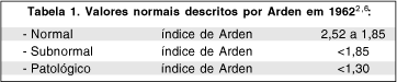
O Índice de Arden que corresponde à razão entre o maior valor do pico da amplitude na fase clara e o menor valor do pico da amplitude na fase escura após o tempo de deslumbramento para cada fase(2) (Figura 1), possui os seguintes valores normais descritos na literatura:
Nos dados obtidos para índice de Arden neste trabalho, temos que o valor do percentil de 2,5% é de 1,73 para ambos os olhos, ou seja, quando analisamos uma amostra de escolha não aleatória, temos um Índice de Arden menor que 1,85, porém corremos o risco de causar um vício de amostra, que faz com que os dados não sejam confiáveis, já se fizermos uma escolha aleatória dos olhos, será a melhor forma de trabalhar evitando um vício de seleção ou até mesmo um erro estatístico. Desta maneira será mais certo que em 95% de nossa amostra, o índice de Arden encontra-se entre 4,00 e 1,92, e que abaixo disto é considerado subnormal para exames realizados em nosso laboratório.
A relação interocular (OE/OD) é um valor que nos mostra a variação do Índice de Arden de um olho para outro. Teoricamente este índice deve ser bem semelhante em ambos os olhos, mas na prática observamos que este valor varia devido ao posicionamento de eletrodos, influência do olho dominante, atenção do indivíduo durante o exame, etc. Esta relação é tão melhor quanto mais próxima de 1,0. No presente estudo a média deste valor se encontra em 1,04 sendo o desvio-padrão igual a 0,16.
O EOG pode ser realizado com as pupilas dilatadas ou não. As pupilas dilatadas fornecem um melhor controle dos níveis de iluminação, mas por outro lado podem causar um desconforto ao paciente. Se o diâmetro pupilar do sujeito estiver aproximadamente igual 3 mm e a iluminação de no mínimo 500 cd/m² não há necessidade de dilatação pupilar(3). Em nosso trabalho os exames foram realizados sem dilatação pupilar. O exame não necessita ser feito com correção óptica, sejam por óculos ou lentes de contato, pois não há influência na resposta do exame, desde que o indivíduo consiga fixar a mira (LED vermelho).
A padronização do exame de eletro-oculografia é importante devido à escassez de estudos e padronização tanto dos valores normais, quanto ao método de realização e procedimentos de exame.
CONCLUSÃO
Os valores normativos obtidos para o índice de Arden num grupo de 33 voluntários normais saudáveis estão de acordo com valores descritos anteriormente na literatura, sendo o valor maior ou igual a 1,92 considerado normal. Estes valores serão úteis para avaliação de pacientes com distúrbios retinianos. Há necessidade da inclusão de outras faixas etárias, para obtenção de dados normativos mais abrangentes.
REFERÊNCIAS
1. Fishman GA. The electro-oculogram. In: Fishman GA, Birch DG, Holder GE, Briggell MG, editors. Electrophysiologic testing in disorders of the retina, optic nerve, and visual pathway. 2nd ed. San Francisco: The Foundation of American Academy of Ophthalmology; 2001. p.157-75.
2. Pacini Neto L. Eletro-oculografia. In: Dantas AM, editor. Eletrofisiologia ocular. Rio de Janeiro: Cultura Médica, 1995. p.112-40.
3. Arden G B, Barrada A, Kelsey JH. New clinical test of retinal function based upon the standing potential of the eye. Br J Ophthalmol 1962;46:449-67.
4. Marmor MF, Zrenner E. Standard for clinical electro-oculography. Doc Ophthalmol 1993;85:115-24.
5. Pereira JM, Mendieta L, Sacai PY, Salomão SR, Berezovsky A. Estudo normativo do eletrorretinograma de campo total em adultos jovens. Arq Bras Oftalmol 2003;66:137-44.
6. Holder HE. Best's disease. In: Heckenlively JR, Arden GB, editors. Principles and practice of clinical eletrophysiology. St Louis: Mosby Year Book; 1991. p.692-99.
7. Kolder HE Electro-oculography. In: Heckenlively JR, Arden GB, editors. Principles and practice of clinical eletrophysiology. St Louis: Mosby Year Book; 1991. p.301-13.
Endereço para correspondência
Solange Rios Salomão
R. Botucatu, 822
São Paulo (SP) Cep 04023-062
E-mail: [email protected]
Recebido para publicação em 02.07.2003
Versão revisada recebida em 19.11.2003
Aprovação em 26.11.2003
Apoio: FAPESP no. 97/11493-3 para Solange R. Salomão; Bolsa PIBIC-CNPq no. 110046/01-3, para Juliana Simões Munhoz.
Nota Editorial: Pela análise deste trabalho e por sua anuência na divulgação desta nota, agradecemos aos Drs. Fernando Oréfice e Flávio da Rocha Lima Paranhos.

How to cite this article:
 ABO is licensed under a Creative Commons Attribution-NonComercial 4.0 Internacional.