Arq. Bras. Oftalmol. 2006; 69 (6): 10.1590/S0004-27492006000600016
Total: 2084
Rodrigo Almeida Vieira Santos1; Hellmann Dantas2; Carlos Teixeira Brandt4; Adélia Almeida1; Daniela Lyra Antunes1; Helder Viana Santana1
DOI: 10.1590/S0004-27492006000600016
ABSTRACT
PURPOSE: To study the retinal nerve fiber layer in young patients suffering from hepatosplenic schistosomiasis mansoni. METHODS: Twenty-three patients with hepatosplenic schistosomiasis mansoni who were submitted, when children, to splenectomy, ligature of the left gastric vein and auto-implantation of spleen tissue in the major omentum underwent GDx Scanning Laser System evaluations. All patients presented with intraocular pressure below 21 mmHg. RESULTS: Only one patient suffering from hepatosplenic schistosomiasis mansoni showed abnormalities on the GDx examination. There were no abnormalities on GDx examination in the control group. CONCLUSION: There was no significant difference between the two groups of this study. Only one patient showed retinal nerve fiber layer reduction.
Keywords: Nerve fibers; Retina; Schistosomiasis; Diagnostic techniques; Microscopy
RESUMO
OBJETIVO: Estudar a camada de fibras nervosas da retina de portadores de esquistossomose mansônica hepatoesplênica. MÉTODOS: Foram submetidos ao exame com o GDx Scanning Laser System, 23 portadores de esquistossomose na forma hepatoesplênica que tinham sido submetidos, quando crianças, a esplenectomia, ligadura da veia gástrica esquerda e auto-implante de tecido esplênico no omento maior. Todos apresentaram pressão intra-ocular menor que 21 mmHg. No grupo controle foram estudados 23 indivíduos com idade e condição socioeconômico-geográfica similar, sem esquistossomose. RESULTADOS: Em apenas um paciente do grupo portador de esquistossomose foram observadas alterações em quatro parâmetros: superior nasal, média superior, média da espessura e número de fibra. Todos os indivíduos do grupo controle apresentaram GDx com parâmetros dentro da normalidade. CONCLUSÃO: No estudo realizado não foi encontrado diferença significante entre os dois grupos. Apenas um paciente mostrou redução da camada de fibras nervosas.
Descritores: Fibras nervosas; Retina; Esquistossomose; Técnicas de diagnóstico oftalmológico; Microscopia eletrônica de varredura
INTRODUÇÃO
A esquistossomose, uma das infecções mais comuns parasitárias do homem, continua endêmica em várias regiões do mundo(1-4). No Brasil, Pernambuco é um dos estados que apresenta maior número de esquistossomóticos, com taxa de prevalência média de 23% na população geral(3,5-7). Ovos, larvas ou depósitos de imunocomplexos antígeno-anticorpo do esquistossoma podem ser encontrados em praticamente qualquer órgão ou tecido do organismo(1-4,8-21). Há descrição da associação do estágio avançado da esquistossomose mansônica na forma hepato-esplênica com lesões oculares(1-3).
O Schistosoma mansoni também é responsável por lesões que repercutem na hemodinâmica da circulação venosa portal, arterial pulmonar e venosa sistêmica(9,22). Há relatos de pacientes portadores da forma cirúrgica da esquistossomose mansônica que apresentavam dilatações e tortuosidade dos vasos retinianos, especialmente do componente venoso(1,9,22).
Recentemente, foi descrito retardo no tempo de chegada do contraste fluoresceínico na retina de pacientes esquistossomóticos, na fase venosa precoce do exame angiofluoresceinográfico, o que sugere dificuldade de irrigação arterial da retina face ao aumento da pressão nos capilares devido à diminuição de drenagem venosa deste órgão para a circulação sistêmica(22).
O padrão de suprimento arterial e de drenagem venosa entre a região peripapilar e a cabeça do nervo óptico pode ter implicações na fisiopatologia da lesão glaucomatosa e, consequentemente, na perda das células ganglionares da retina(23). Uma das formas de avaliação desta estrutura é por meio do analisador da camada de fibras nervosas da retina (GDx)(24). O estudo apresentado por Lopes de Faria et al.(25) que avaliou a camada de fibras nervosas da retina (CFN) por meio do GDx em pacientes com diabetes melito tipo I sem sinais de retinopatia observou a redução da CFN nos pacientes avaliados.
Hoyt e col. foram os primeiros a destacar as alterações na CFN antes do aparecimento de defeitos correspondentes na perimetria manual em pacientes com glaucoma(26).
O uso do laser polarizado para análise da espessura da CFN só foi possível graças à sua capacidade de birrefringência, que provoca um retardo na velocidade da luz ao incidir sobre ela, e este retardo é proporcional à espessura da CFN, sendo possível assim quantificar a espessura da CFN de forma indireta e conseqüentemente a perda nervosa(24). O retardo corresponde a uma redução da CFN como observada em estudo envolvendo macacos(27). O exame apresenta boa reprodutibilidade e índices de especificidade de 91,1% e sensibilidade de 87,3%(28).
Esta pesquisa teve por objetivo estudar a camada de fibras nervosas da retina de pacientes portadores de esquistossomose mansônica hepato-esplênica que haviam sido submetidos, quando crianças, a esplenectomia, ligadura da veia gástrica esquerda e auto-implante de tecido esplênico no omento maior.
MÉTODOS
O presente estudo foi desenvolvido na Fundação Altino Ventura (FAV) e Hospital de Olhos de Pernambuco (HOPE), em conjunto com o Departamento de Cirurgia da Universidade Federal de Pernambuco.
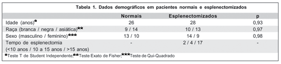
Analisaram-se prospectivamente 46 olhos de 23 pacientes, 13 do sexo masculino e 10 do sexo feminino, portadores de esquistossomose mansônica na forma hepato-esplênica submetidos, quando crianças, a esplenectomia, ligadura da veia gástrica esquerda e auto-implante de tecido esplênico no omento maior. O grupo controle foi constituído por 23 indivíduos da mesma condição socioeconômica e geográfica, com idade e gênero pareados, não portadores de esquistossomose. O protocolo do exame foi previamente submetido à análise de comissão ética tendo obtido sua aprovação.
Os pacientes foram submetidos a exame oftalmológico completo na FAV, que constou de acuidade visual com correção, avaliação da fixação ocular e da motilidade extrínseca, biomicroscopia do segmento anterior, aferição da pressão intra-ocular com tonômetro de aplanação tipo Goldmann, refração, biomicroscopia do segmento posterior com lente de 90 dioptrias.
Os critérios de inclusão neste estudo foram pressão intra-ocular menor que 21 mmHg, ausência de história familiar de glaucoma, acuidade visual corrigida igual ou melhor que 20/40 em cada um dos os olhos, escavação de disco óptico menor ou igual a 0,4 e, ainda, assimetria nas escavações de discos ópticos de um mesmo indivíduo, caso presente, inferior a 0,2.
Foram excluídos aqueles que apresentassem diâmetro pupilar inferior a dois milímetros, erro refracional superior a quatro dioptrias, história de traumatismo ocular ou uso prévio de corticosteróide tópico ou sistêmico, alterações cório-retinianas detectáveis oftalmoscopicamente assim como qualquer outra doença ocular e os que já houvessem sido submetidos a procedimentos a laser ou cirurgia ocular.
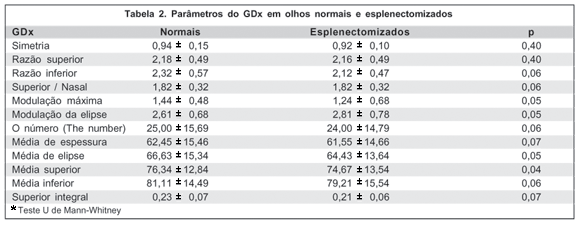
A análise das fibras nervosas da retina foi realizada utilizando-se o polarímetro de varredura a laser GDxTM Glaucoma Scanning Laser System® (Laser Diagnostic Technologies, inc. San Diego, CA, USA) cujo modelo contém o pacote de análise estatística versão 1.0. Obteve-se duas imagens e realizou-se uma imagem média para análise, após ajuste de foco e brilho, e centralização do disco óptico. Utilizando-se essas imagens calculou-se sua média (parte do programa do aparelho). As razões e valores selecionados neste estudo para análise estatística foram aqueles fornecidos pelo aparelho, quando se utilizava o programa completo de comparação, obtidos na impressão. As razões e valores foram: simetria, razão superior, razão inferior, superior/nasal, modulação máxima, modulação da elipse, média da espessura, média da elipse, média superior, média inferior, superior integral e o número (The Number), cujos resultados compreendido entre 0 e 30 é considerado normal, entre 30 e 70 suspeito de glaucoma e acima de 70 possível glaucoma.
A margem do disco óptico era marcada com um círculo ou elipse e a espessura da camada de fibras nervosa da retina era determinada à distância de 1,75 diâmetro papilar para cada quadrante de região peripapilar: superior (120º), inferior (120º), nasal (70º), temporal (50º).
Todos os exames com GDx foram realizados pelo mesmo examinador, que não conhecia os dados clínicos dos pacientes, sendo realizados com luz da sala acesa, sem midríase medicamentosa.
Os resultados foram analisados quanto à freqüência de exames anormais e dos respectivos índices alterados. Considerou-se exame anormal a presença de um ou mais índices considerados fora da normalidade pelo pacote estatístico do equipamento GDx.
RESULTADOS
Nos 46 olhos do grupo com esquistossomose hepato-esplênica analisados, o GDx mostrou-se alterado, no parâmetro "o número" (The Number), em apenas dois olhos de um mesmo paciente. No grupo controle, nenhum exame mostrou-se alterado. A comparação da freqüência de exames alterados não mostrou diferença estatisticamente significante.
DISCUSSÃO
O relato do estudo da camada de fibras nervosas da retina por meio do GDx em pacientes adultos com esquistossomose hepatoesplênica tratados clínica e cirurgicamente, quando crianças, é original na literatura.
A esquistossomose mansônica é responsável por lesões hepáticas que podem alterar a hemodinâmica da circulação venosa portal, arterial pulmonar e venosa sistêmica(5). Os pacientes esquistossomóticos têm comportamento hemodinâmico semelhante aos pacientes cirróticos(11). Foi demonstrado que pacientes com cirrose hepática apresentaram diminuição do fluxo sanguíneo regional cerebral(13). Como conseqüência, é possível que existam alterações hemodinâmicas na circulação ocular desses pacientes.
Um estudo recente verificou tortuosidade e alargamento dos vasos retinianos, especialmente das veias, em 28% dos pacientes com esquistossomose na forma hepatoesplênica tratados clínica e cirurgicamente. Observou-se, também, retardo no tempo de chegada do contraste na retina de pacientes esquistossomóticos, sugerindo retardo no fluxo sanguíneo arterial da retina, o que seria explicado pelo aumento da pressão capilar e retardo da drenagem venosa para circulação sistêmica(22). Essa alteração hemodinâmica na circulação sanguínea retiniana poderia, por sua vez, produzir, a longo prazo, danos nas estruturas oculares da região peripapilar e papila do nervo óptico, danos estes relacionados a padrões distintos tanto do suprimento arterial quanto da drenagem venosa(23).
CONCLUSÃO
Concluindo, em nosso estudo detectamos a presença de um paciente com esquistossomose hepatoesplênica que fora submetido, quando criança, a esplenectomia, ligadura da veia gástrica esquerda e auto-implante de tecido esplênico no omento maior, e que apresentou alterações na camada de fibras nervosas da retina. Apesar de não haver diferença estatisticamente significante quando comparado com o grupo controle, este achado poderia, talvez, dar suporte à hipótese de que repercussões hemodinâmicas encontradas em pacientes com esquistossomose, poderiam, a longo prazo, tornar estes indivíduos mais suscetíveis a alterações estruturais oculares. Portanto, tal fato corrobora para investigações futuras sobre aspectos oculares em indivíduos com esquistossomose.
REFERÊNCIAS
1. Oréfice F, Simal CJ, Pittella JE. Schistosomotic choroiditis. I. Funduscopic changes and differential diagnosis. Br J Ophthalmol. 1985;69(4):294-9.
2. Milligan A, Burns DA. Ectopic cutaneous schistosomiasis and schistosomal ocular inflammatory disease. Br J Dermatol. 1988;119(6):793-8.
3. Salomão MRI. Alterações coriorretinianas em indivíduos de área hiperendêmica de esquistossomose [tese]. Minas Gerais: Faculdade de Medicina. Universidade Federal de Minas Gerais; 1995.
4. Oréfice F, Pittela JEH, Simal CJR, Coscarelli G. Uveíte esquistossomótica: alterações fundoscópicas; achados histológicos do ovo do S. mansoni, abordagem da etiologia e tratamento. Arq Bras Oftalmol. 1988;51(3):123-34.
5. Brandt CT, Sá HP, Caneca OAF, Santana JV, Miranda P, Carvalheira R. Esquistossomose mansônica hepatoesplênica em adolescentes: carga parasitária residual após tratamento clinico-cirúrgico. An Fac Med Univ Fed Pernamb. 1998;43(2):123-6.
6. Brandt CT, Maciel DT, Almeida Filho P, Torres IAA. Cintilografia renal em crianças com esquistossomose mansônica, na forma hepato-esplênica, submetidas a esplenectomia, ligadura da veia gástrica esquerda e auto-implante esplênico. An Fac Med Univ Fed Pernamb. 1998;43(1):13-22.
7. Brandt CT, Maciel DT, Frei Caneca OA. Esplenose associada ao tratamento cirúrgico da hipertensão porta esquistossomótica na criança: avaliação de dez anos. An Fac Med Univ Fed Pernamb. 1999;44(1):15-20.
8. Doumenge JP, Mott KE, Cheung C, Villenave D, Chapuis O, Perrin MF, et al. Atlas of the global distribution of schistosomiasis. Geneva: OMS; 1987.
9. Dickinson AJ, Rosenthal AR, Nicholson KE. Inflammation of the retinal pigment epithelium: a unique presentation of ocular schistosomiasis. Br J Ophthalmol. 1990;74(7):440-2.
10. Moreno RC. Sobre algunas lesiones oculares en la Schistosomiasis Mansoni. Arch Ven Soc Otorrino Laringol Oftalmol Neurol. 1978;1:158-75.
11. Stein PC, Char DH. Intraocular granuloma: a Schistosoma mansoni model of ocular inflammation. Invest Ophthalmol Vis Sci. 1982;23(4):479-88.
12. Pittella JE, Orefice F. Schistosomotic choroiditis. II. Report of first case. Br J Ophthalmol. 1985;69(4):300-2.
13. Neves J, Pedroso ERP, Oréfice F, Souza DWC, Greco D, Rocha MOC, et al. Esquistossomose pulmonar. III: forma crônica extensa com hipertensão pulmonar e na vigência de hipertensão portal associada à provável coroidite e retinite esquistossomótica. Arq Bras Oftalmol. 1978;41:215-20.
14. Lemos E. Alterações retinianas na esquistossomose hepatoesplênica. Rev Bras Oftalmol. 1980;39(3):219-24.
15. Vedy J, Rivaud C, Chanut G, Graveline J. Uvéites et parasitoses intestinales. Bull Soc Ophtalmol Fr. 1982;82(1):97-8.
16. Oréfice F. Enfermidades causadas por helmintos. In: Belfort Jr R, Couto CA, Castro FM. Uveítes: sinopsis diagnóstica y terapêutica. México: Ciba Vision; 1997. p.262-4.
17. Oréfice F, Belfort RJ. Helmintíases: esquistossomose ocular. São Paulo: Roca; 1987.
18. Ferreira JLL, Oréfice F, Katz N, Pittella JEH. Ocular fundoscopic lesions and Schistosomiasis Mansoni. In: Recent advances in uveitis. Amsterdam: Kugler; 1992.
19. Queiroz JM. Aspectos experimentais e clínicos das manifestações oculares da esquistossomose mansoni. Oftalmol Ibero Am. 1961;22:115-78.
20. Mahmoud AAF. Schistosomiasis (Bilharziasis). In: Wyngaarden JB, Cecil RL, Smith LH, editors. Cecil textbook of medicine. 17th ed. Philadelphia: WB Saunders; 1985. p.1809-15.
21. Bac DJ, Teichler MJ, Jonker LC, van der Merwe CF. Schistosomiasis in ectopic or unusual sites. A report of 5 cases. S Afr Med J. 1987;72(10):717-8.
22. Souza AC, Brandt CT, Ventura LO, Oréfice F. Achados oftalmológicos em jovens portadores de esquistossomose hepatoesplênica submetidos a esplenectomia, ligadura da veia gástrica esquerda e auto-implante esplênico. An Fac Med Univ Fed Pernamb. 2001;46(2):89-94.
23. Vasconcelos JPC, Costa VP. Anatomia e fisiologia da irrigação sangüínea do nervo óptico. In: Dias JFP, Almeida HG. Glaucoma. 2a ed. Rio de Janeiro: Cultura Médica; 2000. p.21-2.
24. Lauande-Pimentel R, Costa VP. Introdução à análise da camada de fibras nervosas da retina. In: Lauande-Pimentel R, Costa VP. Análise da camada de fibras nervosas da retina. Um guia para interpretar o exame de polarimetria. Rio de Janeiro: Cultura Médica; 2001. p.9-16.
25. Lopes de Faria JM, Russ H, Costa VP. Retinal nerve fibre layer loss in patients with type 1 diabetes mellitus without retinopathy. Br J Ophthalmol. 2002;86(7):725-8.
26. Hoyt WF, Frisen L, Newman NM. Fundoscopy of nerve fiber layer defects in glaucoma. Invest Ophthalmol. 1973;12(11):814-29.
27. Weinreb RN, Dreher AW, Coleman A, Quigley H, Shaw B, Reiter K. Histopathologic validation of Fourier-ellipsometry measurements of retinal nerve fiber layer thickness. Arch Ophthalmol. 1990;108(4):557-60.
28. Susanna Junior R, Takahashi W, Walter Y, Nakamura NKF. Sensibilidade e especificidade da avaliação da camada de fibras nervosas examinadas através da polarimetria a laser. Rev Bras Oftalmol. 1998;57(1):17-22.
Endereço para correspondência:
Rodrigo Almeida Vieira Santos
SQS 111, Bl-G - Apto. 301
Brasília (DF) CEP 70374-070
E-mail: [email protected]
Recebido para publicação em 15.08.2005
Última versão recebida em 02.03.2006
Aprovação em 05.03.2006
Trabalho realizado no Hospital de Olhos de Pernambuco - Recife (PE) - Brasil.
Nota Editorial: Depois de concluída a análise do artigo sob sigilo editorial e com a anuência do Dr. Marcelo Carvalho da Cunha sobre a divulgação de seu nome como revisor, agradecemos sua participação neste processo.

How to cite this article:
 ABO is licensed under a Creative Commons Attribution-NonComercial 4.0 Internacional.