Arq. Bras. Oftalmol. 2021;84 (3 )
:209-213
| DOI: 10.5935/0004-2749.20210035
Abstract
OBJETIVOS: Identificar vasos linfáticos em espécimes orbitários de cadáveres humanos através de microscopia óptica e análise imunohistoquímica.
MÉTODOS: Um estudo postmortem incluiu dez espécimes orbitários provenientes de dez cadáveres humanos. Todos os espécimes orbitários foram obtidos até 12 horas após a morte com uma técnica cirúrgica de exenteração orbitária e dissecados em glândula lacrimal, nervo óptico, gordura órbitária e músculos extraoculares. Para classificar como um vaso linfático, os critérios histológicos incluíram vasos endoteliais de parede única sem membrana basal bem desenvolvida, irregulares e lúmen sem hemácias, e os critérios imunohistoquímicos incluíram vasos endoteliais de parede única, com formato irregular e lúmen sem hemácias e reagentes a podoplanina D2-40.
RESULTADOS: As lâminas histológicas de glândula lacrimal, nervo óptico, tecido adiposo e músculos extraoculares reagiram positivamente a podoplanina D2-40.
CONCLUSÃO: Este estudo demonstrou vasos linfáticos na órbita humana, mais exatamente, na glândula lacrimal, no nervo óptico, na gordura orbitária e nos músculos extrínsecos extraoculares via microscopia óptica e imunohistoquímica.
Keywords: Vasos linfáticos; Órbita; Nervo óptico; Aparelho lacrimal; Músculos oculomotores; Tecido adiposo; Microscopia
Arq. Bras. Oftalmol. 2023;86 (5 )
:1-7
| DOI: 10.5935/0004-2749.20230066
Abstract
Objetivo: Descrever os resultados anatômicos e visuais associados à injeção intravítrea de perfluoropropano seguida de tratamento a laser para descolamento de retina macular secundário à fosseta do disco óptico.
Métodos: Estudo retrospectivo em um único centro. Foram revisados os prontuários médicos dos pacientes com descolamento macular associado a fosseta do disco óptico congênito em um centro de referência terciário de retina entre 2011 e 2018. Todos receberam como estratégia de tratamento inicial injeção intravítrea de perfluoropropano 100% seguido por fotocoagulação a laser ao longo da margem temporal do disco óptico.
Resultados: Foram identificados seis pacientes com descolamento macular associado a fosseta do disco óptico durante o período do estudo. O seguimento pós-operatório variou de 13 a 52 meses, com média de 28 meses. SD-OCT demonstrou resolução completa do fluido em cindo dos seis casos, sem recorrência. Quatro casos apresentaram reabsorção completa após perfluoropropano intravítreo associado a laser, e um paciente necessitou de procedimento adicional (vitrectomia via pars plana com peeling da membrana limitante interna e inversão do retalho do pedículo sobre a margem temporal do disco óptico) para obter reabsorção completa de fluidos. Um paciente apresentou fluido intrarretiniano persistente e negou tratamentos adicionais. O tempo entre o procedimento inicial e a resolução completa do fluido variou entre 6,5 a 41 meses, com média de 19,5 meses. A acuidade visual corrigida melhorou após a cirurgia, considerando a última consulta de acompanhamento em todos os casos.
Conclusão: A injeção intravítrea de perfluoropropano 100% seguida de fotocoagulação ao longo da margem temporal da margem do disco óptico foi associada à melhora anatômica e visual na maioria dos casos e representa uma abordagem terapêutica alternativa para o descolamento macular associado a fosseta do disco óptico.
Keywords: Disco óptico/anormalidades; Doenças do nervo óptico/complicações; Descolamento retiniano; Terapia a laser; Injeções intravítreas; Fluorcarbonetos/administração & dosagem; Gases/administração & dosagem.
Arq. Bras. Oftalmol. 2025;88 (4 )
:1-5
| DOI: 10.5935/0004-2749.2024-0279
Abstract
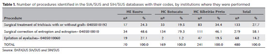
PURPOSE: Trachoma is the major infectious cause of preventable blindness in the world, and its sequelae include the presence of cicatricial entropion and trachomatous trichiasis. Trachoma can be corrected by surgical treatment of the eyelids and, if left untreated, may result in corneal opacification, low vision, and blindness. There are limited data on trachomatous trichiasis in Brazil. This study was conducted to estimate the frequency of entropion and trichiasis surgeries of trachomatous origin based on the records of procedures performed in specialized hospitals that served the Unified Health System (SUS) in the years 2016 and 2017.
METHODS: This was a retrospective study conducted in the oculoplastic sectors of the ophthalmology services of the following three hospitals in the state of São Paulo: Hospital das Clínicas da Faculdade de Medicina de Botucatu (HC Botucatu), Hospital das Clínicas da Faculdade de Medicina de Ribeirão Preto da Universidade de São Paulo (HC Ribeirão Preto), and Hospital Estadual de Bauru (HE Bauru). Medical records corresponding to the codes of interest were evaluated.
RESULTS: In total, 462 medical records were evaluated, including 170 (36.8%) at HC Botucatu, 61 (13.2%) at HE Bauru, and 231 (50.0%) at HC Ribeirão Preto. There were 39 (8.4%) cases of trachomatous trichiasis, ranging from 9 (14.8%) at HE Bauru to 15 (6.5%) at HC Ribeirão Preto.
CONCLUSIONS: The frequency of surgery due to trachoma was low in these oculoplastic services. The state of São Paulo might have reached the goal for trachoma elimination in the surgical component. The questionnaire used for data collection was successfully tested despite some difficulties in collecting data from the medical records. Studies with the same methodology are recommended in other services in the areas of endemic trachoma in the past to understand the frequency of eye lid surgeries performed for treating trachomatous sequelae.
Keywords: Trachoma; Trichiasis; Medical records; Epidemiology; Neglected diseases; Unified Health System; Brazil
Arq. Bras. Oftalmol. 2025;88 (3 )
:1-8
| DOI: 10.5935/0004-2749.2024-0104
Abstract
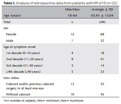
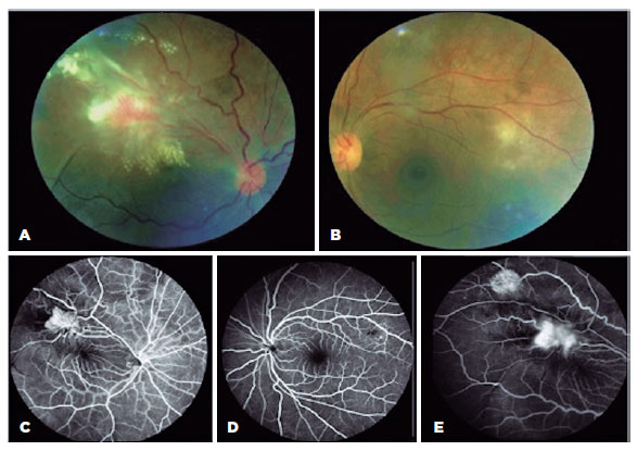
PURPOSE: This study aimed to characterize retinitis pigmentosa associated with the eyes shut homolog gene, which causes hereditary retinal degeneration.
METHODS: The anatomical and functional findings of retinitis pigmentosa in patients with variants of the eyes shut homolog gene were characterized and compared using multimodal imaging and genetic analysis of the variants. Clinical data such as visual acuity, lens status, and refraction were obtained from medical records. Patients underwent an ophthalmic examination, including static visual field, microperimetry, optical coherence tomography, fundus autofluorescence, and fundus photography.
RESULTS: Twenty-two patients were included in the study. Several anatomical and functional characteristics of retinitis pigmentosa-eyes shut homolog were identified, including the presence of cataracts, cystoid macular edema, epiretinal membrane, and a tubular visual field. Genetic results revealed 26 distinct variants in the cohort, with 7 novel variants not previously documented or reported in the scientific literature or databases.
CONCLUSION: The findings demonstrate that eyes shut homolog-retinitis pigmentosa manifests in specific patterns, starting in adolescence with mild progression and advancing with age. The integration of multimodal imaging and genetic analysis has provided a detailed understanding of the anatomical and functional features of retinitis pigmentosa-eyes shut homolog. Seven novel variants of the eyes shut homolog gene have been identified. These findings enhance the understanding of eyes shut homolog-related retinitis pigmentosa characteristics of by detailing the spectrum of mutations in this gene within the Brazilian population.
Keywords: Retinal diseases/diagnostic imaging; Retinitis pigmentosa/genetics; Retinal degeneration; Eye proteins/genetics; Eye diseases, hereditary/genetics; Genes, recessive; Phenotype; Multimodal imaging; Tomography, optical coherence/methods; Fluorescein angiogr
Arq. Bras. Oftalmol. 2024;87 (2 )
:1-6
| DOI: 10.5935/0004-2749.2021-0435
Abstract
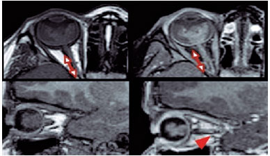
PURPOSE: This study aimed to analyze the association between magnetic resonance imaging apparent diffusion coefficient map value and histopathological differentiation in patients who underwent eye enucleation due to retinoblastomas.
METHODS: An observational chart review study of patients with retinoblastoma that had histopathology of the lesion and orbit magnetic resonance imaging with apparent diffusion coefficient analysis at Hospital de Clínicas de Porto Alegre between November 2013 and November 2016 was performed. The histopathology was reviewed after enucleation. To analyze the difference in apparent diffusion coefficient values between the two major histopathological prognostic groups, Student's t-test was used for the two groups. All statistical analyses were performed using SPSS version 19.0 for Microsoft Windows (SPSS, Inc., Chicago, IL, USA). Our institutional review board approved this retrospective study without obtaining informed consent.
RESULTS: Thirteen children were evaluated, and only eight underwent eye enucleation and were included in the analysis. The others were treated with photocoagulation, embolization, radiotherapy, and chemotherapy and were excluded due to the lack of histopathological results. When compared with histopathology, magnetic resonance imaging demonstrated 100% accuracy in retinoblastoma diagnosis. Optic nerve invasion detection on magnetic resonance imaging showed a 66.6% sensitivity and 80.0% specificity. Positive and negative predictive values were 66.6% and 80.0%, respectively, with an accuracy of 75%. In addition, the mean apparent diffusion coefficient of the eight eyes was 0.615 × 103 mm2/s. The mean apparent diffusion coefficient value of poorly or undifferentiated retinoblastoma and differentiated tumors were 0.520 × 103 mm2/s and 0.774 × 103 mm2/s, respectively.
CONCLUSION: This study revealed that magnetic resonance imaging is useful in the diagnosis of retinoblastoma and detection of optic nerve infiltration, with a sensitivity of 66.6% and specificity of 80%. Our results also showed lower apparent diffusion coefficient values in poorly differentiated retinoblastomas with a mean of 0.520 ×
103 mm2/s, whereas in well and moderately differentiated, the mean was 0.774 × 103 mm2/s.
Keywords: Retinoblastoma; Prognosis; Retinal neoplasms; Orbit; Diffusion magnetic resonance imaging
 ABO is licensed under a Creative Commons Attribution-NonComercial 4.0 Internacional.
02-fig01.jpg)
08-tab01Atb.jpg)