Arq. Bras. Oftalmol. 2025;88 (2 )
:1-7
| DOI: 10.5935/0004-2749.2024-0029
Abstract
PURPOSE: To evaluate the effect of upper eyelid ptosis repair with Muller muscle-conjunctival resection on meibomian gland function and ocular surface parameters.
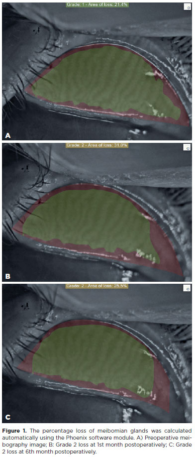
METHODS: Thirty-eight patients who underwent ptosis repair with Muller muscle-conjunctival resection were retrospectively reviewed. Meibomian gland loss, Ocular Surface Disease Index OXFORD score, meiboscore, and noninvasive keratograph break-up time were measured preoperatively and at 1st, 3rd, and 6th months postoperatively.
RESULTS: Noninvasive keratograph break-up time values decreased significantly at 1st and 3rd months postoperatively compared to the preoperative level, but were similar to the preoperative level at 6th months postoperatively (p<0.001 and p=0.628, respectively). Ocular surface disease index, OXFORD score, meibomian gland loss, and meiboscore values increased significantly in the 1st and 3rd postoperative months compared to the preoperative period, but these values decreased to preoperative levels in the 6th postoperative month (p<0.001 and p>0.05, respectively).
CONCLUSION: There is a transient deterioration in meibography findings and OSDI score in the early postoperative period after Muller muscle-conjunctival resection. Patients undergoing Muller muscle-conjunctival resection may require topical lubricants, especially in the first 3 postoperative months.
Keywords: Meibomian glands; Blepharoptosis; Preoperative period; Conjunctiva; Muscles; Eyelid diseases; Diagnostic techniques, ophthalmological
Arq. Bras. Oftalmol. 2026;89 (4 )
:1-6
| DOI: 10.5935/0004-2749.2025-0373
Abstract
PURPOSE: The purpose of this study was to assess the clinical factors associated with dacryocystitis and the need for surgical intervention in infants with congenital dacryocystocele.
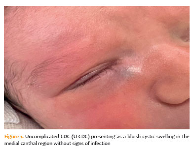
METHODS: This retrospective study included 26 infants diagnosed with congenital dacryocystocele and divided them into two groups: complicated-congenital dacryocystocele (with dacryocystitis or preseptal cellulitis) and uncomplicated-congenital dacryocystocele (without infection). Demographic and perinatal characteristics such as age at diagnosis, gender, birth weight, gestational age, delivery method, and laterality were compared between the groups. For an uncomplicated-congenital dacryocystocele, treatment included conservative management and intravenous antibiotics, followed by probing with intraoperative nasal endoscopy and endonasal marsupialization for a complicated-congenital dacryocystocele.
RESULTS: Of the 26 infants, 14 (53.8%) had complicated-congenital dacryocystocele, while 12 (46.2%) had uncomplicated-congenital dacryocystocele. There were no significant differences between the groups in terms of demographic or perinatal characteristics (p>0.05). Surgical intervention was necessary for all complicated-congenital dacryocystocele cases (100%) and two uncomplicated-congenital dacryocystocele cases (16.7%; p<0.001). During a 6-month median follow-up period, all patients demonstrated complete clinical recovery with no intraoperative complications.
CONCLUSION: In conclusion, approximately half of infants with congenital dacryocystocele developed infection-related complications. While perinatal factors were similar across groups, infectious presentation was linked to the need for surgical intervention. These findings suggest that early detection, prompt conservative management, and close follow-up can help reduce the risk of dacryocystitis and the need for surgery.
Keywords: Dacryocystocele; Dacryocystitis; Lacrimal duct obstruction; Marsupialization; Postoperative complication; Gestational age


 ABO is licensed under a Creative Commons Attribution-NonComercial 4.0 Internacional.