Arq. Bras. Oftalmol. 2025;88 (3 )
:1-8
| DOI: 10.5935/0004-2749.2023-0115
Abstract
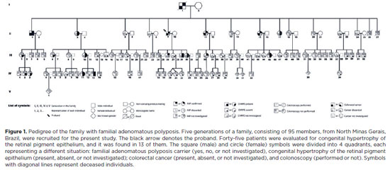
PURPOSE: To evaluate the presence of congenital hypertrophy of the retinal pigment epithelium in a large family affected by familial adenomatous polyposis and identify the causative mutation in the adenomatous polyposis coli gene. Thus, we aimed to determine the significance of congenital hypertrophy of the retinal pigment epithelium as a phenotypic marker of the disease.
METHODS: A family consisting of 95 individuals was evaluated. Among these, 45 individuals were randomly selected by convenience sampling method to undergo ophthalmological evaluation. A funduscopic exam, including slit lamp and indirect ophthalmoscopy, were performed in the selected patients. In those with retinal lesions, a retinography was obtained. The adenomatous polyposis coli gene was sequenced in one affected family member to identify the pathogenic mutation. Once the variant was identified, six undiagnosed family members were tested for the mutation via capillary electrophoresis sequencing.
RESULTS: Congenital hypertrophy of the retinal pigment epithelium was observed in 13 (28.9%) of the 45 individuals evaluated. Of these, nine patients were confirmed to have familial adenomatous polyposis (via colonoscopy or molecular testing). However, four patients had not been investigated. Of the 32 (71.1%) family members without the lesion, 14 did not have familial adenomatous polyposis and 18 were yet to be evaluated. The lesions were bilaterally present and exhibited a peculiar fish-tail shape in all the evaluated individuals. Adenomatous polyposis coli gene sequencing revealed a pathogenic variant c.4031del. (Ser1344*), in heterozygosity (49.27%), in exon 16.
CONCLUSIONS: The study findings confirmed the significance of congenital hypertrophy of the retinal pigment epithelium as a phenotypic marker for familial adenomatous polyposis. Furthermore, it is an effective first-line screening method for at risk family members of such patients. The novel mutation identified in our study participants, which is yet to be described in the literature, causes an aggressive form of the disease.
Keywords: Retinal diseases/congenital; Retinal pigment epithelium; Hypertrophy/congenital; Adenomatous polyposis coli / genetics; Phenotype; Optical coherence tomography
Arq. Bras. Oftalmol. 2024;87 (6 )
:1-7
| DOI: 10.5935/0004-2749.2022-0252
Abstract
Objetivo: Determinar as taxas de fechamento de buracos maculares idiopáticos grandes tratados com vitrectomia posterior e técnica de flap invertido 360 graus pediculado de membrana limitante interna, sem posicionamento de cabeça pós-operatório e definir melhora visual, tipos de fechamento do buraco macular e integridade das camadas retinianas externas como objetivo secundário.
Métodos: Este estudo foi uma série retrospectiva de casos. Todos os pacientes foram submetidos a vitrectomia com flap invertido 360 graus pediculado de membrana limitante interna e tamponamento com gás, sem posição de cabeça no pós-operatório. Idade, gênero, tempo de redução da acuidade visual, outras patologias oculares e status do cristalino foram compilados. Medida de melhor acuidade visual corrigida e tomografia de coerência óptica foram registradas durante as visitas de pré e pós-operatório (15 dias e 2 meses após cirurgia).
Resultados: Vinte olhos de 19 pacientes foram incluídos neste estudo. A idade média foi de sessenta e seis anos. Um total de 19 olhos (95%) atingiu fechamento do buraco, observado através das imagens de tomografia de coerência óptica após 2 meses de cirurgia. Melhor acuidade visual corrigida média aumentou +1,08 pré-operatória para +0,66 LogMAR em 2 meses de cirurgia (p<0,001), com média de 20 letras de melhora visual (0,4 LogMAR) na tabela do Early Treatment Diabetic Retinopathy Study. Dois tipos de fechamento do buraco foram observados: V (47,36%) e U (52,63%).
Conclusão: A técnica de flap invertido 360 graus pediculado de membrana limitante interna, sem posicionamento de cabeça no pós-operatório promoveu elevada taxa de fechamento (95%), reestabelecimento das camadas retinianas externas, fechamento com contorno foveal dos tipos V e U, além de melhora visual na maioria dos casos de BMI grandes (mesmo nos buracos maiores que 650 μm). Esta técnica pode representar uma alternativa para o tratamento de buracos maculares grandes em pacientes impossibilitados de cumprir o tradicional posicionamento de cabeça pós-operatório.
Keywords: Perfurações retinianas; Cuidados pós-operatórios; Vitrectomia; Cirurgia vitreorretiniana
Arq. Bras. Oftalmol. 2026;89 (3 )
:1-14
| DOI: 10.5935/0004-2749.2025-0248
Abstract
PURPOSE: This study aimed to identify barriers to diabetic retinopathy screening among a socioeconomically vulnerable urban population in northeast Brazil.
METHODS: A cross-sectional study was conducted during a diabetic retinopathy screening campaign at primary healthcare units. Ninety-five patients with diabetes underwent retinal examinations and completed a structured interview. Clinical, demographic, and socioeconomic data were collected.
RESULTS: The study population consisted predominantly of older adults (mean age: 60.7 ± 10.5 years), with a high prevalence of type 2 diabetes (99.0%) and low educational attainment. Most participants were economically inactive (81.1%) and reported low income (83.2%). Diabetic retinopathy and maculopathy were highly prevalent, affecting 50.0% and 22.9% of participants, respectively. Longer duration of diabetes was significantly associated with greater awareness of diabetic retinopathy (p=0.035), higher HbA1c levels (p<0.001), and increased prevalence of diabetic retinopathy (p=0.013) and maculopathy (p=0.002). Notably, 33.3% of participants reported difficulties attending medical appointments for diabetes management. In addition, 78.1% experienced challenges scheduling ophthalmologic evaluations, and 76.3% reported that no ophthalmologist was available in their city through the public healthcare system. Financial constraints also limited adherence to recommended dietary practices (90.4%) and impaired glycemic control, with more than half of participants reporting difficulty maintaining target glucose levels.
CONCLUSION: Major barriers to diabetic retinopathy screening included limited awareness of the importance of screening, financial hardship, and transportation challenges. Targeted educational initiatives and structural interventions such as expanded screening programs incorporating telemedicine and subsidized transportation—may improve screening adherence among vulnerable populations.
Keywords: Diabetic retinopathy; Mass screening; Health services accessibility; Health knowledge, attitudes, practices; Socioeconomic factors
 ABO is licensed under a Creative Commons Attribution-NonComercial 4.0 Internacional.
04-fig01tb.jpg)