Arq. Bras. Oftalmol. 2017;80 (1 )
:41-45
| DOI: 10.5935/0004-2749.20170011
Abstract
Objetivo: Determinar os resultados da ceratoplastia penetrante (PK) para o tratamento da cicatriz da córnea consequente à ceratite por Herpes simplex vírus (HSV), e se o tipo de cicatriz na córnea afeta o resultado cirúrgico. Métodos: Foi realizada análise retrospectiva dos pacientes, submetidos à PK para a cicatriz da córnea relacionados com o HSV entre janeiro de 2008 e julho de 2011. Os pacientes foram divididos em dois grupos. Grupo 1 consistiu de pacientes que tiveram cicatriz corneana herpética quiescente e grupo 2 consistiu de pacientes que desenvolveram descemetocele ou perfuração córnea secundária a defeitos epiteliais persistentes sem inflamação estromal ativa. O seguimento médio foi de 21,30 ± 14,59 meses. Os principais parâmetros avaliados foram recorrência de ceratite herpética, rejeição de enxerto, falência do enxerto, acuidade visual e taxa de sobrevida do enxerto. Resultados: Foram avaliados 42 pacientes do grupo 1 e 13 doentes do grupo 2. Acuidade visual pré-operatória variou de movimentos das mãos (HM) para 0,7 logMAR. No pós-operatório, 34 pacientes (61,8%) atingiram acuidade visual de 0,6 logMAR ou melhor. Doze olhos (28,57%) tiveram recorrência de HSV ceratite no grupo 1, e quatro olhos (30,76%) tiveram recorrência no grupo 2 (p=0,40). A rejeição do enxerto ocorreu em 4 olhos (9,52%) no grupo 1, e em 3 olhos do grupo 2 (23,07%; p=0,58), taxa de sobrevivência do enxerto foi de 91,9% a 1 ano, 76,0% aos 2 anos e 65,1% aos 3 anos no grupo 1, e 89,5% a 1 ano, 76,0% aos 2 anos e 63,6% aos 3 anos no grupo 2 (p=0,91). Conclusões: Embora diferentes taxas de recorrência e de rejeição do enxerto foram encontradas nos dois grupos, a taxa de sobrevida do enxerto em 3 anos foi semelhantes nos dois grupos. De acordo com nossos resultados, em casos sem inflamação, a cicatriz herpética da córnea com descemetocele ou perfuração demonstra as taxas de sobrevivência do enxerto semelhantes às da cicatriz corneana herpética quiescente.
Keywords: Aciclovir/uso terapêutico; Ceratite herpética; Ceratoplastia penetrante; Córnea; Sobrevivência de enxerto
Arq. Bras. Oftalmol. 2025;88 (2 )
:1-6
| DOI: 10.5935/0004-2749.2023-0229
Abstract
PURPOSE: To compare the outcomes of intravitreal dexamethasone implant used as either an adjuvant or a switching therapy for diabetic macular edema in patients with poor anatomic response after three consecutive monthly injections of ranibizumab.
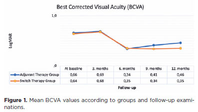
METHODS: This retrospective study included patients with diabetic macular edema who received three consecutive doses of ranibizumab as initial therapy and demonstrated poor response. A single dose of intravitreal dexamethasone implant was administered to these patients. The patients were divided into two groups according to the treatment modalities: the adjuvant therapy group, consisting of patients who continued treatment with ranibizumab injection after receiving intravitreal dexamethasone implant, and the switch therapy group, consisting of patients who were switched from ranibizumab treatment to intravitreal dexamethasone implant as needed. The main outcome measurements were best corrected visual acuity and central retinal thickness at baseline and at 3, 6, 9, and 12 months of follow-up.
RESULTS: In this study that included 64 eyes of 64 patients, the best corrected visual acuity and central retinal thickness values did not significantly differ between the groups at baseline and at 6 months of follow-up (p>0.05). However, at 12 months, the best corrected visual acuity values in the adjuvant and switch therapy groups were 0.46 and 0.35 LogMAR, respectively (p=0.012), and the central retinal thickness values were 344.8 and 270.9, respectively (p=0.007).
CONCLUSIONS: In a real-world setting, it seems more reasonable to use intravitreal dexamethasone implant as a switch therapy rather than an adjuvant therapy for diabetic macula edema refractory to ranibizumab despite three consecutive monthly injections of ranibizumab. Patients switched to intravitreal dexamethasone implant were found to have better anatomic and visual outcomes at 12 months than those who continued ranibizumab therapy despite their less-than-optimal responses.
Keywords: Diabetic retinopathy; Macular edema/drug therapy; Dexamethasone/administration & dosage; Drug implants; Intravitreal injections; Ranibizumab/administration & dosage; Tomography, optical coherence; Endothelial growth factors
 ABO is licensed under a Creative Commons Attribution-NonComercial 4.0 Internacional.