

Acacio Lima Filho2; Paulo Schor1; Maria Cecília Zorat Yu1; Ana Luisa Hofling de Lima1
DOI: 10.1590/S0004-27491999000600018
ABSTRACT
We describe a new methods to produce an instrument to scrap the cornea. It is made from either a fine needle or a cannula. The material is flattened by means of a laminator or a hammer and a flat metal surface. A sandpaper is used to rough the front end and round it. The tip is slightly bent and the device is submitted to ethylene oxide for sterilization. A handle may be used to hold the instrument, which may be reusable, after washing and sterilization using local heat at the tip, or gas.
Keywords: Spatula; Microbiology; Diagnosis; Bioenginee-ring; Cornea
RESUMO
Os autores descrevem um novo método para produzir um instrumento para colheita de material de córnea. Tal instrumento pode ser manufaturado com uma cânula ou agulha fina. O material é moldado de forma plana com o auxílio de um laminador, ou de um martelo e placa de metal. Lixa fina é utilizada para arredondar as bordas e a ponta é ligeiramente dobrada. Um cabo pode ser adicionado de modo a facilitar a manipulação do instrumento, que pode ser reutilizado após lavagem e esterilização local na ponta do metal, ou por óxido de etileno.
Descritores: Espátula; Microbiologia; Diagnóstico; Bioengenharia; Córnea
INTRODUCTION
Spatulas are used to scrap the corneal stroma and epithelium, specially in infectious diagnostic procedures. They need to be flexible, small and reusable instruments, useful to produce cultures, and direct examinations of fresh material extracted from (mainly) an infected lesion at the corneal surface. These devices were made of platinum, carved into, a thin and curve shape. Heat may be applied for the sterilization of these devices. Since the original concept of Kimura's spatula 1, 2, few modifications were added to the device and technique. In fact the material was replaced by an ordinary metal (i.e. iron), that often degrades after several heatings, leading to a dirty smear. In addition, this metal did not have the useful flexibility of platinum.
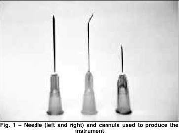
With the advent of less expensive resistant disposable materials, i.e. stainless steel needles and cannulas ( Figure 1), it is possible to manufacture a new instrument, that preserves the classic concepts, that is, being flexible, thin, small and reusable.
METHODS
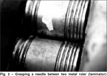
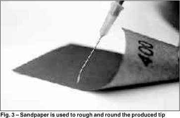
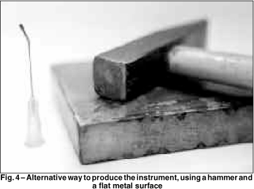
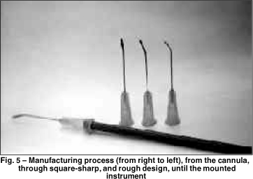
Two ways of producing the device are presented: with the aid of a laminator, or a hammer and a metal surface. The first consists of grasping the needle (or cannula) between two flat hard metal rollers ( Figure 2). It is rolled half an inch and rolled back. The needle is therefore flattened. Sandpaper is gently applied to the sharp tip (of the needle) to rough and round it ( Figure 3). The tip is bent to the desired angle, and sent to gas sterilization. The second way replaces the laminator with a hammer and a flat metal surface ( Figure 4). The needle is successively hit to produce a flat surface. Sandpaper and oxide sterilization follow in the same manner as described above. A handle may be used to ease the manipulation. Figure 5 shows the whole process from the cannula to the mounted instrument.
RESULTS
Although the sharper quality of the new device might interfere in the spreading of the material, and care must be taken not to cut the agar, this limitation did not interfere with the culture results. Moreover the microscopic smear appearance was similar to current technique.
DISCUSSION
We have been using this instrument for almost 2 years in the Ophthalmology Department of the Federal University of São Paulo and the Ophthalmos Microbiology Laboratory. Several hundred corneal lesions were cultured and submitted to direct observation, resulting in adequate smears.
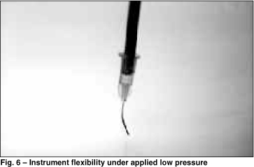
The devices are being sent to satellite laboratories and offices, that may scrap the cornea at a low coast, and due to the easy availability and physical properties (i.e. flexibility and delicate manipulation ¾ Figure 6), it may be used to remove foreign bodies, keratoconus scars, scrap the epithelium before PRK and lift the LASIK flap.
REFERENCES
1. Hyndiuk RA, Seideman S. Clinical and laboratory techniques in external ocular disease. In: HB Fedukowicz (ed.), External Infections of the Eye (2nd ed.). New York: Appleton-Century-Crofts chapter 5, 1978.
2. Kimura SJ, Thygeson MD. The cytology of external ocular disease. Am J Ophthalmol 1955;39:137-45.
From the Bioengineering ( 1) and Laboratory ( 2) Sections - Ophthalmology Department - Federal University of São Paulo, and Ophthalmos Microbiology Laboratory ( 3).
Endereço para correspondência: Acacio Lima Filho - Universidade Federal de São Paulo - UNIFESP - Departamento de Oftalmologia - Rua Botucatú, 822 - CEP 04023-062 - São Paulo-SP.
© 2024 - All rights reserved - Conselho Brasileiro de Oftalmologia
![]()
 English PDF
 Print
Print
 Send this article by email
 How to cite this article
 Submit a comment
 Mendeley
 Scielo
 Pocket
