

Cíntia Maria Felix Medrado Parcero1; Bruno de Paula Freitas1; Eduardo Ferrari Marback1; Otacílio de Oliveira Maia Júnior1; Roberto Lorens Marback1
DOI: 10.1590/S0004-27492010000200019
ABSTRACT
PURPOSE: to report a case of branch retinal artery occlusion in the acute phase, using optical coherence tomography to evaluate the morphologic changes. A 27 year-old man had a sudden superior scotoma in the right eye, with fundus examination compatible with inferior temporal branch retinal artery obstruction. The optical coherence tomography revealed increase in thickness and hyper-reflectivity of the inner retinal layers in affected area, with decreased reflectivity of photoreceptor and retinal pigment epithelial layers. The optical coherence tomography findings are consistent with intracellular edema, and not with secondary vascular leakage of extracellular fluid, according to histopathological theories of retinal ischemia and necrosis that occurs after retinal artery occlusion.
Keywords: Retinal artery; Retinal artery occlusion; Retinal vessels; Visual acuity; Optical coherence tomography; Human; Male; Adult; Case reports
RESUMO
O objetivo é demonstrar alterações morfológicas retinianas por meio da tomografia de coerência óptica na oclusão de ramo de artéria central da retina na fase aguda. Homem de 27 anos apresentando escotoma súbito no campo superior de olho direito, com exame fundoscópico compatível com oclusão de ramo ínfero-temporal da artéria central da retina. A tomografia de coerência óptica revelou aumento da espessura e hiperrefletividade das camadas internas da retina, com redução da refletividade das camadas de fotorreceptores e epitélio pigmentar da retina. Os achados na tomografia de coerência óptica são compatíveis com edema intracelular, e não com fluido extracelular secundário a extravasamento vascular, reforçando teorias histopatológicas da isquemia e necrose retiniana, que ocorrem após oclusão arterial retiniana.
Descritores: Artéria retiniana; Oclusão da artéria retiniana; Vasos retinianos; Acuidade visual; Tomografia de coerência óptica; Humanos; Masculino; Adulto; Relatos de casos
RELATOS DE CASOS
Optical coherence tomography findings in acute phase of branch retinal artery occlusion: case report
Achados da tomografia de coerência óptica na fase aguda da oclusão de ramo da artéria central da retina: relato de caso
Cíntia Maria Felix Medrado ParceroI; Bruno de Paula FreitasII; Eduardo Ferrari MarbackIII; Otacílio de Oliveira Maia JúniorIV; Roberto Lorens MarbackV
IRetina Fellow of the Department of Ophthalmology, São Rafael Hospital, Monte Tabor Foundation, Salvador (BA) - Brazil
IIRetina Fellow of the Department of Ophthalmology, São Rafael Hospital, Monte Tabor Foundation, Salvador (BA) - Brazil
IIIOphthalmic Pathology, and Cataract Specialist of the Department of Ophthalmology, São Rafael Hospital, Monte Tabor Foundation, Salvador (BA) - Brazil. Doctorate of the Department of Ophthalmology, Federal University of São Paulo - UNIFESP - São Paulo (SP) -Brazil
IVVitreoretinal Specialist of the Department of Ophthalmology, São Rafael Hospital, Monte Tabor Foundation, Salvador (BA) - Brazil. Posdoctorate of the Department of Ophthalmology, São Paulo University - USP - São Paulo (SP) - Brazil
VHead of the Department of Ophthalmology, São Rafael Hospital, Monte Tabor Foundation, Salvador (BA) -Brazil. Chairman of the Department of Ophthalmology, Federal University of Bahia - UFBA - Salvador (BA) -Brazil
ABSTRACT
PURPOSE: to report a case of branch retinal artery occlusion in the acute phase, using optical coherence tomography to evaluate the morphologic changes. A 27 year-old man had a sudden superior scotoma in the right eye, with fundus examination compatible with inferior temporal branch retinal artery obstruction. The optical coherence tomography revealed increase in thickness and hyper-reflectivity of the inner retinal layers in affected area, with decreased reflectivity of photoreceptor and retinal pigment epithelial layers. The optical coherence tomography findings are consistent with intracellular edema, and not with secondary vascular leakage of extracellular fluid, according to histopathological theories of retinal ischemia and necrosis that occurs after retinal artery occlusion.
Keywords: Retinal artery/abnormalities; Retinal artery occlusion; Retinal vessels; Visual acuity; Optical coherence tomography; Human; Male; Adult; Case reports
RESUMO
O objetivo é demonstrar alterações morfológicas retinianas por meio da tomografia de coerência óptica na oclusão de ramo de artéria central da retina na fase aguda. Homem de 27 anos apresentando escotoma súbito no campo superior de olho direito, com exame fundoscópico compatível com oclusão de ramo ínfero-temporal da artéria central da retina. A tomografia de coerência óptica revelou aumento da espessura e hiperrefletividade das camadas internas da retina, com redução da refletividade das camadas de fotorreceptores e epitélio pigmentar da retina. Os achados na tomografia de coerência óptica são compatíveis com edema intracelular, e não com fluido extracelular secundário a extravasamento vascular, reforçando teorias histopatológicas da isquemia e necrose retiniana, que ocorrem após oclusão arterial retiniana.
Descritores: Artéria retiniana/anormalidades; Oclusão da artéria retiniana; Vasos retinianos; Acuidade visual; Tomografia de coerência óptica; Humanos; Masculino; Adulto; Relatos de casos
INTRODUCTION
Acute retinal arterial obstruction usually occurs with a sudden, painless unilateral loss of vision and/or visual field defect that can involve the central retinal artery or an arterial branch, most commonly secondary to arterial occlusive disease in older adults(1). Retinal arterial occlusions in young adults occurs as result of diverse group of pathologic conditions. In contrast to retinal artery occlusion in the elderly, when the commonest mechanism of occlusion involves embolic disease from the carotid artery, cardiac embolic sources and many other hematological diseases leading to thrombosis seems to be more common in younger patients(1).
Branch retinal artery occlusion (BRAO) occurs when the emboli lodges in a more distal branch of the retinal artery, causing inner retinal ischemia and leading to permanent inner retinal dysfunction and loss of only a portion of the visual field in the area of supply of the artery(2).
Optical coherence tomography (OCT) allows in vivo images visualization that matches retinal (histological) structures, as well as its morphological changes, including macular diseases(3). This case report describes OCT findings in the acute phase of BRAO in a young adult.
CASE REPORT
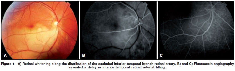
A 27 year-old man had a sudden superior scotoma in the right eye for 3 days. His best-corrected visual acuity was 20/20 in both eyes. Slit-lamp examination and intraocular pressure were normal in both eyes. Fundus examination of the right eye was notable for retinal whitening and associated edema along the distribution of the inferior temporal branch retinal artery (Figure 1), without evidence of embolic material in the retinal arterioles. Ophthalmologic examination of the left eye was unremarkable. The left eye was normal. Fluorescein angiography revealed a delay in inferior temporal retinal arterial filling compatible with inferior temporal branch retinal artery occlusion (Figure 1).
The patient underwent clinical investigation, transthoracic echocardiography and carotid dupplex scan, but no risk factors and systemic diseases were identified.
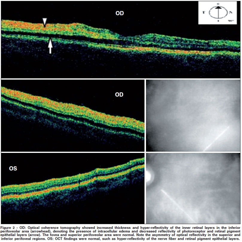
In the acute phase, the patient underwent scanning by the optical coherence tomography (Stratus OCTTM, Model 3000, Version 4.0.1, Carl Zeiss Ophthalmic System Inc. Humphrey Division. Dublin, CA, USA), using the line scan protocol (6.0 mm) in the macular area and in the inferior branch retinal artery area. OCT revealed increased thickness and hyper-reflectivity of the inner retinal layers in the inferior perifoveolar area, denoting the presence of intracellular edema, with decreased reflectivity of photoreceptor and retinal pigment epithelial layers. The asymmetry of optical reflectivity in perifoveal region was an important finding (Figure 2). OCT findings in the fovea and superior perifoveolar area in the right eye and fundus in the fellow eye were normal.
DISCUSSION
In retinal artery occlusive diseases, the central retinal artery is affected in 57% of occlusions, the branch retinal artery is involved in 38% of occlusions, and cilioretinal artery obstructions occur in 5% of occlusions(4). The visual prognosis is substantially better with branch retinal artery obstruction than with central retinal artery obstruction(2).
The high prevalence of underlying systemic disease in young patients with retinal arterial occlusion requires a thorough, aggressive examination to rule out potential lifethreatening embolic and hypercoagulable conditions, induced by various factors such as migraines, deficiency of protein C or S, antiphospholipid antibody syndromes, Susac syndrome, cigarette smoking, pregnancy, and oral contraceptives(1,5). The present case had no associated risk factor or systemic complications identified. However, clinical segment is necessary to early diagnosis of further complications.
Retinal artery occlusion causes ischemia of the inner layers of the retina, leading to intracellular edema as a result of cellular injury and ischemic necrosis. This intracellular edema in a branch retinal artery obstruction has the ophthalmoscopic appearance as a localized region of superficial retinal whitening. The whitening is most prominent in the posterior pole, along the distribution of the obstructed vessel(2).
Electroretinography typically discloses a decrease in the amplitude of the b-wave (corresponding to the function of the Muller and/or bipolar cells) secondary to inner layer retinal isquemia. The a-wave, which corresponds to photoreceptor function, is generally unaffected(2). These findings are compatible with the inner retinal layers injury observed through the optical coherence tomography.
Because central retinal artery occlusion may be rarely followed by immediate enucleation, there is little opportunity to study the histopatology of the acute changes in these vessels and in the retinal layers before such changes are obscured by the more chronic processes of scarring and fibrosis(6). In 1965, Dahrling described diffuse edema of the inner retinal layers with marked ''cloudy swelling'' shown by the ganglion cells(6). OCT findings as increased thickness and hyper-reflectivity of the inner retinal layers in the affected area confirm the presence of intracellular edema as acute histopathological changes of retinal ischemia secondary to occlusion of retinal artery(6). Hypo-reflective cystic spaces in the retinal structure were not observed, showing this whitening is due to ischemia and not to extracellular edema secondary to retinal capillary leakage. These findings are consistent with those reported in other studies(7-10). In the present case, although the OCT showed that inferior perifoveal area was affected (Figure 2), the visual acuity remained 20/20 surprisingly. One of the advantages of the documentation of these findings by OCT in the present case of branch retinal artery occlusion is exactly this possibility of comparison of the asymmetry in the same OCT cross-section of macular area showing both the area affected by the branch occlusion and normal area (Figure 2).
In conclusion, optical coherence tomography findings reported in this patient are consistent with intracellular edema, and not with extracellular fluid secondary to vascular leakage, according to histopathological theories of retinal ischemia and necrosis that occurs after retinal artery occlusion.
REFERENCES
1. Greven CM, Slusher MM, Weaver RG. Retinal arterial occlusions in young adults. Am J Ophthalmol. 1995;120(6):776-83. Comment in: Am J Ophthalmol. 1996;122(1):134. Am J Ophthalmol. 1996;122(1):134-6.
2. Ryan SJ, Schachat AP, Wilkinson P, Hinton DR, editors. Retina. Philadelphia: Mosby; 2006.
3. Sander B, Larsen M, Thrane L, Hougaard JL, Jørgensen TM. Enhanced optical coherence tomography imaging by multiple scan averaging. Br J Ophthalmol. 2005;89(2):207-12.
4. Brown GC, Reber R. An unusual presentation of branch retinal artery obstruction in association with ocular neovascularization. Can J Ophthalmol. 1986; 21(3):103-6.
5. Brown GC, Margargal LE, Shields JA, Goldberg RE, Walsh PN. Retinal arterial obstruction in children and young adults. Ophthalmology. 1981; 88(1):18-25.
6. Dahrling BE 2nd. The histopathology of early central retinal artery occlusion. Arch Ophthalmol. 1965;73:506-10.
7. Karacorlu M, Ozdemir H, Karacorlu SA. Optical Coherence Tomography findings in branch retinal artery occlusion Eur J of Ophthalmol. 2006:352-3.
8. Asefzadeh B, Ninyo K. Longitudinal analysis of retinal changes after branch retinal artery occlusion using optical coherence tomography. Optometry. 2008; 79(2):85-9.
9. Cella W, Avila M. Optical coherence tomography as a means of evaluating acute ischaemic retinopathy in branch retinal artery occlusion. Acta Ophthalmol Scand. 2007;85(7):799-801.
10. Falkenberry SM, Ip MS, Blodi BA, Gunther JB. Optical coherence tomography findings in central retinal artery occlusion. Ophthalmic Surg Lasers Imaging. 2006;37(6):502-5.
 Correspondence address:
Otacilio O. Maia Jr. Retina
Vitreous Service, Department of Ophthalmology
São Rafael Hospital - Monte Tabor Foundation
Av. São Rafael - 2.142 - Salvador (BA)
CEP 41253-190
E-mail: otacilio.maia@hsr.com.br
Recebido para publicação em 31.05.2009
Última versão recebida em 20.08.2009
Aprovação em 08.10.2009
From the Retina and Vitreous Service, São Rafael Hospital - Monte Tabor Foundation - Salvador (BA) - Brazil.
© 2025 - All rights reserved - Conselho Brasileiro de Oftalmologia
![]()
 English PDF
 Print
Print
 Send this article by email
 How to cite this article
 Submit a comment
 Mendeley
 Scielo
 Pocket
